Wojtasik Magdalena, Bludau Sebastian, Eickhoff Simon B, Mohlberg Hartmut, Gerboga Fatma, Caspers Svenja, Amunts Katrin
Cécile and Oskar Vogt-Institute for Brain Research, Medical Faculty, Heinrich-Heine-University Düsseldorf, Düsseldorf, Germany.
Institute of Neuroscience and Medicine 1 (INM-1), Research Center Jülich, Jülich, Germany.
Front Neuroanat. 2020 Feb 5;14:2. doi: 10.3389/fnana.2020.00002. eCollection 2020.
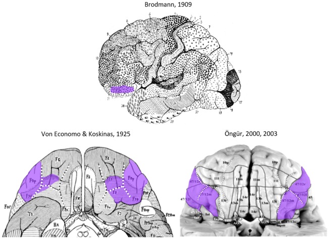
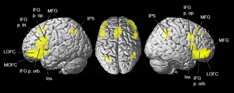
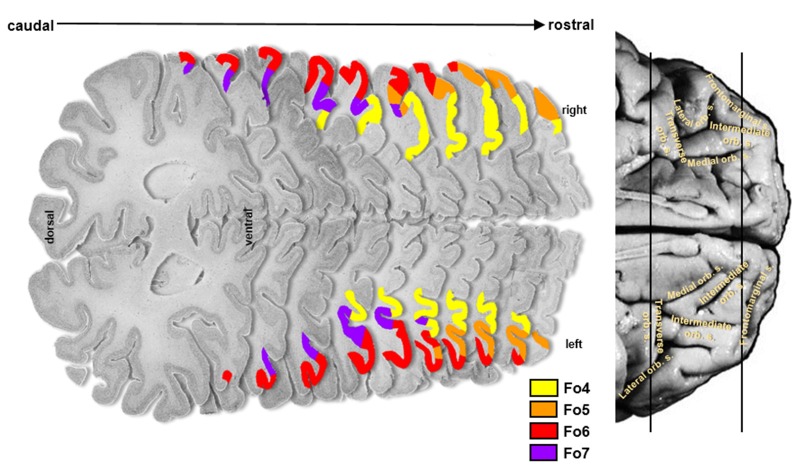
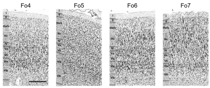
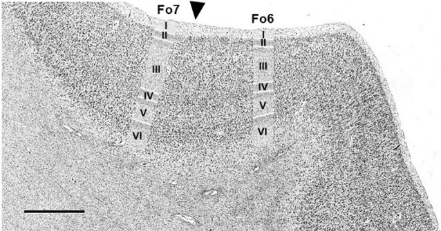
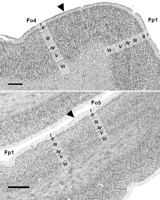
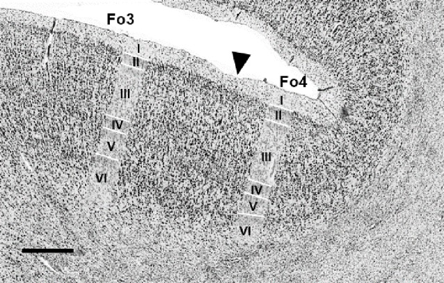
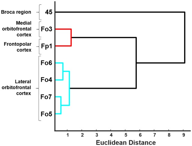
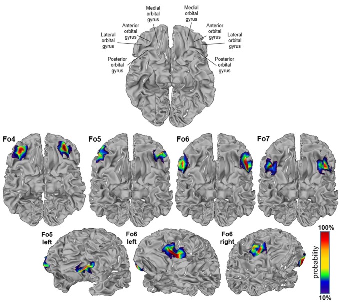
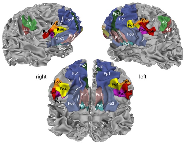
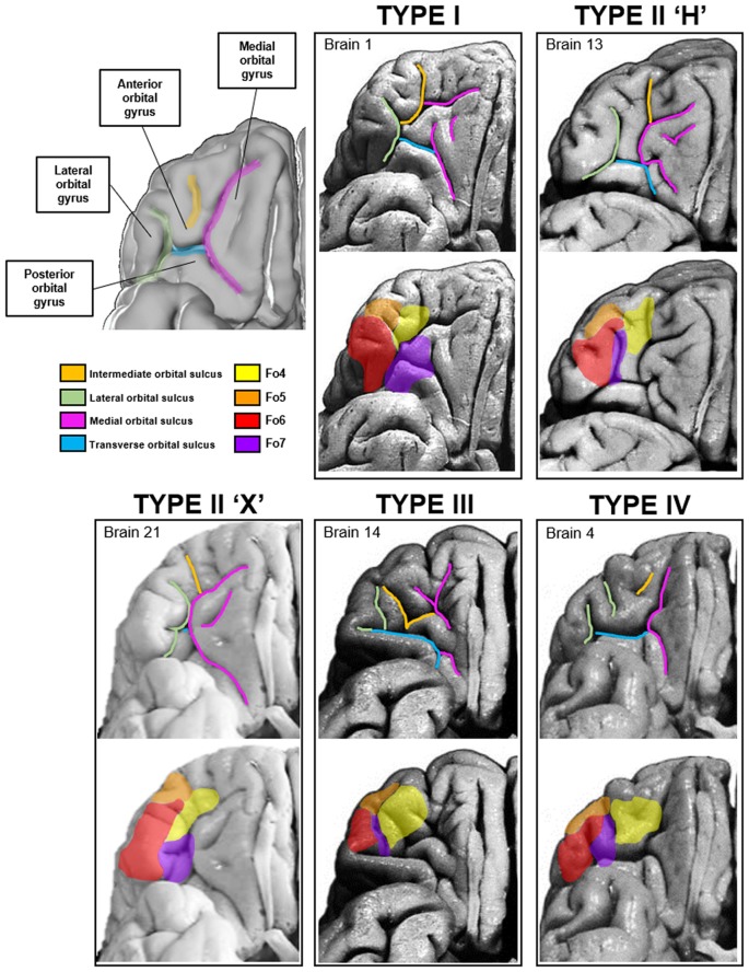
A comprehensive concept of the biological basis of reward, social and emotional behavior, and language requires a deeper understanding of the microstructure and connectivity of the underlying brain regions. Such understanding could provide deeper insights into their role in functional networks, and form the anatomical basis of the functional segregation of this region as shown in recent imaging studies. Here, we investigated the cytoarchitecture of the lateral orbitofrontal cortex (lateral OFC) in serial histological sections of 10 human postmortem brains by image analysis and a statistically reproducible approach to detect borders between cortical areas. Profiles of the volume fraction of cell bodies were therefore extracted from digitized histological images, describing laminar changes from the layer I/layer II boundary to the white matter. As a result, four new areas, Fo4-7, were identified. Area Fo4 was mainly found in the anterior orbital gyrus (AOG), Fo5 anteriorly in the inferior frontal gyrus (IFG), Fo6 in the lateral orbital gyrus (LOG), and Fo7 in the lateral orbital sulcus. Areas differed in cortical thickness, abundance and size of pyramidal cells in layer III and degree of granularity in layer IV. A hierarchical cluster analysis was used to quantify cytoarchitectonic differences between them. The 3D-reconstructed areas were transformed into the single-subject template of the Montreal Neurological Institute (MNI), where probabilistic maps and a maximum probability map (MPM) were calculated as part of the JuBrain Cytoarchitectonic Atlas. These maps served as reference data to study the functional properties of the areas using the BrainMap database. The type of behavioral tasks that activated them was analyzed to get first insights of co-activation patterns of the lateral OFC and its contribution to cognitive networks. Meta-analytic connectivity modeling (MACM) showed that functional decoding revealed activation in gustatory perception in Fo4; reward and somesthetic perception in Fo5; semantic processing and pain perception in Fo6; and emotional processing and covert reading in Fo7. Together with existing maps of the JuBrain Cytoarchitectonic Atlas, the new maps can now be used as an open-source reference for neuroimaging studies, allowing to further decode brain function.
要全面理解奖赏、社会和情感行为以及语言的生物学基础,需要更深入地了解相关脑区的微观结构和连接性。这种理解能够更深入地洞察它们在功能网络中的作用,并形成该区域功能分离的解剖学基础,正如最近的影像学研究所显示的那样。在此,我们通过图像分析和一种具有统计学可重复性的方法来检测皮质区域之间的边界,对10例人类尸检大脑的连续组织学切片中的外侧眶额皮质(外侧OFC)的细胞结构进行了研究。因此,从数字化的组织学图像中提取了细胞体体积分数的剖面图,描述了从第I层/第II层边界到白质的分层变化。结果,确定了四个新区域,即Fo4 - 7。区域Fo4主要位于眶前回(AOG),Fo5位于额下回(IFG)前部,Fo6位于眶外侧回(LOG),Fo7位于外侧眶沟。这些区域在皮质厚度、第III层锥体细胞的丰度和大小以及第IV层的颗粒度方面存在差异。使用层次聚类分析来量化它们之间的细胞结构差异。将三维重建的区域转换为蒙特利尔神经病学研究所(MNI)的单受试者模板,在该模板中,作为JuBrain细胞结构图谱的一部分,计算了概率图谱和最大概率图谱(MPM)。这些图谱作为参考数据,用于使用BrainMap数据库研究这些区域的功能特性。分析激活它们的行为任务类型,以初步了解外侧OFC的共激活模式及其对认知网络的贡献。元分析连接建模(MACM)表明,功能解码显示Fo4在味觉感知中激活;Fo5在奖赏和躯体感觉感知中激活;Fo6在语义处理和疼痛感知中激活;Fo7在情绪处理和隐蔽阅读中激活。与JuBrain细胞结构图谱的现有图谱一起,新图谱现在可作为神经影像学研究的开源参考,有助于进一步解码脑功能。