Laboratories of Animal Pathology, Department of Veterinary Preventive Medicine, Universidade Estadual de Londrina, Paraná, Brazil.
Tissue Processing Department of Veterinary Preventive Medicine, Universidade Estadual de Londrina, Paraná, Brazil.
Sci Rep. 2018 Sep 7;8(1):13477. doi: 10.1038/s41598-018-31540-0.
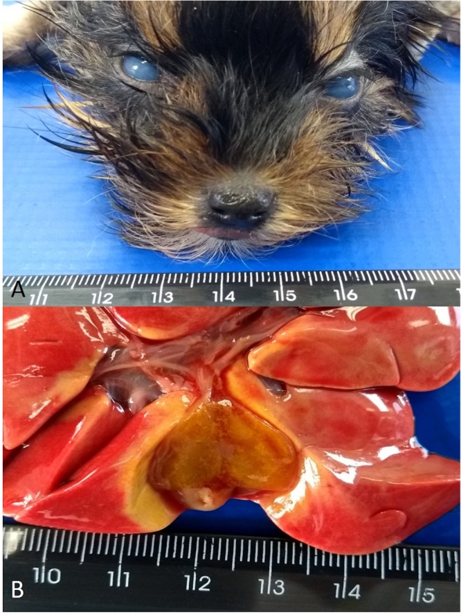
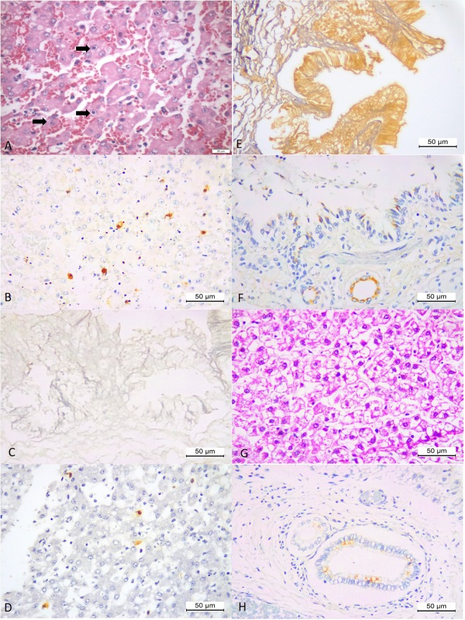
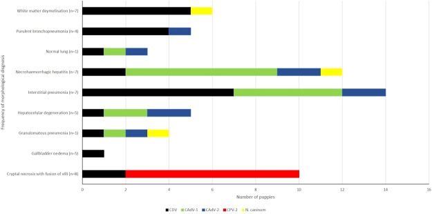
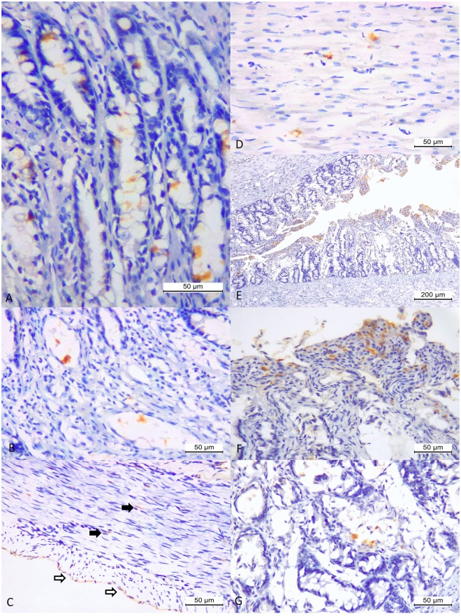
A retrospective immunohistochemical study was designed to investigate the frequency of concomitant traditional infectious disease pathogens in puppies that died suddenly and review the aspects of associated pathogenesis. Fifteen puppies were evaluated; the pathology reports and histopathologic slides of these animals were reviewed to determine the pattern of histopathologic lesions. The intralesional identification of antigens of canine (distemper) morbillivirus (CDV), canine adenovirus-1 and -2 (CAdV-1 and -2), canine parvovirus-2 (CPV-2), Toxoplasma gondii, and Neospora caninum was evaluated by IHC within the histopathologic patterns observed. All puppies contained CDV nucleic acid by molecular testing. The most frequent histopathologic patterns were intestinal crypt necrosis (n = 8), white matter cerebellar demyelination (n = 7), necrohaemorrhagic hepatitis (n = 7), interstitial pneumonia (n = 7), and gallbladder oedema (n = 5). All puppies contained intralesional antigens of CDV in multiple tissues resulting in singular (n = 3), and concomitant dual (n = 3), triple (n = 5) and quadruple (n = 4) infections by CAdV-1, and -2, CPV-2, and N. caninum; T. gondii was not identified. Concomitant infections by CDV was observed with N. caninum (100%; 1/1), CPV-2 (100%; 8/8), CAdV-1 (100%; 8/8), and CAdV-2 (100%; 8/8). Intralesional antigens of CDV and not CAdV-1 were identified in cases of gallbladder oedema. The "blue eye" phenomenon was histologically characterized by corneal oedema and degenerative lesions to the corneal epithelium, without inflammatory reactions.
一项回顾性免疫组织化学研究旨在调查幼犬突然死亡时同时存在传统传染病病原体的频率,并回顾相关发病机制的各个方面。评估了 15 只幼犬;回顾这些动物的病理学报告和组织病理学切片,以确定组织病理学病变模式。通过免疫组织化学在观察到的组织病理学模式内评估犬(麻疹)副粘病毒(CDV)、犬腺病毒-1 和 -2(CAdV-1 和 -2)、犬细小病毒-2(CPV-2)、刚地弓形虫和新孢子虫的抗原的体内鉴定。所有幼犬的分子检测均含有 CDV 核酸。最常见的组织病理学模式是肠隐窝坏死(n = 8)、小脑白质脱髓鞘(n = 7)、坏死性出血性肝炎(n = 7)、间质性肺炎(n = 7)和胆囊水肿(n = 5)。所有幼犬的多个组织中均存在 CDV 抗原,导致单一感染(n = 3)、双重感染(n = 3)、三重感染(n = 5)和四重感染(n = 4),由 CAdV-1 和 -2、CPV-2 和 N. caninum 引起;未鉴定出刚地弓形虫。观察到 CDV 与 N. caninum(100%;1/1)、CPV-2(100%;8/8)、CAdV-1(100%;8/8)和 CAdV-2(100%;8/8)同时感染。在胆囊水肿的情况下,鉴定出 CDV 的体内抗原,而不是 CAdV-1。“蓝眼病”现象的组织学特征是角膜水肿和角膜上皮退行性病变,无炎症反应。