Garnavos Christos
Orthopaedic Department, "Evangelismos" General Hospital, Athens, Greece.
JB JS Open Access. 2017 Apr 18;2(2):e0017. doi: 10.2106/JBJS.OA.16.00017. eCollection 2017 Jun 26.
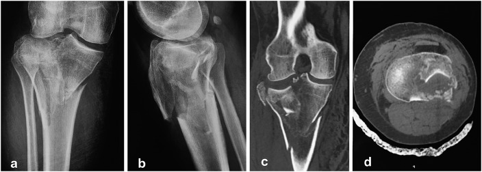
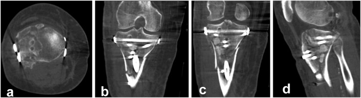
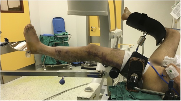
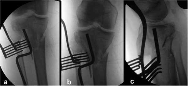
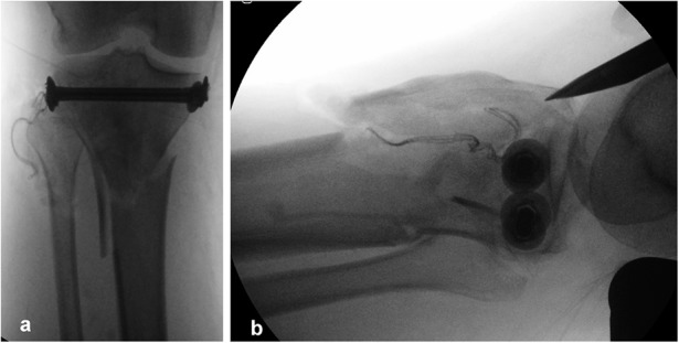
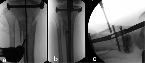
Bicondylar tibial plateau fractures have been treated with either plating or external fixation techniques, with conflicting results. A recently introduced technique involving the combined use of intramedullary nailing via a suprapatellar approach and condylar bolts could represent a new pathway toward better treatment of this severe injury.
The present report describes a retrospective and prospective study of all 17 patients (age range, 25 to 75 years) who were admitted under the author's care for the treatment of a closed, bicondylar tibial plateau fracture between 2013 and 2015. All patients consented to undergo fixation of the fracture with intramedullary nailing through a suprapatellar approach and with use of condylar bolts. The reconstructed articular surface was supported with freeze-dried allograft that had been previously soaked in concentrated bone marrow. The patients were followed at regular intervals, and the results were assessed with the Knee injury and Osteoarthritis Outcome Score (KOOS).
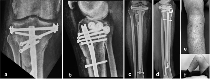
All patients were followed for at least 1 year (average and standard deviation, 25.23 ± 8.95 months; range, 12 to 46 months). All fractures united clinically and radiographically between 10 and 22 weeks (average, 15.1 ± 2.91 weeks), with no instances of neurovascular complication, infection, or implant failure. One patient underwent early revision of the fixation because of unsatisfactory reduction of the articular surface, and 1 patient had secondary fracture displacement. One condylar bolt was removed after fracture healing because of irritation at the insertion site. However, all patients regained knee motion without physiotherapy and all were fully weight-bearing by the fifth postoperative month.
The short and intermediate-term results associated with the use of the proposed technique appear to be satisfactory. However, the effectiveness of the technique should be reassessed with long-term studies as well as comparative studies involving other fixation techniques.
Therapeutic Level IV. See Instructions for Authors for a complete description of levels of evidence.
双髁胫骨平台骨折的治疗方法有钢板固定或外固定技术,但结果存在争议。最近引入的一种技术,即通过髌上入路联合使用髓内钉和髁螺栓,可能为更好地治疗这种严重损伤开辟一条新途径。
本报告描述了一项回顾性和前瞻性研究,研究对象为2013年至2015年间在作者治疗下因闭合性双髁胫骨平台骨折入院的所有17例患者(年龄范围25至75岁)。所有患者均同意采用髌上入路髓内钉固定并使用髁螺栓固定骨折。用预先浸泡在浓缩骨髓中的冻干同种异体骨支撑重建的关节面。定期对患者进行随访,并采用膝关节损伤和骨关节炎疗效评分(KOOS)评估结果。
所有患者均随访至少1年(平均和标准差,25.23±8.95个月;范围,12至46个月)。所有骨折在10至22周(平均,15.1±2.91周)临床和影像学上均愈合,无神经血管并发症、感染或植入物失败的情况。1例患者因关节面复位不满意而早期进行了内固定翻修,1例患者出现二次骨折移位。1枚髁螺栓在骨折愈合后因插入部位刺激而取出。然而,所有患者未经物理治疗即恢复了膝关节活动,且在术后第五个月时均完全负重。
与所提出技术相关的短期和中期结果似乎令人满意。然而,该技术的有效性应通过长期研究以及涉及其他固定技术的比较研究进行重新评估。
治疗性IV级。有关证据水平的完整描述,请参阅作者须知。