Koh Uyen, Janda Monika, Aitken Joanne F, Duffy David L, Menzies Scott, Sturm Richard A, Schaider Helmut, Betz-Stablein Brigid, Prow Tarl, Soyer H Peter, Green Adele C
Centre of Health Services Research, Faculty of Medicine, The University of Queensland, Brisbane, Queensland, Australia.
School of Public Health and Social Work, Institute for Health and Biomedical Innovation, Queensland University of Technology, Brisbane, Queensland, Australia.
BMJ Open. 2018 Sep 19;8(9):e025857. doi: 10.1136/bmjopen-2018-025857.
Having many melanocytic naevi or 'moles' on the skin is the strongest predictor of melanoma; thus, much can be learnt from investigating naevi in the general population. We aim to improve the understanding of the epidemiology and biology of naevi by conducting a 3-year prospective study of melanocytic naevi in adults.
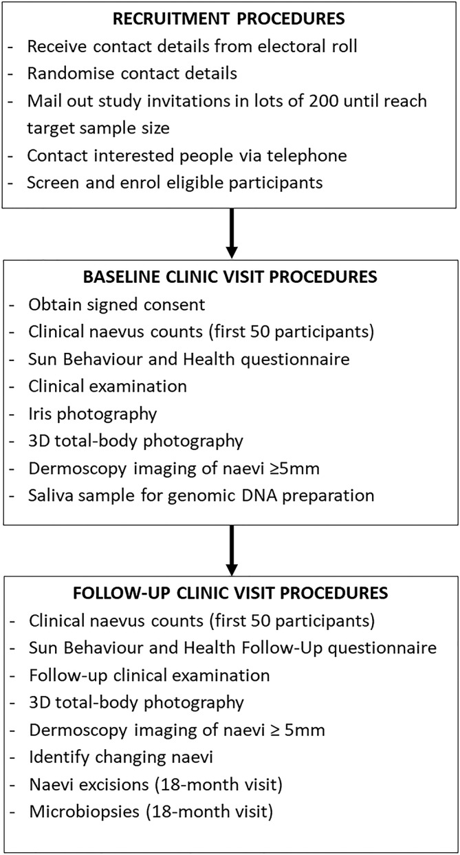
This is a population-based cohort study of melanocytic naevi in 200 adults aged 20-69 years recruited via the Australian electoral roll. At baseline, participants will complete a questionnaire on their sun behaviour and health and undergo a clinical examination. Three-dimensional (3D) total-body photography will be used to record the images of skin lesions. Pigmented naevi will be analysed in terms of number, diameter, colour and border irregularity using automated analysis software (excluding scalp, beneath underwear and soles of feet). All naevi ≥5 mm will be recorded using the integrated dermoscopy photographic system. A saliva sample will be obtained at baseline for genomic DNA analysis of pigmentation, naevus and melanoma-associated genes using the Illumina HumanCoreExome platform. The sun behaviour and health follow-up questionnaire, clinical examination and 3D total-body photography will be repeated every 6 months for 3 years. The first 50 participants will also undergo manual counts of naevi ≥2 mm and ≥5 mm at baseline, 6-month and 12-month follow-ups. Microbiopsy and excision of naevi of research interest is planned to commence at the 18-month time point among those who agree to donate samples for detailed histopathological and molecular assessment.
This study was approved by the Metro South Health Human Research Ethics Committee in April 2016 (approval number: HREC/16/QPAH/125). The findings will be disseminated through peer-reviewed and non-peer-reviewed publications and presentations at conferences.
皮肤上有许多黑素细胞痣或“痣”是黑色素瘤最强的预测指标;因此,通过对普通人群中的痣进行调查可以了解很多信息。我们旨在通过对成年人的黑素细胞痣进行为期3年的前瞻性研究,来增进对痣的流行病学和生物学的理解。
这是一项基于人群的队列研究,研究对象为通过澳大利亚选民名册招募的200名年龄在20至69岁之间的成年人的黑素细胞痣。在基线时,参与者将完成一份关于其阳光暴露行为和健康状况的问卷,并接受临床检查。将使用三维(3D)全身摄影来记录皮肤病变的图像。使用自动分析软件(不包括头皮、内衣下方和脚底)对色素痣的数量、直径、颜色和边界不规则性进行分析。所有直径≥5毫米的痣将使用集成皮肤镜摄影系统进行记录。在基线时采集唾液样本,使用Illumina HumanCoreExome平台对色素沉着、痣和黑色素瘤相关基因进行基因组DNA分析。在3年时间里,每6个月重复进行一次阳光暴露行为和健康状况随访问卷、临床检查和3D全身摄影。前50名参与者还将在基线、6个月和12个月随访时对手动计数直径≥2毫米和≥5毫米的痣。对于同意捐赠样本进行详细组织病理学和分子评估的参与者,计划在18个月时间点开始对感兴趣的痣进行微活检和切除。
本研究于2016年4月获得南布里斯班健康人类研究伦理委员会批准(批准号:HREC/16/QPAH/125)。研究结果将通过同行评审和非同行评审的出版物以及在会议上的报告进行传播。