Dermatology Service, Department of Medicine, Memorial Sloan Kettering Cancer Center, New York, New York, USA.
Canfield Scientific, Inc., Parsippany, New Jersey, USA.
Sci Data. 2024 Aug 14;11(1):884. doi: 10.1038/s41597-024-03743-w.
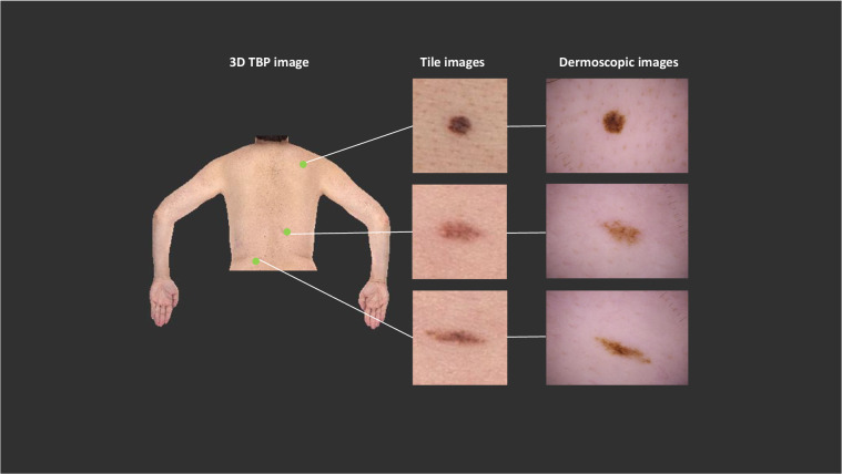
AI image classification algorithms have shown promising results when applied to skin cancer detection. Most public skin cancer image datasets are comprised of dermoscopic photos and are limited by selection bias, lack of standardization, and lend themselves to development of algorithms that can only be used by skilled clinicians. The SLICE-3D ("Skin Lesion Image Crops Extracted from 3D TBP") dataset described here addresses those concerns and contains images of over 400,000 distinct skin lesions from seven dermatologic centers from around the world. De-identified images were systematically extracted from sensitive 3D Total Body Photographs and are comparable in optical resolution to smartphone images. Algorithms trained on lower quality images could improve clinical workflows and detect skin cancers earlier if deployed in primary care or non-clinical settings, where photos are captured by non-expert physicians or patients. Such a tool could prompt individuals to visit a specialized dermatologist. This dataset circumvents many inherent limitations of prior datasets and may be used to build upon previous applications of skin imaging for cancer detection.
人工智能图像分类算法在皮肤癌检测中显示出了有前景的结果。大多数公开的皮肤癌图像数据集都由皮肤镜照片组成,受到选择偏差、缺乏标准化的限制,并且倾向于开发只能由熟练的临床医生使用的算法。这里描述的 SLICE-3D(“从 3D TBP 提取的皮肤病变图像裁剪”)数据集解决了这些问题,其中包含来自世界各地七个皮肤科中心的超过 40 万个不同皮肤病变的图像。从敏感的 3D 全身照片中系统地提取出已去识别的图像,其光学分辨率可与智能手机图像相媲美。如果在初级保健或非临床环境中部署,这些算法可以在质量较低的图像上进行训练,从而改善临床工作流程并更早地检测皮肤癌,在这些环境中,照片由非专业医生或患者拍摄。这样的工具可以促使个人去看皮肤科专家。该数据集规避了先前数据集的许多固有局限性,并可用于在癌症检测中建立以前的皮肤成像应用。