Translational and Molecular Imaging Institute, Icahn School of Medicine at Mount Sinai, New York, NY, United States; Department of Radiology, Icahn School of Medicine at Mount Sinai, New York, NY, United States.
Translational and Molecular Imaging Institute, Icahn School of Medicine at Mount Sinai, New York, NY, United States; Department of Radiology, Icahn School of Medicine at Mount Sinai, New York, NY, United States.
Seizure. 2018 Nov;62:3-10. doi: 10.1016/j.seizure.2018.09.005. Epub 2018 Sep 17.
MRI-negative epilepsy patients could benefit from advanced imaging techniques such as high-resolution diffusion magnetic resonance imaging (dMRI). Our aim was to perform hippocampal subfield-specific tractography and quantify connectivity of the subfields in MRI-negative patients. Abnormal connectivity of the hippocampal subfields may help inform seizure focus hypothesis and provide information to guide surgical intervention.
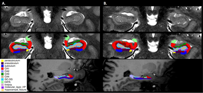
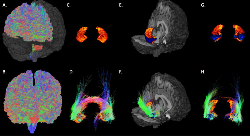
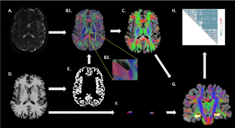
Hippocampal structural imaging and dMRI was acquired in 25 drug resistant MRI-negative patients and 25 healthy volunteers. The hippocampi of each subject was segmented on high-resolution structural images and dMRI-based probabilistic tractography was performed in each subfield. The degrees of connectivity and fiber densities of the hippocampal subfields were quantified and compared between epilepsy patients and healthy volunteers.
We were able to perform subfield-specific hippocampal tractography in each subject that participated in this study. These methods identified some hippocampal subfields that are abnormally connected in MRI-negative patients. In particular patients suspected of left temporal seizure focus exhibited increased connectivity of certain ipsilateral subfields, especially the subiculum, presubiculum, and parasubiculum, and reduced connectivity of some contralateral subfields, such as CA1 and subiculum.
Our data suggest that the hippocampal subfields are connected in distinct ways in different types of epilepsy. These results may provide important information that could help inform seizure focus hypothesis and in the surgical treatment of MRI-negative patients. These observations suggest that high-resolution dMRI-based tractography of the hippocampal subfields can detect subtle abnormalities in otherwise normal-appearing MRI-negative patients.
MRI 阴性的癫痫患者可能受益于高级成像技术,如高分辨率弥散磁共振成像(dMRI)。我们的目的是对 MRI 阴性患者进行海马亚区特异性束追踪,并定量分析亚区的连接。海马亚区的异常连接可能有助于告知癫痫灶假说,并提供信息以指导手术干预。
在 25 例耐药性 MRI 阴性患者和 25 例健康志愿者中采集了海马结构成像和 dMRI。对每个受试者的海马进行高分辨率结构图像分割,并在每个亚区进行基于 dMRI 的概率束追踪。定量比较了癫痫患者和健康志愿者之间海马亚区的连接程度和纤维密度。
我们能够对参与这项研究的每个受试者进行特定于亚区的海马束追踪。这些方法识别出 MRI 阴性患者中一些异常连接的海马亚区。特别是怀疑左颞叶癫痫灶的患者表现出某些同侧亚区的连接增加,特别是下托、前下托和副下托,以及某些对侧亚区的连接减少,如 CA1 和下托。
我们的数据表明,不同类型的癫痫中,海马亚区以不同的方式连接。这些结果可能提供重要信息,有助于告知癫痫灶假说,并为 MRI 阴性患者的手术治疗提供参考。这些观察结果表明,基于高分辨率 dMRI 的海马亚区束追踪可以检测到在其他方面正常的 MRI 阴性患者中的细微异常。