Pujol Sonia, Baldwin Michael, Nassiri Joshua, Kikinis Ron, Shaffer Kitt
Department of Radiology, Brigham and Women's Hospital, Harvard Medical School, 75 Francis Street, Boston, MA 02115.
Department of Radiology, Brigham and Women's Hospital, Harvard Medical School, 75 Francis Street, Boston, MA 02115.
Acad Radiol. 2016 Apr;23(4):507-16. doi: 10.1016/j.acra.2015.12.012. Epub 2016 Feb 17.
Anatomy is an essential component of medical education as it is critical for the accurate diagnosis in organs and human systems. The mental representation of the shape and organization of different anatomical structures is a crucial step in the learning process. The purpose of this pilot study is to demonstrate the feasibility and benefits of developing innovative teaching modules for anatomy education of first-year medical students based on three-dimensional (3D) reconstructions from actual patient data.
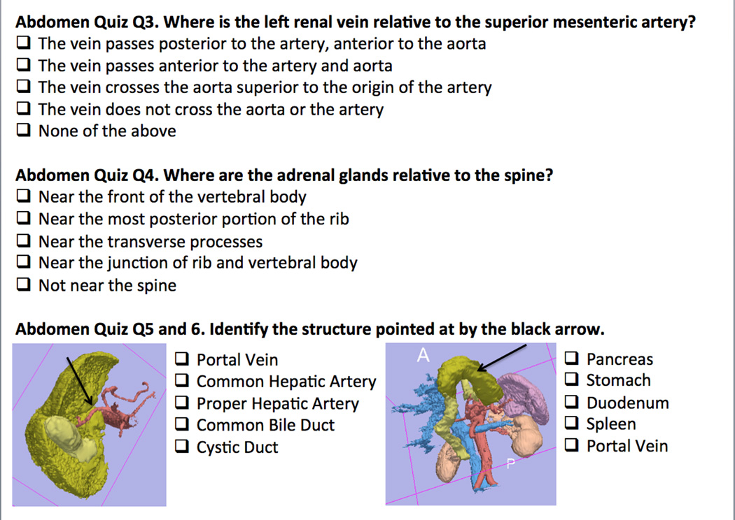
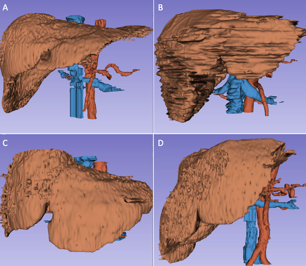
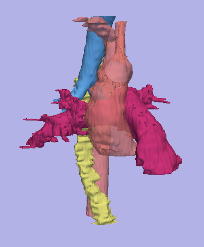
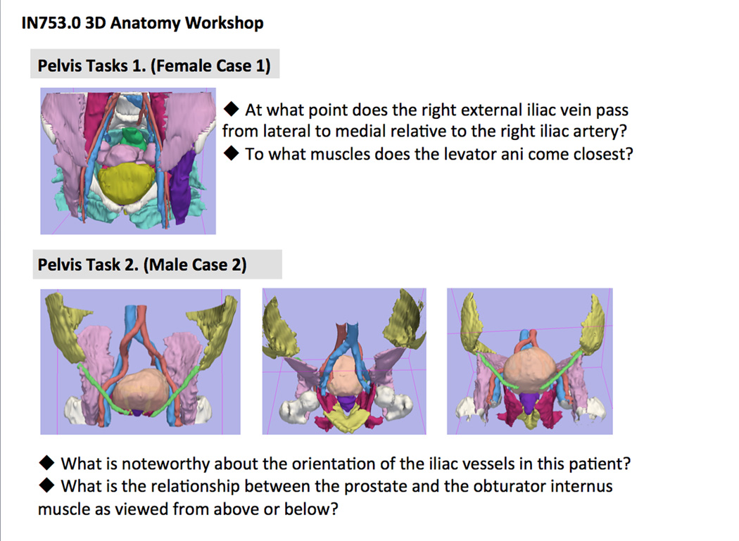
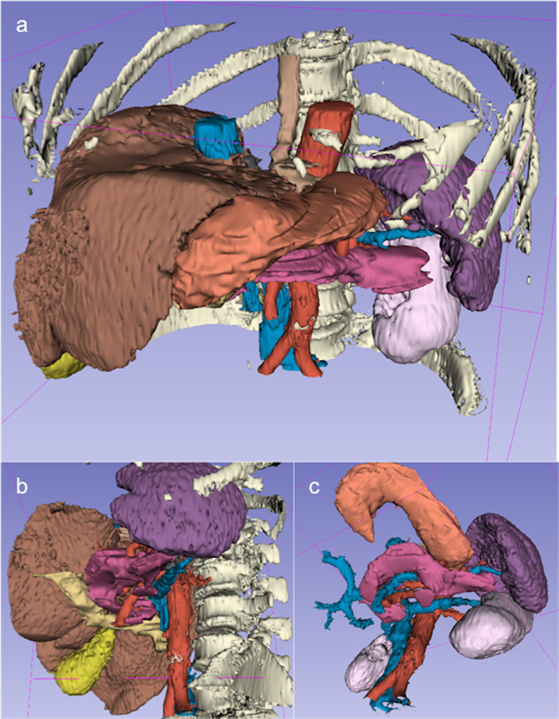
A total of 196 models of anatomical structures from 16 anonymized computed tomography datasets were generated using the 3D Slicer open-source software platform. The models focused on three anatomical areas: the mediastinum, the upper abdomen, and the pelvis. Online optional quizzes were offered to first-year medical students to assess their comprehension in the areas of interest. Specific tasks were designed for students to complete using the 3D models.
Scores of the quizzes confirmed a lack of understanding of 3D spatial relationships of anatomical structures despite standard instruction including dissection. Written task material and qualitative review by students suggested that interaction with 3D models led to a better understanding of the shape and spatial relationships among structures, and helped illustrate anatomical variations from one body to another.
The study demonstrates the feasibility of one possible approach to the generation of 3D models of the anatomy from actual patient data. The educational materials developed have the potential to supplement the teaching of complex anatomical regions and help demonstrate the anatomical variation among patients.
解剖学是医学教育的重要组成部分,因为它对于准确诊断器官和人体系统至关重要。不同解剖结构的形状和组织的心理表征是学习过程中的关键一步。这项初步研究的目的是证明基于实际患者数据的三维(3D)重建为一年级医学生开发解剖学教育创新教学模块的可行性和益处。
使用3D Slicer开源软件平台从16个匿名计算机断层扫描数据集中生成了总共196个解剖结构模型。这些模型聚焦于三个解剖区域:纵隔、上腹部和骨盆。为一年级医学生提供了在线选修测验,以评估他们在感兴趣领域的理解情况。设计了特定任务让学生使用3D模型完成。
测验分数证实,尽管进行了包括解剖在内的标准教学,但学生对解剖结构的三维空间关系仍缺乏理解。书面任务材料和学生的定性评价表明,与3D模型的互动使学生对结构的形状和空间关系有了更好的理解,并有助于说明个体之间的解剖变异。
该研究证明了一种从实际患者数据生成解剖学3D模型的可行方法。所开发的教育材料有潜力补充复杂解剖区域的教学,并有助于展示患者之间的解剖变异。