• 文献检索
  • 文档翻译
  • 深度研究
  • 学术资讯
  • Suppr Zotero 插件Zotero 插件
  • 邀请有礼
  • 套餐&价格
  • 历史记录
应用&插件
Suppr Zotero 插件Zotero 插件浏览器插件Mac 客户端Windows 客户端微信小程序
定价
高级版会员购买积分包购买API积分包
服务
文献检索文档翻译深度研究API 文档MCP 服务
关于我们
关于 Suppr公司介绍联系我们用户协议隐私条款
关注我们

Suppr 超能文献

核心技术专利:CN118964589B侵权必究
粤ICP备2023148730 号-1Suppr @ 2026

文献检索

告别复杂PubMed语法,用中文像聊天一样搜索,搜遍4000万医学文献。AI智能推荐,让科研检索更轻松。

立即免费搜索

文件翻译

保留排版,准确专业,支持PDF/Word/PPT等文件格式,支持 12+语言互译。

免费翻译文档

深度研究

AI帮你快速写综述,25分钟生成高质量综述,智能提取关键信息,辅助科研写作。

立即免费体验

在清醒成年小鼠中利用静息态功能磁共振成像评估脊髓损伤后的皮质可塑性。

Assessing cortical plasticity after spinal cord injury by using resting-state functional magnetic resonance imaging in awake adult mice.

机构信息

Department of Orthopaedic Surgery, Keio University School of Medicine, Tokyo, Japan.

Department of Physiology, Keio University School of Medicine, Tokyo, Japan.

出版信息

Sci Rep. 2018 Sep 26;8(1):14406. doi: 10.1038/s41598-018-32766-8.

DOI:10.1038/s41598-018-32766-8
PMID:30258091
原文链接:https://pmc.ncbi.nlm.nih.gov/articles/PMC6158265/
Abstract

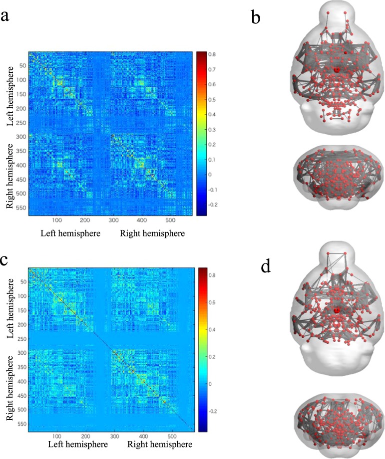
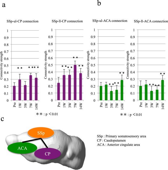
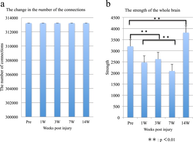
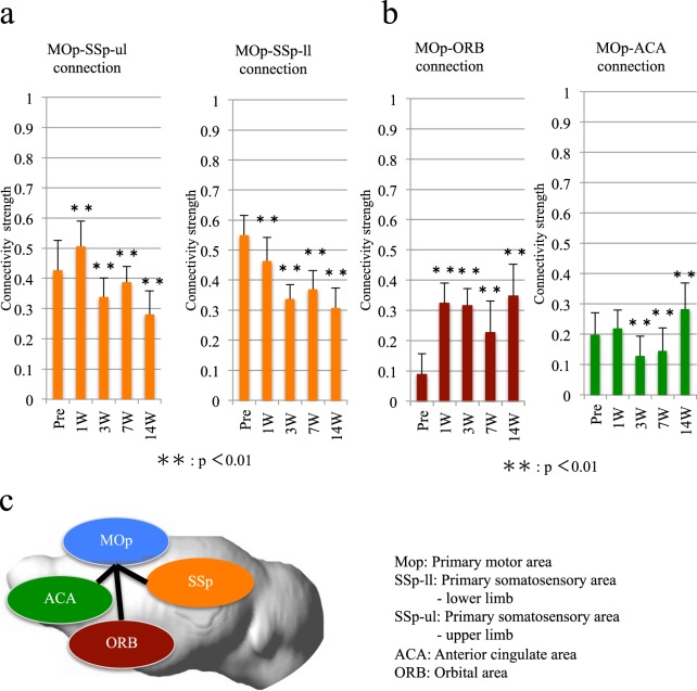
Neural connectivity has recently been shown to be altered after spinal cord injury (SCI) not only in the spinal cord but also in the brain. However, to date, no studies have analyzed the functional alterations after SCI in various areas of the cerebral cortex over time. To examine the plasticity of the neural connectivity in the brain after SCI, we performed resting-state functional magnetic resonance imaging (rs-fMRI) in awake adult mice pre- and post-SCI. After a complete thoracic SCI, the functional connectivity between the primary motor (MOp) and primary sensory (SSp) areas was significantly decreased during the chronic phase. In contrast, the connectivity between the MOp and motivation area was increased. Thus, impairments in sensory and motor connections after SCI led to a time-dependent compensatory upregulation of "motor functional motivation". Moreover, the functional connectivity between the SSp and pain-related areas, such as the caudoputamen (CP) and the anterior cingulate area (ACA), was strengthened during the chronic phase, thus suggesting that rs-fMRI can indicate the presence of neuropathic pain after SCI. Therefore, rs-fMRI is a useful tool for revealing the pathological changes that occur in the brain after SCI.

摘要

神经连接最近已经被证明在脊髓损伤 (SCI) 后不仅在脊髓中,而且在大脑中发生改变。然而,迄今为止,尚无研究分析 SCI 后大脑各个皮质区域随时间推移的功能变化。为了研究 SCI 后大脑中神经连接的可塑性,我们在 SCI 前和后对清醒的成年小鼠进行了静息态功能磁共振成像 (rs-fMRI)。在完全胸段 SCI 后,在慢性期初级运动 (MOp) 和初级感觉 (SSp) 区之间的功能连接显著降低。相比之下,MOp 和动机区之间的连接增加了。因此,SCI 后感觉和运动连接的损伤导致了“运动功能动机”的时间依赖性代偿上调。此外,在慢性期,SSp 和与疼痛相关的区域(如尾壳核 (CP) 和前扣带皮层区 (ACA))之间的功能连接增强,这表明 rs-fMRI 可以指示 SCI 后存在神经性疼痛。因此,rs-fMRI 是揭示 SCI 后大脑发生病理变化的有用工具。

https://cdn.ncbi.nlm.nih.gov/pmc/blobs/13ea/6158265/92c6bd2d3595/41598_2018_32766_Fig4_HTML.jpg
https://cdn.ncbi.nlm.nih.gov/pmc/blobs/13ea/6158265/a0f1f7719366/41598_2018_32766_Fig1_HTML.jpg
https://cdn.ncbi.nlm.nih.gov/pmc/blobs/13ea/6158265/927e958991f2/41598_2018_32766_Fig2_HTML.jpg
https://cdn.ncbi.nlm.nih.gov/pmc/blobs/13ea/6158265/cd03ee9a5d9e/41598_2018_32766_Fig3_HTML.jpg
https://cdn.ncbi.nlm.nih.gov/pmc/blobs/13ea/6158265/92c6bd2d3595/41598_2018_32766_Fig4_HTML.jpg

相似文献

1
Assessing cortical plasticity after spinal cord injury by using resting-state functional magnetic resonance imaging in awake adult mice.在清醒成年小鼠中利用静息态功能磁共振成像评估脊髓损伤后的皮质可塑性。
Sci Rep. 2018 Sep 26;8(1):14406. doi: 10.1038/s41598-018-32766-8.
2
Alteration of Resting-State Brain Sensorimotor Connectivity following Spinal Cord Injury: A Resting-State Functional Magnetic Resonance Imaging Study.脊髓损伤后静息态脑感觉运动连接的改变:一项静息态功能磁共振成像研究
J Neurotrauma. 2015 Sep 15;32(18):1422-7. doi: 10.1089/neu.2014.3661. Epub 2015 Jun 25.
3
Alterations in Cortical Sensorimotor Connectivity following Complete Cervical Spinal Cord Injury: A Prospective Resting-State fMRI Study.完全性颈脊髓损伤后皮质感觉运动连接的改变:一项前瞻性静息态功能磁共振成像研究
PLoS One. 2016 Mar 8;11(3):e0150351. doi: 10.1371/journal.pone.0150351. eCollection 2016.
4
Evaluation of Whole-Brain Resting-State Functional Connectivity in Spinal Cord Injury: A Large-Scale Network Analysis Using Network-Based Statistic.脊髓损伤中全脑静息态功能连接性评估:基于网络统计的大规模网络分析
J Neurotrauma. 2017 Mar 15;34(6):1278-1282. doi: 10.1089/neu.2016.4649. Epub 2017 Jan 27.
5
Spinal Cord Injury Disrupts Resting-State Networks in the Human Brain.脊髓损伤会扰乱人脑的静息态网络。
J Neurotrauma. 2018 Mar 15;35(6):864-873. doi: 10.1089/neu.2017.5212. Epub 2018 Jan 11.
6
Motor recovery at 6 months after admission is related to structural and functional reorganization of the spine and brain in patients with spinal cord injury.入院后6个月时的运动恢复与脊髓损伤患者脊柱和大脑的结构及功能重组有关。
Hum Brain Mapp. 2016 Jun;37(6):2195-209. doi: 10.1002/hbm.23163. Epub 2016 Mar 3.
7
Longitudinal evaluation of functional connectivity variation in the monkey sensorimotor network induced by spinal cord injury.脊髓损伤诱导的猴子感觉运动网络功能连接变化的纵向评估。
Acta Physiol (Oxf). 2016 Jun;217(2):164-73. doi: 10.1111/apha.12645. Epub 2016 Jan 13.
8
Specific Brain Reorganization Underlying Superior Upper Limb Motor Function After Spinal Cord Injury: A Multimodal MRI Study.脊髓损伤后上肢运动功能优异的特定脑重组:一项多模态 MRI 研究。
Neurorehabil Neural Repair. 2021 Mar;35(3):220-232. doi: 10.1177/1545968321989347. Epub 2021 Jan 29.
9
Resting-State Functional Connectivity of the Thalamus in Complete Spinal Cord Injury.完全性脊髓损伤患者丘脑的静息态功能连接。
Neurorehabil Neural Repair. 2020 Feb;34(2):122-133. doi: 10.1177/1545968319893299. Epub 2020 Jan 6.
10
Supraspinal Sensorimotor and Pain-Related Reorganization after a Hemicontusion Rat Cervical Spinal Cord Injury.半脱位型颈脊髓损伤后脊髓以上感觉运动和痛觉相关重组。
J Neurotrauma. 2021 Dec;38(24):3393-3405. doi: 10.1089/neu.2021.0190.

引用本文的文献

1
Spiral volumetric optoacoustic tomography of reduced oxygen saturation in the spinal cord of M83 mouse model of Parkinson's disease.帕金森病M83小鼠模型脊髓中氧饱和度降低的螺旋体积光声断层扫描
Eur J Nucl Med Mol Imaging. 2025 Jan;52(2):427-443. doi: 10.1007/s00259-024-06938-w. Epub 2024 Oct 9.
2
Region-Specific Variation in the Electrophysiological Responses of Spodoptera frugiperda (Lepidoptera: Noctuidae) to Synthetic Sex Pheromone Compounds.斜纹夜蛾(鳞翅目:夜蛾科)对合成性信息素化合物的电生理反应的区域特异性变化。
J Chem Ecol. 2024 Nov;50(11):631-642. doi: 10.1007/s10886-024-01479-w. Epub 2024 Feb 29.
3

本文引用的文献

1
Functional Connectivity Mapping in the Animal Model: Principles and Applications of Resting-State fMRI.动物模型中的功能连接映射:静息态功能磁共振成像的原理与应用
Front Neurol. 2017 May 10;8:200. doi: 10.3389/fneur.2017.00200. eCollection 2017.
2
Functional brain mapping using specific sensory-circuit stimulation and a theoretical graph network analysis in mice with neuropathic allodynia.使用特定感觉回路刺激和理论图网络分析在神经病理性痛觉过敏的小鼠中进行功能性脑映射。
Sci Rep. 2016 Nov 29;6:37802. doi: 10.1038/srep37802.
3
Physiological effects of a habituation procedure for functional MRI in awake mice using a cryogenic radiofrequency probe.
Rehabilitation Training after Spinal Cord Injury Affects Brain Structure and Function: From Mechanisms to Methods.
脊髓损伤后的康复训练对脑结构和功能的影响:从机制到方法
Biomedicines. 2023 Dec 22;12(1):41. doi: 10.3390/biomedicines12010041.
4
Where do we stand on fMRI in awake mice?在清醒小鼠的 fMRI 研究方面,我们目前处于什么阶段?
Cereb Cortex. 2024 Jan 14;34(1). doi: 10.1093/cercor/bhad478.
5
A shift of brain network hub after spinal cord injury.脊髓损伤后脑网络枢纽的转移。
Front Mol Neurosci. 2023 Oct 17;16:1245902. doi: 10.3389/fnmol.2023.1245902. eCollection 2023.
6
Habilitation Improves Mouse Gait Development Following Neonatal Brain Injury.功能训练可改善新生小鼠脑损伤后的步态发育。
Prog Rehabil Med. 2022 Dec 1;7:20220061. doi: 10.2490/prm.20220061. eCollection 2022.
7
Functional connectivity and amplitude of low-frequency fluctuations changes in people with complete subacute and chronic spinal cord injury.完全亚急性和慢性脊髓损伤患者的功能连接和低频波动振幅变化。
Sci Rep. 2022 Dec 3;12(1):20874. doi: 10.1038/s41598-022-25345-5.
8
Effects of chronic caffeine intake and withdrawal on neural activity assessed via resting-state functional magnetic resonance imaging in mice.长期摄入咖啡因及戒断对通过静息态功能磁共振成像评估的小鼠神经活动的影响。
Heliyon. 2022 Nov 19;8(11):e11714. doi: 10.1016/j.heliyon.2022.e11714. eCollection 2022 Nov.
9
imaging in experimental spinal cord injury - Techniques and trends.实验性脊髓损伤的影像学——技术与趋势
Brain Spine. 2021 Dec 29;2:100859. doi: 10.1016/j.bas.2021.100859. eCollection 2022.
10
Neural regeneration therapy after spinal cord injury induces unique brain functional reorganizations in rhesus monkeys.脊髓损伤后的神经再生疗法会在恒河猴的大脑中引起独特的功能重组。
Ann Med. 2022 Dec;54(1):1867-1883. doi: 10.1080/07853890.2022.2089728.
使用低温射频探头对清醒小鼠进行功能磁共振成像的习惯化程序的生理效应。
J Neurosci Methods. 2016 Dec 1;274:38-48. doi: 10.1016/j.jneumeth.2016.09.013. Epub 2016 Oct 1.
4
Multimodal MRI Evaluation of the MitoPark Mouse Model of Parkinson's Disease.帕金森病MitoPark小鼠模型的多模态磁共振成像评估
PLoS One. 2016 Mar 22;11(3):e0151884. doi: 10.1371/journal.pone.0151884. eCollection 2016.
5
Alterations in Cortical Sensorimotor Connectivity following Complete Cervical Spinal Cord Injury: A Prospective Resting-State fMRI Study.完全性颈脊髓损伤后皮质感觉运动连接的改变:一项前瞻性静息态功能磁共振成像研究
PLoS One. 2016 Mar 8;11(3):e0150351. doi: 10.1371/journal.pone.0150351. eCollection 2016.
6
Function of the nucleus accumbens in motor control during recovery after spinal cord injury.伏隔核在脊髓损伤后恢复过程中运动控制中的作用。
Science. 2015 Oct 2;350(6256):98-101. doi: 10.1126/science.aab3825. Epub 2015 Oct 1.
7
Change of Brain Functional Connectivity in Patients With Spinal Cord Injury: Graph Theory Based Approach.脊髓损伤患者脑功能连接的变化:基于图论的方法
Ann Rehabil Med. 2015 Jun;39(3):374-83. doi: 10.5535/arm.2015.39.3.374. Epub 2015 Jun 30.
8
Altered contralateral sensorimotor system organization after experimental hemispherectomy: a structural and functional connectivity study.实验性大脑半球切除术后对侧感觉运动系统组织的改变:一项结构和功能连接性研究。
J Cereb Blood Flow Metab. 2015 Aug;35(8):1358-67. doi: 10.1038/jcbfm.2015.101. Epub 2015 May 13.
9
Alteration of Resting-State Brain Sensorimotor Connectivity following Spinal Cord Injury: A Resting-State Functional Magnetic Resonance Imaging Study.脊髓损伤后静息态脑感觉运动连接的改变:一项静息态功能磁共振成像研究
J Neurotrauma. 2015 Sep 15;32(18):1422-7. doi: 10.1089/neu.2014.3661. Epub 2015 Jun 25.
10
Identifying patients with Alzheimer's disease using resting-state fMRI and graph theory.利用静息态功能磁共振成像和图论识别阿尔茨海默病患者。
Clin Neurophysiol. 2015 Nov;126(11):2132-41. doi: 10.1016/j.clinph.2015.02.060. Epub 2015 Apr 1.