Department of Periodontics, College of Dentistry, University of Illinois at Chicago, Chicago, Illinois, United States of America.
Undergraduate Program, University of Illinois at Chicago, Chicago, Illinois, United States of America.
PLoS One. 2018 Oct 3;13(10):e0204941. doi: 10.1371/journal.pone.0204941. eCollection 2018.
The results from cross sectional and longitudinal studies show that periodontitis is closely associated with cognitive impairment (CI) and Alzhemer's Disease (AD). Further, studies using animal model of periodontitis and human post-mortem brain tissues from subjects with AD strongly suggest that a gram-negative periodontal pathogen, Porphyromonas gingivalis (Pg) and/or its product gingipain is/are translocated to the brain. However, neuropathology resulting from Pg oral application is not known. In this work, we tested the hypothesis that repeated exposure of wild type C57BL/6 mice to orally administered Pg results in neuroinflammation, neurodegeneration, microgliosis, astrogliosis and formation of intra- and extracellular amyloid plaque and neurofibrillary tangles (NFTs) which are pathognomonic signs of AD.
Experimental chronic periodontitis was induced in ten wild type 8-week old C57BL/6 WT mice by repeated oral application (MWF/week) of Pg/gingipain for 22 weeks (experimental group). Another 10 wild type 8-week old C57BL/6 mice received vehicle alone (control group) MWF per week for 22 weeks. Brain tissues were collected and the presence of Pg/gingipain was determined by immunofluorescence (IF) microscopy, confocal microscopy, and quantitative PCR (qPCR). The hippocampi were examined for the signs of neuropathology related to AD: TNFα, IL1β, and IL6 expression (neuroinflammation), NeuN and Fluoro Jade C staining (neurodegeneration) and amyloid beta1-42 (Aβ42) production and phosphorylation of tau protein at Ser396 were assessed by IF and confocal microscopy. Further, gene expression of amyloid precursor protein (APP), beta-site APP cleaving enzyme 1 (BACE1), a disintegrin and metalloproteinase domain-containing protein10 (ADAM10) for α-secretase and presenilin1 (PSEN1) for ɣ-secretase, and NeuN (rbFox3) were determined by RT-qPCR. Microgliosis and astrogliosis were also determined by IF microscopy.
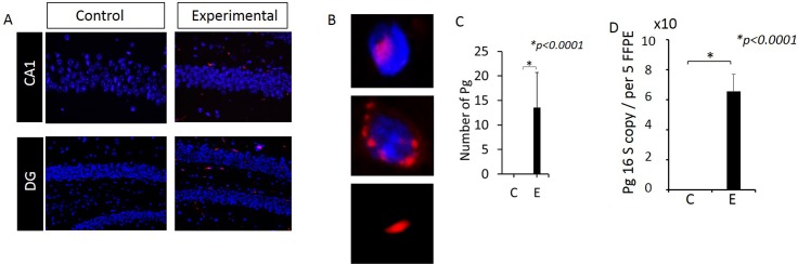
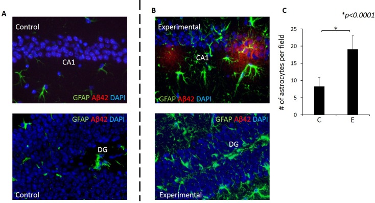
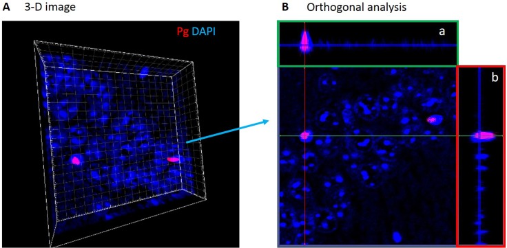
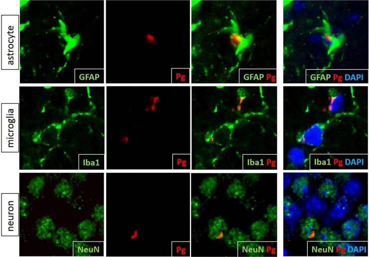
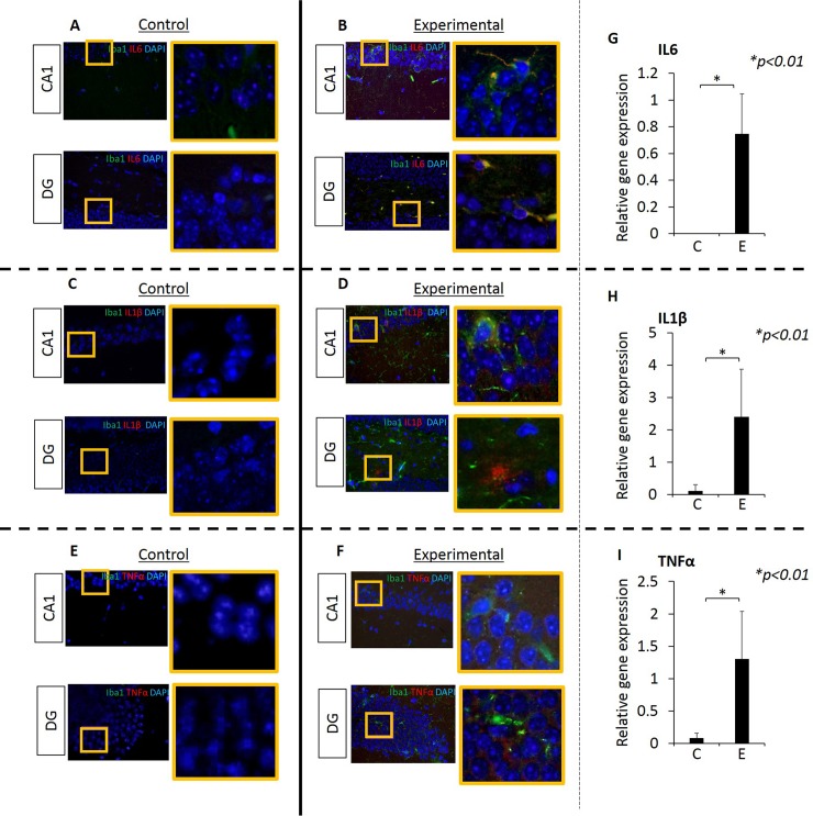
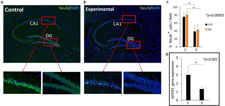
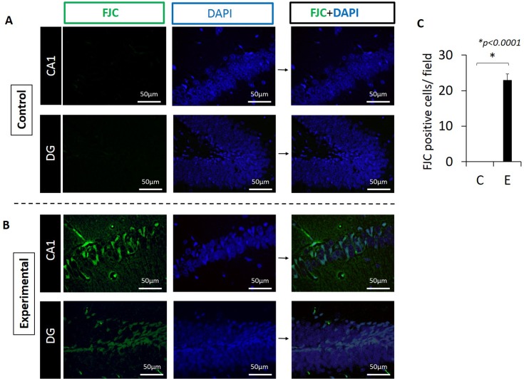
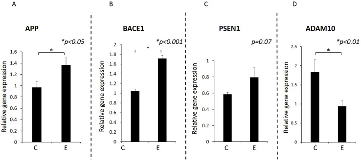
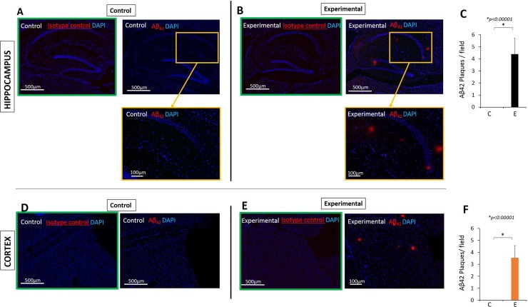
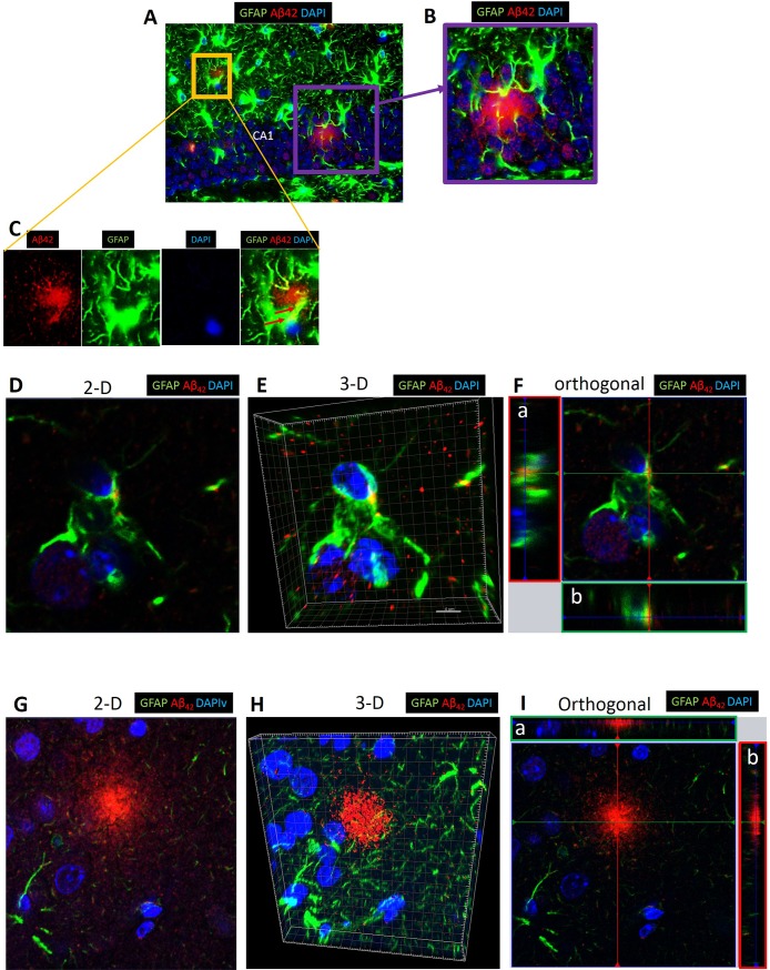
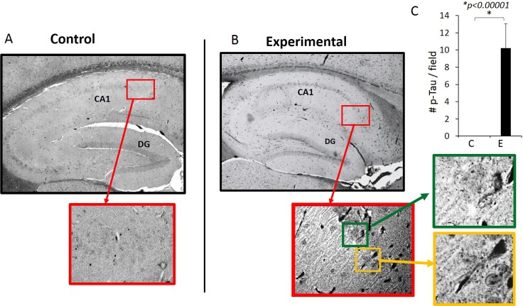
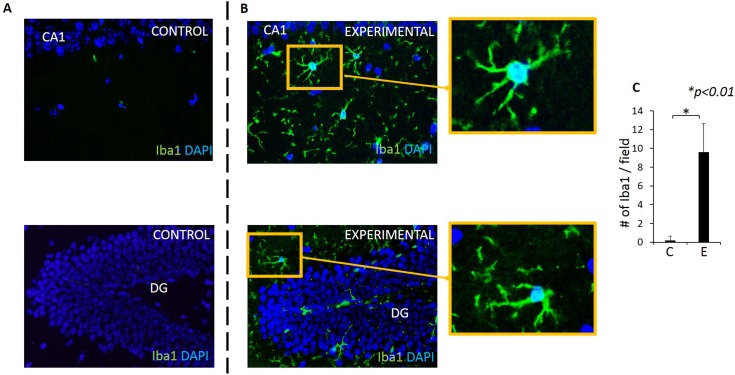
Pg/gingipain was detected in the hippocampi of mice in the experimental group by immunohistochemistry, confocal microscopy, and qPCR confirming the translocation of orally applied Pg to the brain. Pg/gingipain was localized intra-nuclearly and peri-nuclearly in microglia (Iba1+), astrocytes (GFAP+), neurons (NeuN+) and was evident extracellularly. Significantly greater levels of expression of IL6, TNFα and IL1β were evident in experimental as compared to control group (p<0.01, p<0.00001, p<0.00001 respectively). In addition, microgliosis and astrogliosis were evident in the experimental but not in control group (p <0.01, p<0.0001 respectively). Neurodegeneration was evident in the experimental group based on a fewer number of intact neuronal cells assessed by NeuN positivity and rbFOX3 gene expression, and there was a greater number of degenerating neurons in the hippocampi of experimental mice assessed by Fluoro Jade C positivity. APP and BACE1 gene expression were increased in experimental group compared with control group (p<0.05, p<0.001 respectively). PSEN1 gene expression was higher in experimental than control group but the difference was not statistically significant (p = 0.07). ADAM10 gene expression was significantly decreased in experimental group compared with control group (p<0.01). Extracellular Aβ42 was detected in the parenchyma in the experimental but not in the control group (p< 0.00001). Finally, phospho-Tau (Ser396) protein was detected and NFTs were evident in experimental but not in the control group (p<0.00001).
This study is the first to show neurodegeneration and the formation of extracellular Aβ42 in young adult WT mice after repeated oral application of Pg. The neuropathological features observed in this study strongly suggest that low grade chronic periodontal pathogen infection can result in the development of neuropathology that is consistent with that of AD.
横断面和纵向研究的结果表明,牙周炎与认知障碍(CI)和阿尔茨海默病(AD)密切相关。此外,使用牙周炎动物模型和 AD 患者死后脑组织的研究强烈表明,革兰氏阴性牙周病原体牙龈卟啉单胞菌(Pg)和/或其产物牙龈蛋白酶被/被转移到大脑。然而,Pg 口服应用引起的神经病理学尚不清楚。在这项工作中,我们检验了这样一个假设,即重复给予野生型 C57BL/6 小鼠口服 Pg 会导致神经炎症、神经退行性变、小胶质细胞增生、星形胶质细胞增生以及形成细胞内和细胞外淀粉样斑块和神经原纤维缠结(NFTs),这些都是 AD 的特征性标志。
通过重复口服应用(MWF/周)Pg/牙龈蛋白酶 22 周,在 10 只 8 周龄野生型 C57BL/6 WT 小鼠中诱导实验性慢性牙周炎(实验组)。另 10 只 8 周龄野生型 C57BL/6 小鼠接受单独的载体(MWF/周)22 周(对照组)。收集脑组织,并通过免疫荧光(IF)显微镜、共聚焦显微镜和定量 PCR(qPCR)确定 Pg/牙龈蛋白酶的存在。通过 IF 和共聚焦显微镜评估与 AD 相关的神经病理学迹象:TNFα、IL1β 和 IL6 表达(神经炎症)、NeuN 和 Fluoro Jade C 染色(神经退行性变)以及β淀粉样蛋白 1-42(Aβ42)的产生和 tau 蛋白丝氨酸 396 磷酸化。此外,通过 RT-qPCR 测定淀粉样前体蛋白(APP)、β-位点 APP 切割酶 1(BACE1)、含金属蛋白酶域蛋白 10(ADAM10)的基因表达α-分泌酶和早老素 1(PSEN1)的γ-分泌酶,以及 NeuN(rbFox3)。通过 IF 显微镜还测定了小胶质细胞和星形胶质细胞的增生。
通过免疫组化、共聚焦显微镜和 qPCR 检测到实验组小鼠海马区有 Pg/牙龈蛋白酶,证实了口服应用的 Pg 向大脑的转移。Pg/牙龈蛋白酶在小胶质细胞(Iba1+)、星形胶质细胞(GFAP+)和神经元(NeuN+)的核内和核周定位,并在细胞外明显存在。实验组中 IL6、TNFα 和 IL1β 的表达明显高于对照组(p<0.01、p<0.00001、p<0.00001)。此外,实验组有明显的小胶质细胞和星形胶质细胞增生,但对照组没有(p<0.01,p<0.0001)。实验组神经退行性变明显,通过 NeuN 阳性和 rbFOX3 基因表达评估,神经元数量减少,实验组海马神经元变性数量增多,通过 Fluoro Jade C 阳性评估。与对照组相比,实验组 APP 和 BACE1 基因表达增加(p<0.05,p<0.001)。PSEN1 基因表达实验组高于对照组,但差异无统计学意义(p=0.07)。ADAM10 基因表达实验组明显低于对照组(p<0.01)。实验组脑实质中检测到细胞外 Aβ42(p<0.00001)。最后,在实验组中检测到磷酸化 Tau(Ser396)蛋白,形成 NFT(p<0.00001)。
本研究首次表明,在重复口服应用 Pg 后,年轻野生型 WT 小鼠会发生神经退行性变和细胞外 Aβ42 的形成。本研究观察到的神经病理学特征强烈表明,低度慢性牙周病原体感染可导致与 AD 一致的神经病理学发展。