Wong-Sam Andres, Wang Yuan-Fang, Zhang Ying, Ghosh Arun K, Harrison Robert W, Weber Irene T
Department of Biology, Molecular Basis of Disease Program, Department of Computer Science, and Department of Chemistry, Georgia State University, Atlanta, Georgia 30303, United States.
RNA Therapeutics Institute and Department of Biochemistry and Molecular Pharmacology, University of Massachusetts Medical School, Worcester, Massachusetts 01605, United States.
ACS Omega. 2018 Sep 30;3(9):12132-12140. doi: 10.1021/acsomega.8b01683. Epub 2018 Sep 27.
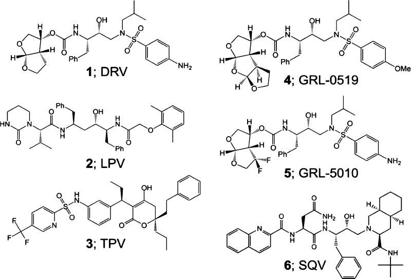
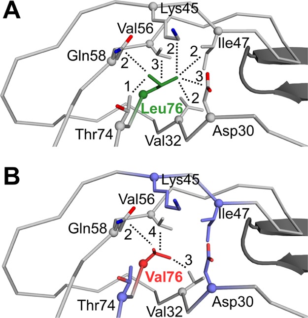
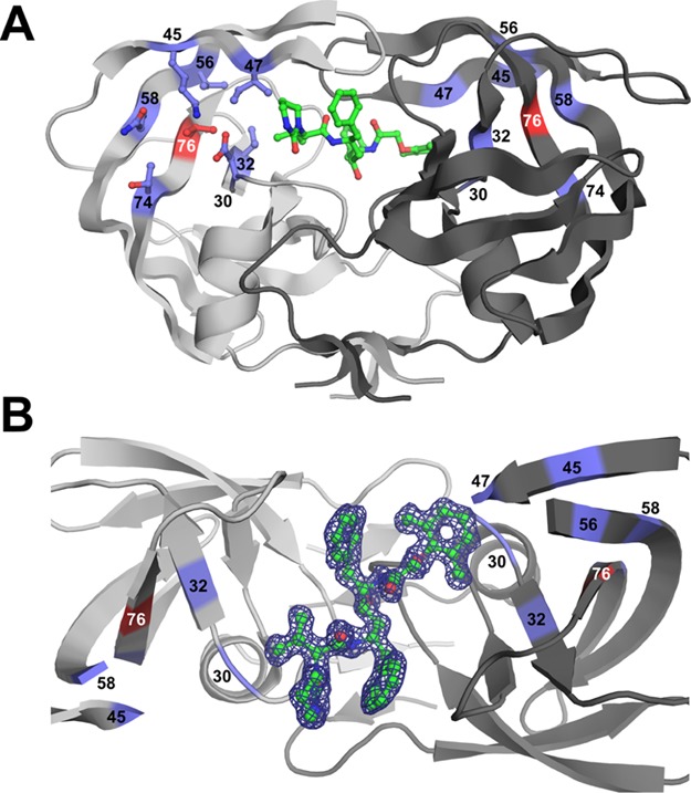
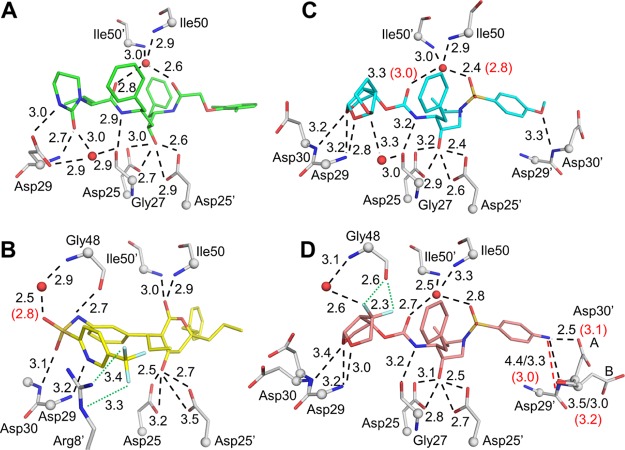
Four HIV-1 protease (PR) inhibitors, clinical inhibitors lopinavir and tipranavir, and two investigational compounds and , were studied for their effect on the structure and activity of PR with drug-resistant mutation L76V (PR). Compound exhibited the best value of 1.9 nM for PR, whereas the other three inhibitors had values of 4.5-7.6 nM, 2-3 orders of magnitude worse than for wild-type enzymes. Crystal structures showed only minor differences in interactions of inhibitors with PR compared to wild-type complexes. The shorter side chain of Val76 in the mutant lost hydrophobic interactions with Lys45 and Ile47 in the flap, and with Asp30 and Thr74 in the protein core, consistent with decreased stability. Inhibitors forming additional polar interactions with the flaps or dimer interface of PR were unable to compensate for the decrease in internal hydrophobic contacts. These structures provide insights for inhibitor design.
研究了四种HIV-1蛋白酶(PR)抑制剂,临床抑制剂洛匹那韦和替拉那韦,以及两种研究性化合物 和 ,考察它们对具有耐药性突变L76V的PR(PR)的结构和活性的影响。化合物 对PR表现出最佳的 值,为1.9 nM,而其他三种抑制剂的 值为4.5 - 7.6 nM,比野生型酶差2 - 3个数量级。晶体结构显示,与野生型复合物相比,抑制剂与PR的相互作用仅有微小差异。突变体中Val76较短的侧链失去了与瓣片中Lys45和Ile47以及蛋白质核心中Asp30和Thr74的疏水相互作用,这与稳定性降低一致。与PR的瓣片或二聚体界面形成额外极性相互作用的抑制剂无法弥补内部疏水接触的减少。这些结构为抑制剂设计提供了见解。