• 文献检索
  • 文档翻译
  • 深度研究
  • 学术资讯
  • Suppr Zotero 插件Zotero 插件
  • 邀请有礼
  • 套餐&价格
  • 历史记录
应用&插件
Suppr Zotero 插件Zotero 插件浏览器插件Mac 客户端Windows 客户端微信小程序
定价
高级版会员购买积分包购买API积分包
服务
文献检索文档翻译深度研究API 文档MCP 服务
关于我们
关于 Suppr公司介绍联系我们用户协议隐私条款
关注我们

Suppr 超能文献

核心技术专利:CN118964589B侵权必究
粤ICP备2023148730 号-1Suppr @ 2026

文献检索

告别复杂PubMed语法,用中文像聊天一样搜索,搜遍4000万医学文献。AI智能推荐,让科研检索更轻松。

立即免费搜索

文件翻译

保留排版,准确专业,支持PDF/Word/PPT等文件格式,支持 12+语言互译。

免费翻译文档

深度研究

AI帮你快速写综述,25分钟生成高质量综述,智能提取关键信息,辅助科研写作。

立即免费体验

通过OPTiSPIM校正的光片荧光显微镜中的衰减伪影。

Attenuation artifacts in light sheet fluorescence microscopy corrected by OPTiSPIM.

作者信息

Mayer Jürgen, Robert-Moreno Alexandre, Sharpe James, Swoger Jim

机构信息

1Centre for Genomic Regulation (CRG), The Barcelona Institute of Science and Technology, Dr. Aiguader 88, 08003 Barcelona, Spain.

2Universitat Pompeu Fabra (UPF), Barcelona, Spain.

出版信息

Light Sci Appl. 2018 Oct 3;7:70. doi: 10.1038/s41377-018-0068-z. eCollection 2018.

DOI:10.1038/s41377-018-0068-z
PMID:30302241
原文链接:https://pmc.ncbi.nlm.nih.gov/articles/PMC6168557/
Abstract

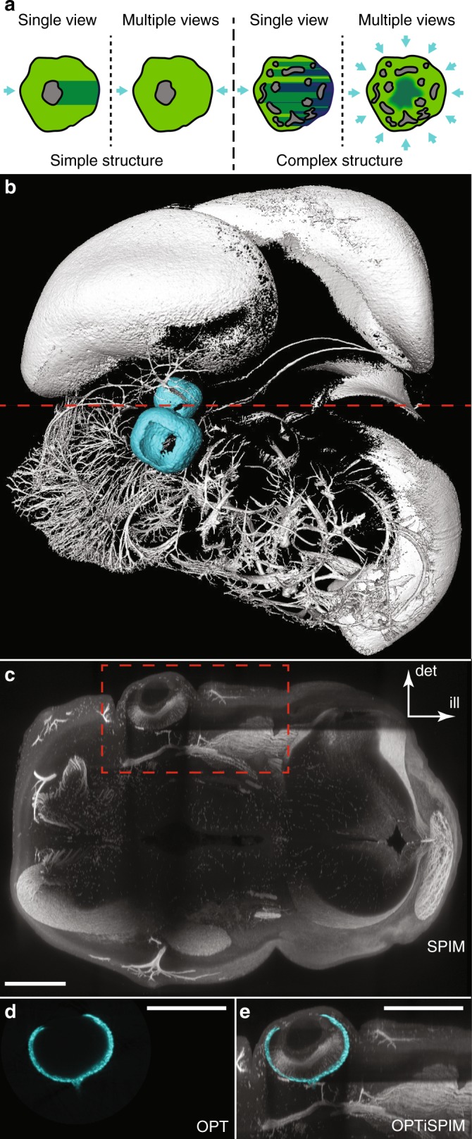
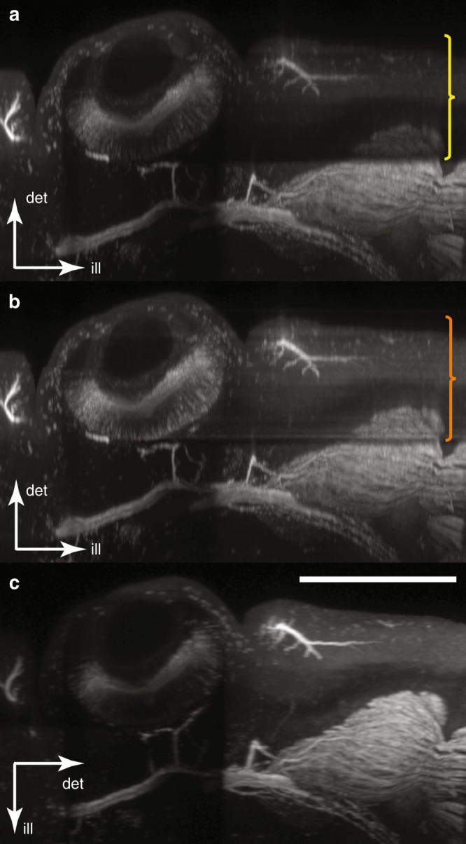
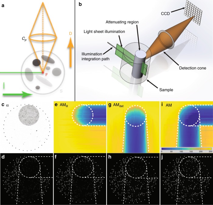
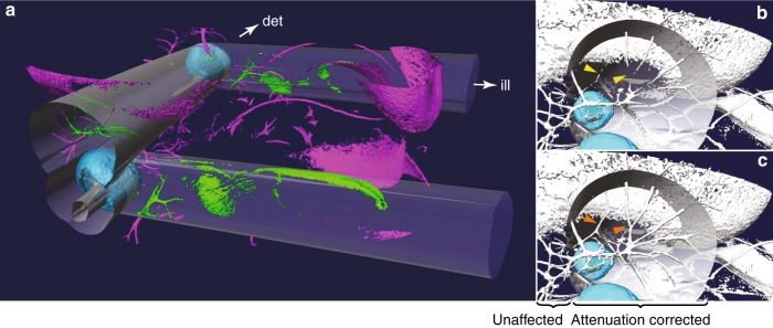
Light sheet fluorescence microscopy (LSFM) is rapidly becoming an essential technology for mesoscopic imaging of samples such as embryos and adult mouse organs. However, LSFM can suffer from optical artifacts for which there is no intrinsic solution. The attenuation of light due to absorbing material causes "shadow" artifacts along both the illumination and detection paths. Several approaches have been introduced to reduce this problem, including scanning illumination and multi-view imaging. However, neither of these approaches completely eliminates the problem. If the distribution of the absorbing material is complex, shadows cannot be avoided. We introduce a new approach that relies on multi-modal integration of two very different mesoscopic techniques. Unlike LSFM, optical projection tomography (OPT) can operate in transmission mode to create a voxel map of the 3D distribution of the sample's optical attenuation. Here, we demonstrate a hybrid instrument (OPTiSPIM) that can quantify this attenuation and use the information to correct the shadow artifacts of LSFM.

摘要

光片荧光显微镜(LSFM)正迅速成为用于对胚胎和成年小鼠器官等样本进行介观成像的一项关键技术。然而,LSFM可能会出现光学伪像,对此尚无内在的解决办法。由于吸收材料导致的光衰减会在照明路径和检测路径上产生“阴影”伪像。已经引入了几种方法来减少这个问题,包括扫描照明和多视图成像。然而,这些方法都不能完全消除该问题。如果吸收材料的分布复杂,阴影就无法避免。我们引入了一种新方法,该方法依赖于两种截然不同的介观技术的多模态整合。与LSFM不同,光学投影断层扫描(OPT)可以在透射模式下运行,以创建样本光学衰减三维分布的体素图。在此,我们展示了一种混合仪器(OPTiSPIM),它可以量化这种衰减,并利用该信息校正LSFM的阴影伪像。

https://cdn.ncbi.nlm.nih.gov/pmc/blobs/5761/6168557/cde18d4afe80/41377_2018_68_Fig4_HTML.jpg
https://cdn.ncbi.nlm.nih.gov/pmc/blobs/5761/6168557/d33dd7a60b5a/41377_2018_68_Fig1_HTML.jpg
https://cdn.ncbi.nlm.nih.gov/pmc/blobs/5761/6168557/c4a7d0758099/41377_2018_68_Fig2_HTML.jpg
https://cdn.ncbi.nlm.nih.gov/pmc/blobs/5761/6168557/64bbfab20c03/41377_2018_68_Fig3_HTML.jpg
https://cdn.ncbi.nlm.nih.gov/pmc/blobs/5761/6168557/cde18d4afe80/41377_2018_68_Fig4_HTML.jpg

相似文献

1
Attenuation artifacts in light sheet fluorescence microscopy corrected by OPTiSPIM.通过OPTiSPIM校正的光片荧光显微镜中的衰减伪影。
Light Sci Appl. 2018 Oct 3;7:70. doi: 10.1038/s41377-018-0068-z. eCollection 2018.
2
Comparison of optical projection tomography and light-sheet fluorescence microscopy.光学投影断层成像术与光片荧光显微镜的比较。
J Microsc. 2019 Jul;275(1):3-10. doi: 10.1111/jmi.12796. Epub 2019 Apr 30.
3
OPTiSPIM: integrating optical projection tomography in light sheet microscopy extends specimen characterization to nonfluorescent contrasts.OPTiSPIM:将光学投影断层成像集成到光片显微镜中,将样品特征扩展到非荧光对比度。
Opt Lett. 2014 Feb 15;39(4):1053-6. doi: 10.1364/OL.39.001053.
4
Removing striping artifacts in light-sheet fluorescence microscopy: a review.消除光片荧光显微镜中的条纹伪影:综述。
Prog Biophys Mol Biol. 2022 Jan;168:52-65. doi: 10.1016/j.pbiomolbio.2021.07.003. Epub 2021 Jul 15.
5
Light Sheet Fluorescence Microscopy (LSFM).光片荧光显微镜(LSFM)。
Curr Protoc Cytom. 2015 Jan 5;71:12.37.1-12.37.15. doi: 10.1002/0471142956.cy1237s71.
6
Fast multi-directional DSLM for confocal detection without striping artifacts.用于共聚焦检测且无条纹伪影的快速多向数字扫描激光显微镜。
Biomed Opt Express. 2020 May 18;11(6):3111-3124. doi: 10.1364/BOE.390916. eCollection 2020 Jun 1.
7
Demonstrating Improved Multiple Transport-Mean-Free-Path Imaging Capabilities of Light Sheet Microscopy in the Quantification of Fluorescence Dynamics.演示光片显微镜在荧光动力学定量中的多重传输平均自由路径成像能力的改善。
Biotechnol J. 2018 Jan;13(1). doi: 10.1002/biot.201700419. Epub 2017 Dec 11.
8
Flexible Multi-Beam Light-Sheet Fluorescence Microscope for Live Imaging Without Striping Artifacts.用于无条纹伪影实时成像的柔性多光束光片荧光显微镜。
Front Neuroanat. 2019 Feb 8;13:7. doi: 10.3389/fnana.2019.00007. eCollection 2019.
9
Illumination angle correction during image acquisition in light-sheet fluorescence microscopy using deep learning.在光片荧光显微镜的图像采集过程中使用深度学习进行照明角度校正。
Biomed Opt Express. 2022 Jan 21;13(2):888-901. doi: 10.1364/BOE.447392. eCollection 2022 Feb 1.
10
Light sheet microscopy and live imaging of plants.植物的光片显微镜技术与活体成像
J Microsc. 2016 Aug;263(2):158-64. doi: 10.1111/jmi.12393. Epub 2016 Mar 28.

引用本文的文献

1
Cytoskeleton imaging of colorectal and lung cancer spheroids using light sheet microscopy.使用光片显微镜对结直肠癌和肺癌球体进行细胞骨架成像。
BJC Rep. 2025 Jun 18;3(1):45. doi: 10.1038/s44276-025-00144-3.
2
Deep learning enhanced light sheet fluorescence microscopy for in vivo 4D imaging of zebrafish heart beating.深度学习增强光片荧光显微镜用于斑马鱼心脏跳动的体内4D成像。
Light Sci Appl. 2025 Feb 25;14(1):92. doi: 10.1038/s41377-024-01710-z.
3
A practical guide to light-sheet microscopy for nanoscale imaging: Looking beyond the cell.

本文引用的文献

1
Versatile reconstruction framework for diffraction tomography with intensity measurements and multiple scattering.用于具有强度测量和多次散射的衍射层析成像的通用重建框架。
Opt Express. 2018 Feb 5;26(3):2749-2763. doi: 10.1364/OE.26.002749.
2
A genome-wide resource for the analysis of protein localisation in Drosophila.用于分析果蝇蛋白质定位的全基因组资源。
Elife. 2016 Feb 20;5:e12068. doi: 10.7554/eLife.12068.
3
Light sheet fluorescence microscopy for in situ cell interaction analysis in mouse lymph nodes.用于小鼠淋巴结原位细胞相互作用分析的光片荧光显微镜技术
用于纳米级成像的光片显微镜实用指南:超越细胞的观察
J Appl Phys. 2024 Sep 7;136(9):091101. doi: 10.1063/5.0218262. Epub 2024 Sep 4.
4
Segmentation of Tissues and Proliferating Cells in Light-Sheet Microscopy Images of Mouse Embryos Using Convolutional Neural Networks.使用卷积神经网络对小鼠胚胎光片显微镜图像中的组织和增殖细胞进行分割
IEEE Access. 2022;10:105084-105100. doi: 10.1109/access.2022.3210542. Epub 2022 Sep 28.
5
Light-sheet mesoscopy with the Mesolens provides fast sub-cellular resolution imaging throughout large tissue volumes.使用中尺度透镜的光片显微镜可以在整个大组织体积内提供快速的亚细胞分辨率成像。
iScience. 2022 Jul 21;25(9):104797. doi: 10.1016/j.isci.2022.104797. eCollection 2022 Sep 16.
6
Benchmarking of deep learning algorithms for 3D instance segmentation of confocal image datasets.深度学习算法在共聚焦图像数据集 3D 实例分割中的基准测试。
PLoS Comput Biol. 2022 Apr 14;18(4):e1009879. doi: 10.1371/journal.pcbi.1009879. eCollection 2022 Apr.
7
Engineering a better light sheet in an axicon-based system using a flattened Gaussian beam of low order.使用低阶平顶高斯光束在基于轴棱锥的系统中设计更好的光片。
J Biophotonics. 2022 Jun;15(6):e202100342. doi: 10.1002/jbio.202100342. Epub 2022 Feb 25.
8
High resolution optical projection tomography platform for multispectral imaging of the mouse gut.用于小鼠肠道多光谱成像的高分辨率光学投影断层扫描平台。
Biomed Opt Express. 2021 May 26;12(6):3619-3629. doi: 10.1364/BOE.423284. eCollection 2021 Jun 1.
9
Three-dimensional bright-field microscopy with isotropic resolution based on multi-view acquisition and image fusion reconstruction.基于多角度采集和图像融合重建的各向同性分辨率三维明场显微镜。
Sci Rep. 2020 Jul 29;10(1):12771. doi: 10.1038/s41598-020-69730-4.
10
Fast multi-directional DSLM for confocal detection without striping artifacts.用于共聚焦检测且无条纹伪影的快速多向数字扫描激光显微镜。
Biomed Opt Express. 2020 May 18;11(6):3111-3124. doi: 10.1364/BOE.390916. eCollection 2020 Jun 1.
J Immunol Methods. 2016 Apr;431:1-10. doi: 10.1016/j.jim.2016.01.015. Epub 2016 Feb 2.
4
High-resolution in-depth imaging of optically cleared thick samples using an adaptive SPIM.使用自适应光片显微镜对光学透明厚样品进行高分辨率深度成像。
Sci Rep. 2015 Nov 18;5:16898. doi: 10.1038/srep16898.
5
Mesoscopic in vivo 3-D tracking of sparse cell populations using angular multiplexed optical projection tomography.使用角度复用光学投影断层扫描对稀疏细胞群体进行介观体内三维追踪。
Biomed Opt Express. 2015 Mar 12;6(4):1253-61. doi: 10.1364/BOE.6.001253. eCollection 2015 Apr 1.
6
Swept confocally-aligned planar excitation (SCAPE) microscopy for high speed volumetric imaging of behaving organisms.用于行为生物体高速体积成像的扫描共焦对齐平面激发(SCAPE)显微镜。
Nat Photonics. 2015 Feb;9(2):113-119. doi: 10.1038/nphoton.2014.323.
7
Optical tomography complements light sheet microscopy for in toto imaging of zebrafish development.光学断层扫描技术可辅助光片显微镜对斑马鱼发育进行整体成像。
Development. 2015 Mar 1;142(5):1016-20. doi: 10.1242/dev.116970.
8
Guide to light-sheet microscopy for adventurous biologists.给勇于探索的生物学家的光片显微镜指南。
Nat Methods. 2015 Jan;12(1):30-4. doi: 10.1038/nmeth.3222.
9
In-vivo optical tomography of small scattering specimens: time-lapse 3D imaging of the head eversion process in Drosophila melanogaster.小散射样本的体内光学层析成像:黑腹果蝇头部外翻过程的延时三维成像
Sci Rep. 2014 Dec 4;4:7325. doi: 10.1038/srep07325.
10
Advanced CLARITY for rapid and high-resolution imaging of intact tissues.先进的CLARITY技术用于完整组织的快速高分辨率成像。
Nat Protoc. 2014 Jul;9(7):1682-97. doi: 10.1038/nprot.2014.123. Epub 2014 Jun 19.