• 文献检索
  • 文档翻译
  • 深度研究
  • 学术资讯
  • Suppr Zotero 插件Zotero 插件
  • 邀请有礼
  • 套餐&价格
  • 历史记录
应用&插件
Suppr Zotero 插件Zotero 插件浏览器插件Mac 客户端Windows 客户端微信小程序
定价
高级版会员购买积分包购买API积分包
服务
文献检索文档翻译深度研究API 文档MCP 服务
关于我们
关于 Suppr公司介绍联系我们用户协议隐私条款
关注我们

Suppr 超能文献

核心技术专利:CN118964589B侵权必究
粤ICP备2023148730 号-1Suppr @ 2026

文献检索

告别复杂PubMed语法,用中文像聊天一样搜索,搜遍4000万医学文献。AI智能推荐,让科研检索更轻松。

立即免费搜索

文件翻译

保留排版,准确专业,支持PDF/Word/PPT等文件格式,支持 12+语言互译。

免费翻译文档

深度研究

AI帮你快速写综述,25分钟生成高质量综述,智能提取关键信息,辅助科研写作。

立即免费体验

迈向 DW-MRI 肿瘤微结构模型的“分辨率极限”:一项探究区分微结构变化可行性的模拟研究

Towards a 'resolution limit' for DW-MRI tumor microstructural models: A simulation study investigating the feasibility of distinguishing between microstructural changes.

机构信息

Division of Informatics, Imaging and Data Sciences, The University of Manchester, Manchester, United Kingdom.

CRUK and EPSRC Cancer Imaging Centre in Cambridge and Manchester, United Kingdom.

出版信息

Magn Reson Med. 2019 Apr;81(4):2288-2301. doi: 10.1002/mrm.27551. Epub 2018 Oct 19.

DOI:10.1002/mrm.27551
PMID:30338871
原文链接:https://pmc.ncbi.nlm.nih.gov/articles/PMC6492139/
Abstract

PURPOSE

To determine the feasibility of extracting sufficiently precise estimates of cell radius, R, and intracellular volume fraction, f , from DW-MRI data in order to distinguish between specific microstructural changes tissue may undergo, specifically focusing on cell death in tumors.

METHODS

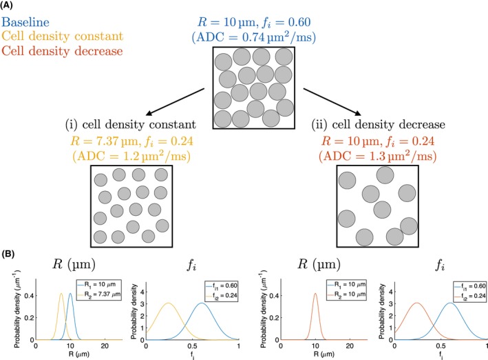
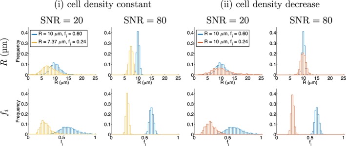
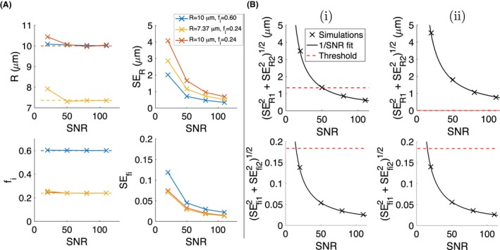
Simulations with optimized and non-optimized clinical acquisitions were performed for a range of microstructures, using a two-compartment model. The ability to distinguish between (i) cell shrinkage with cell density constant, mimicking apoptosis, and (ii) cell size constant with cell density decreasing, mimicking loss of cells, was evaluated based on the precision of simulated parameter estimates. Relationships between parameter precision, SNR, and the magnitude of specific parameter changes, were used to infer SNR requirements for detecting changes.

RESULTS

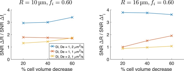
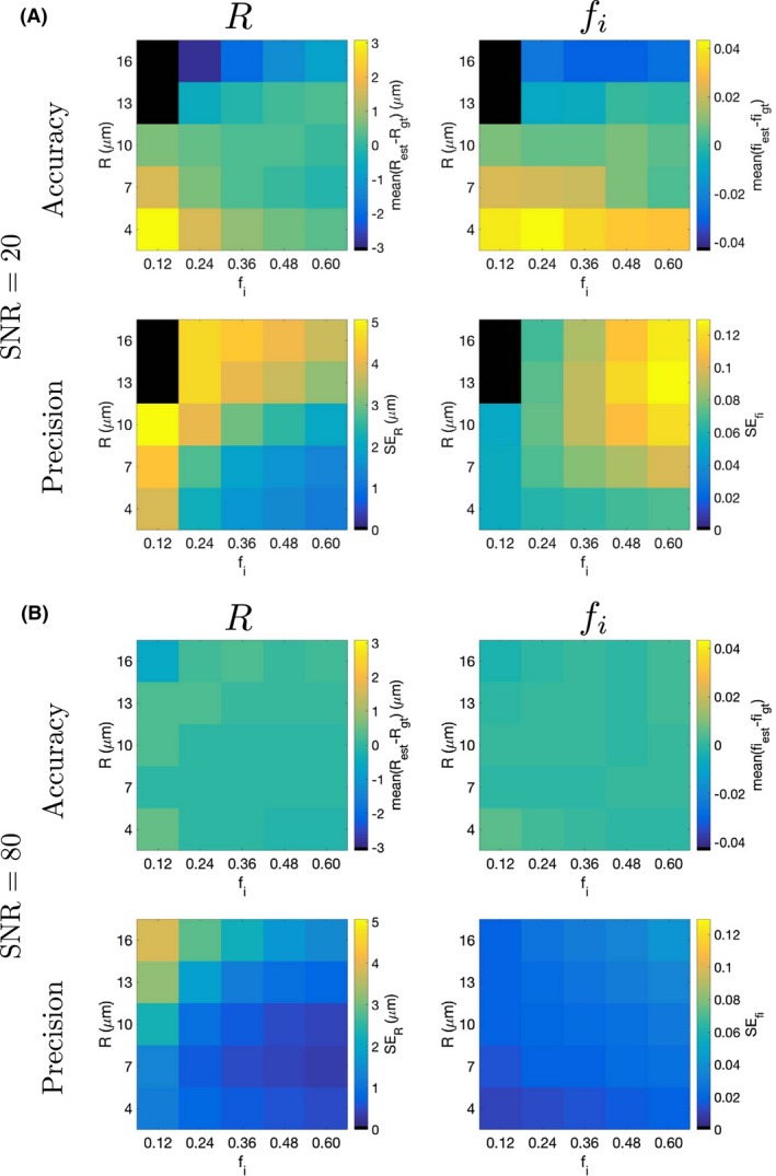
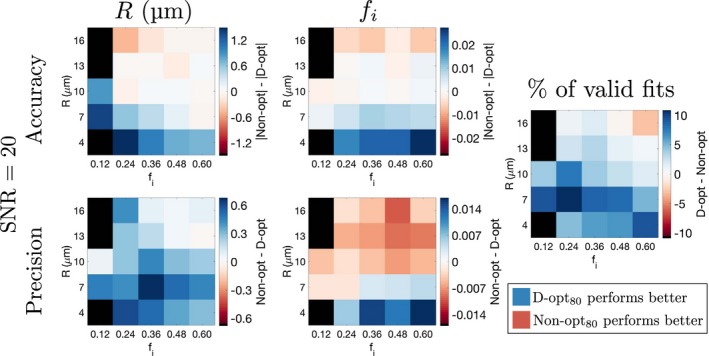
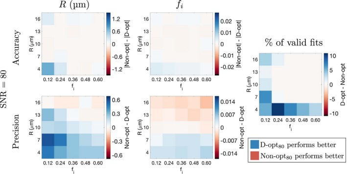
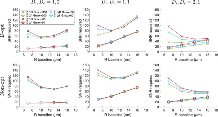
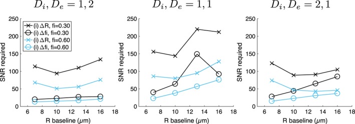
Accuracy and precision depended on microstructural properties, SNR, and the acquisition protocol. The main benefit of optimized acquisitions tended to be improved accuracy and precision of R, particularly for small cells. In most cases considered, higher SNR was required for detecting changes in R than for changes in f .

CONCLUSIONS

Given the relative changes in R and f due to apoptosis, simulations indicate that, for a range of microstructures, detecting changes in R require higher SNR than detecting changes in f , and that such SNR is typically not achieved in clinical data. This suggests that if apoptotic cell size decreases are to be detected in clinical settings, improved SNR is required. Comparing measurement precision with the magnitude of expected biological changes should form part of the validation process for potential biomarkers.

摘要

目的

确定从 DW-MRI 数据中提取足够精确的细胞半径(R)和细胞内体积分数(f)估计值的可行性,以便区分组织可能经历的特定微观结构变化,特别是聚焦于肿瘤中的细胞死亡。

方法

针对一系列微观结构,使用两室模型对优化和非优化的临床采集进行了模拟。基于模拟参数估计的精度,评估了区分(i)细胞密度不变时的细胞收缩,模拟细胞凋亡,和(ii)细胞大小不变时的细胞密度降低,模拟细胞丢失的能力。参数精度、信噪比(SNR)与特定参数变化幅度之间的关系用于推断检测变化所需的 SNR 要求。

结果

准确性和精密度取决于微观结构特性、SNR 和采集协议。优化采集的主要优势往往是提高 R 的准确性和精密度,特别是对于小细胞。在考虑的大多数情况下,检测 R 变化所需的 SNR 高于检测 f 变化。

结论

鉴于细胞凋亡引起的 R 和 f 的相对变化,模拟表明,对于一系列微观结构,检测 R 的变化所需的 SNR 高于检测 f 的变化,而在临床数据中通常无法达到这种 SNR。这表明,如果要在临床环境中检测到凋亡细胞大小的减小,则需要提高 SNR。将测量精度与预期的生物学变化幅度进行比较,应成为潜在生物标志物验证过程的一部分。

https://cdn.ncbi.nlm.nih.gov/pmc/blobs/ea50/6492139/9c300745bb6b/MRM-81-2288-g009.jpg
https://cdn.ncbi.nlm.nih.gov/pmc/blobs/ea50/6492139/b2a66fb79ae7/MRM-81-2288-g001.jpg
https://cdn.ncbi.nlm.nih.gov/pmc/blobs/ea50/6492139/5130a5c0a07b/MRM-81-2288-g002.jpg
https://cdn.ncbi.nlm.nih.gov/pmc/blobs/ea50/6492139/0c9cf1e4a092/MRM-81-2288-g003.jpg
https://cdn.ncbi.nlm.nih.gov/pmc/blobs/ea50/6492139/004f04c0afb6/MRM-81-2288-g004.jpg
https://cdn.ncbi.nlm.nih.gov/pmc/blobs/ea50/6492139/3d393c725099/MRM-81-2288-g005.jpg
https://cdn.ncbi.nlm.nih.gov/pmc/blobs/ea50/6492139/76a4ad7b13cc/MRM-81-2288-g006.jpg
https://cdn.ncbi.nlm.nih.gov/pmc/blobs/ea50/6492139/959987392f59/MRM-81-2288-g007.jpg
https://cdn.ncbi.nlm.nih.gov/pmc/blobs/ea50/6492139/4d50ec6b1f46/MRM-81-2288-g008.jpg
https://cdn.ncbi.nlm.nih.gov/pmc/blobs/ea50/6492139/9c300745bb6b/MRM-81-2288-g009.jpg

相似文献

1
Towards a 'resolution limit' for DW-MRI tumor microstructural models: A simulation study investigating the feasibility of distinguishing between microstructural changes.迈向 DW-MRI 肿瘤微结构模型的“分辨率极限”:一项探究区分微结构变化可行性的模拟研究
Magn Reson Med. 2019 Apr;81(4):2288-2301. doi: 10.1002/mrm.27551. Epub 2018 Oct 19.
2
A simulation study of cell size and volume fraction mapping for tissue with two underlying cell populations using diffusion-weighted MRI.基于扩散加权 MRI 对具有两种潜在细胞群体的组织进行细胞大小和体积分数映射的模拟研究。
Magn Reson Med. 2021 Aug;86(2):1029-1044. doi: 10.1002/mrm.28694. Epub 2021 Feb 28.
3
Assessing tumor microstructure with time-dependent diffusion imaging: Considerations and feasibility on clinical MRI and MRI-Linac.利用时间依赖性扩散成像评估肿瘤微观结构:临床MRI和MRI直线加速器的考量与可行性
Med Phys. 2025 Jan;52(1):346-361. doi: 10.1002/mp.17453. Epub 2024 Oct 10.
4
Towards microstructure fingerprinting: Estimation of tissue properties from a dictionary of Monte Carlo diffusion MRI simulations.面向微观结构特征分析:从蒙特卡罗扩散 MRI 模拟字典中估计组织特性。
Neuroimage. 2019 Jan 1;184:964-980. doi: 10.1016/j.neuroimage.2018.09.076. Epub 2018 Sep 30.
5
High-resolution diffusion-weighted imaging at 7 Tesla: Single-shot readout trajectories and their impact on signal-to-noise ratio, spatial resolution and accuracy.7特斯拉高分辨率扩散加权成像:单次读出轨迹及其对信噪比、空间分辨率和准确性的影响。
Neuroimage. 2023 Jul 1;274:120159. doi: 10.1016/j.neuroimage.2023.120159. Epub 2023 May 5.
6
Assessing the accuracy of using oscillating gradient spin echo sequences with AxCaliber to infer micron-sized axon diameters.评估使用AxCaliber振荡梯度自旋回波序列推断微米级轴突直径的准确性。
MAGMA. 2017 Feb;30(1):1-14. doi: 10.1007/s10334-016-0575-y. Epub 2016 Jul 13.
7
Reliable estimation of incoherent motion parametric maps from diffusion-weighted MRI using fusion bootstrap moves.基于融合引导移动的扩散加权 MRI 非相干运动参数图的可靠估计。
Med Image Anal. 2013 Apr;17(3):325-36. doi: 10.1016/j.media.2012.12.001. Epub 2013 Jan 3.
8
Mapping axonal density and average diameter using non-monotonic time-dependent gradient-echo MRI.使用非单调时间依赖梯度回波MRI绘制轴突密度和平均直径图。
J Magn Reson. 2017 Apr;277:117-130. doi: 10.1016/j.jmr.2017.02.017. Epub 2017 Feb 28.
9
Determination of optimized set of b-values for Apparent Diffusion Coefficient mapping in liver Diffusion-Weighted MRI.肝脏磁共振弥散加权成像中表观弥散系数图的优化 b 值组的确定。
J Magn Reson. 2020 Jan;310:106634. doi: 10.1016/j.jmr.2019.106634. Epub 2019 Oct 31.
10
Microstructural characterization of normal and malignant human prostate tissue with vascular, extracellular, and restricted diffusion for cytometry in tumours magnetic resonance imaging.通过肿瘤磁共振成像中的血管、细胞外和受限扩散对正常和恶性人类前列腺组织进行微观结构表征以用于细胞计数。
Invest Radiol. 2015 Apr;50(4):218-27. doi: 10.1097/RLI.0000000000000115.

引用本文的文献

1
Diffusion phase-imaging in anisotropic media using non-linear gradients for diffusion encoding.各向异性介质中扩散相位成像的非线性梯度扩散编码。
PLoS One. 2023 Mar 30;18(3):e0281332. doi: 10.1371/journal.pone.0281332. eCollection 2023.
2
Diffusion MRI signal cumulants and hepatocyte microstructure at fixed diffusion time: Insights from simulations, 9.4T imaging, and histology.固定扩散时间下的扩散 MRI 信号累积量和肝细胞微观结构:来自模拟、9.4T 成像和组织学的见解。
Magn Reson Med. 2022 Jul;88(1):365-379. doi: 10.1002/mrm.29174. Epub 2022 Feb 18.
3
Initial Condition Assessment for Reaction-Diffusion Glioma Growth Models: A Translational MRI-Histology (In)Validation Study.

本文引用的文献

1
Microstructure Characterization of Bone Metastases from Prostate Cancer with Diffusion MRI: Preliminary Findings.前列腺癌骨转移的扩散磁共振成像微观结构特征:初步研究结果
Front Oncol. 2018 Feb 16;8:26. doi: 10.3389/fonc.2018.00026. eCollection 2018.
2
On modeling.关于建模。
Magn Reson Med. 2018 Jun;79(6):3172-3193. doi: 10.1002/mrm.27101. Epub 2018 Mar 1.
3
Imaging brain microstructure with diffusion MRI: practicality and applications.用弥散 MRI 成像大脑微观结构:实用性和应用。
反应扩散脑胶质瘤生长模型的初始条件评估:转化 MRI-组织学(在)验证研究。
Tomography. 2021 Oct 29;7(4):650-674. doi: 10.3390/tomography7040055.
4
The impeded diffusion fraction quantitative imaging assay demonstrated in multi-exponential diffusion phantom and prostate cancer.多指数扩散体模和前列腺癌中受限扩散分数定量成像检测。
Magn Reson Med. 2022 Apr;87(4):2053-2062. doi: 10.1002/mrm.29075. Epub 2021 Nov 14.
5
MR cell size imaging with temporal diffusion spectroscopy.采用时扩散光谱法的磁共振细胞大小成像。
Magn Reson Imaging. 2021 Apr;77:109-123. doi: 10.1016/j.mri.2020.12.010. Epub 2020 Dec 16.
6
Diffusion model comparison identifies distinct tumor sub-regions and tracks treatment response.扩散模型比较可识别不同的肿瘤亚区域并追踪治疗反应。
Magn Reson Med. 2020 Sep;84(3):1250-1263. doi: 10.1002/mrm.28196. Epub 2020 Feb 14.
NMR Biomed. 2019 Apr;32(4):e3841. doi: 10.1002/nbm.3841. Epub 2017 Nov 29.
4
TE dependent Diffusion Imaging (TEdDI) distinguishes between compartmental T relaxation times.TE 依赖性扩散成像(TEdDI)可区分隔室 T 弛豫时间。
Neuroimage. 2018 Nov 15;182:360-369. doi: 10.1016/j.neuroimage.2017.09.030. Epub 2017 Sep 19.
5
Low frequency oscillating gradient spin-echo sequences improve sensitivity to axon diameter: An experimental study in viable nerve tissue.低频振荡梯度回波序列提高了对轴径的敏感性:活神经组织的实验研究。
Neuroimage. 2018 Nov 15;182:314-328. doi: 10.1016/j.neuroimage.2017.07.060. Epub 2017 Aug 1.
6
Resolution limit of cylinder diameter estimation by diffusion MRI: The impact of gradient waveform and orientation dispersion.扩散磁共振成像估计圆柱直径的分辨率极限:梯度波形和方向离散度的影响。
NMR Biomed. 2017 Jul;30(7). doi: 10.1002/nbm.3711. Epub 2017 Mar 20.
7
Imaging biomarker roadmap for cancer studies.癌症研究的影像生物标志物路线图。
Nat Rev Clin Oncol. 2017 Mar;14(3):169-186. doi: 10.1038/nrclinonc.2016.162. Epub 2016 Oct 11.
8
In vivo imaging of cancer cell size and cellularity using temporal diffusion spectroscopy.利用时间扩散光谱对癌细胞大小和细胞密度进行体内成像。
Magn Reson Med. 2017 Jul;78(1):156-164. doi: 10.1002/mrm.26356. Epub 2016 Aug 6.
9
Pulsed and oscillating gradient MRI for assessment of cell size and extracellular space (POMACE) in mouse gliomas.用于评估小鼠胶质瘤细胞大小和细胞外间隙的脉冲与振荡梯度磁共振成像(POMACE)
NMR Biomed. 2016 Oct;29(10):1350-63. doi: 10.1002/nbm.3577. Epub 2016 Jul 22.
10
In vivo quantification of demyelination and recovery using compartment-specific diffusion MRI metrics validated by electron microscopy.使用电子显微镜验证的基于体素的弥散 MRI 指标对脱髓鞘和恢复进行体内定量评估。
Neuroimage. 2016 May 15;132:104-114. doi: 10.1016/j.neuroimage.2016.02.004. Epub 2016 Feb 11.