Nielsen Emil Østergaard, Chen Li, Hansen Jonas Overgaard, Degn Matilda, Overgaard Søren, Ding Ming
Orthopaedic Research Laboratory, Department of Orthopaedic Surgery and Traumatology, Odense University Hospital, Department of Clinical Research, University of Southern Denmark, Sdr. Boulevard 29, 5000 Odense, Denmark.
Department of Endocrinology and Metabolism, Molecular Endocrinology Laboratory (KMEB), Odense University Hospital, University of Southern Denmark, J. B. Winsløws Vej 25.1, 5000 Odense, Denmark.
Stem Cells Int. 2018 Sep 26;2018:9781393. doi: 10.1155/2018/9781393. eCollection 2018.
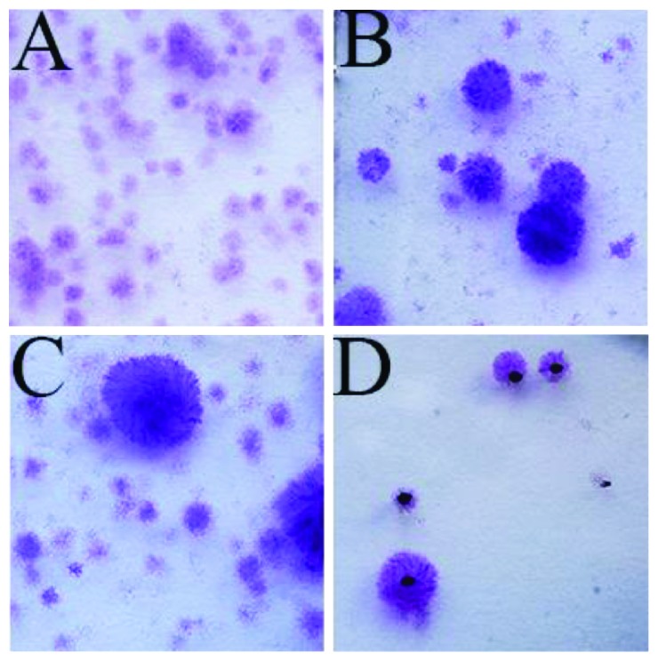
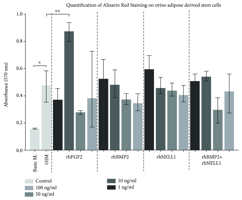
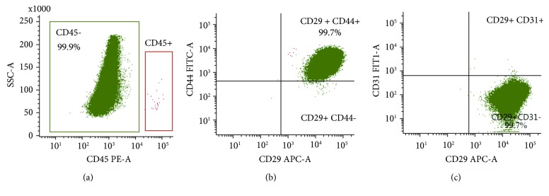
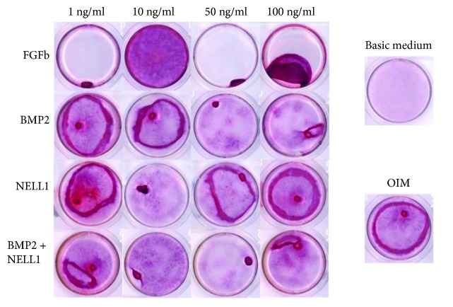
Although adipose-derived stromal cells (ADSCs) have been a major focus as an alternative to autologous bone graft in orthopedic surgery, bone formation potential of ADSCs is not well known and cytokines as osteogenic inducers on ADSCs are being investigated. This study aimed at isolating ADSCs from ovine adipose tissue (AT) and optimizing osteogenic differentiation of ovine ADSCs (oADSC) by culture medium and growth factors. Four AT samples were harvested from two female ovine (Texel/Gotland breed), and oADSCs were isolated and analyzed by flow cytometry for surface markers CD29, CD44, CD31, and CD45. Osteogenic differentiation was made in vitro by seeding oADSCs in osteogenic induction medium (OIM) containing fibroblast growth factor basic (FGFb), bone morphogenetic protein 2 (BMP2), or NEL-like molecule 1 (NELL1) in 4 different dosages (1, 10, 50, and 100 ng/ml, respectively). Basic medium (DMEM) was used as control. Analysis was made after 14 days by Alizarin red staining (ARS) and quantification. This study successfully harvested AT from ovine and verified isolated cells for minimal criteria for adipose stromal cells which suggests a feasible method for isolation of oADSCs. OIM showed significantly higher ARS to basic medium, and FGFb 10 ng/ml revealed significantly higher ARS to OIM alone after 14 days.
尽管脂肪来源的间充质干细胞(ADSCs)作为骨科手术中自体骨移植的替代物一直是主要研究焦点,但ADSCs的骨形成潜力尚不明确,目前正在研究将细胞因子作为ADSCs的成骨诱导剂。本研究旨在从绵羊脂肪组织(AT)中分离ADSCs,并通过培养基和生长因子优化绵羊ADSCs(oADSCs)的成骨分化。从两只雌性绵羊(特克塞尔/哥特兰品种)采集了4个AT样本,通过流式细胞术对分离出的oADSCs进行表面标志物CD29、CD44、CD31和CD45的分析。将oADSCs接种于含有不同剂量(分别为1、10、50和100 ng/ml)碱性成纤维细胞生长因子(FGFb)、骨形态发生蛋白2(BMP2)或NEL样分子1(NELL1)的成骨诱导培养基(OIM)中进行体外成骨分化。以基础培养基(DMEM)作为对照。14天后通过茜素红染色(ARS)和定量分析。本研究成功从绵羊身上采集了AT,并验证了分离出的细胞符合脂肪间充质干细胞的最低标准,这表明oADSCs的分离方法可行。14天后,OIM组的ARS明显高于基础培养基组,单独使用10 ng/ml FGFb组的ARS明显高于单独使用OIM组。