School of Optometry, University of Alabama at Birmingham, Birmingham, AL, 35233, USA.
Department of Ophthalmology, University of Alabama at Birmingham, Birmingham, AL, 35233, USA.
Biomed Eng Online. 2018 Nov 1;17(1):164. doi: 10.1186/s12938-018-0597-y.
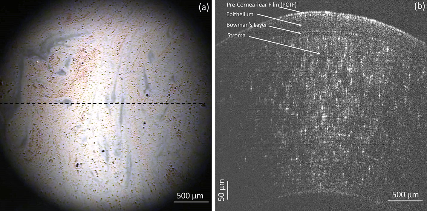
The outermost layer of the tear film consists of a thin lipid layer (LL). The lipid layer serves as a barrier against evaporation of the aqueous component of the tear film. The ability to simultaneously image both the lipid layer thickness and overall tear film thickness is novel, and will help further understandings of mechanisms of how the lipid layer assembles and interacts with the full tear film thickness.
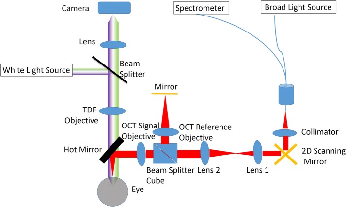
We developed a system that combines simultaneous optical coherence tomography (OCT) and thickness dependent fringes (TDF) interferometry for in vivo imaging of the tear film. The OCT possesses an axial resolution of 1.38 µm in tear film, providing an accurate measurement of the thickness of the overall tear film. The TDF can detect a minimal change of approximately 15 nm in LL thickness. In addition, the spatial resolution of TDF images in x-y plane is 5 µm.
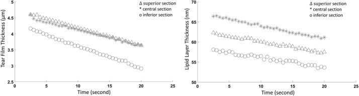
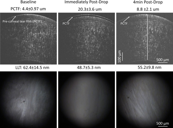
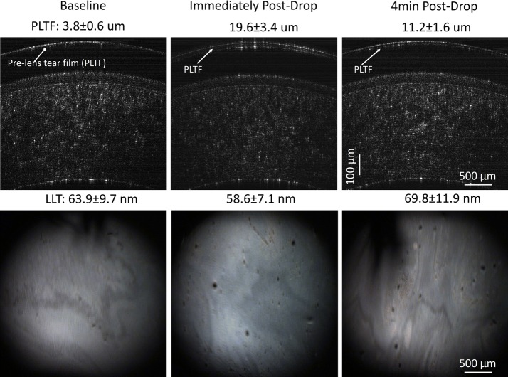
The effect of instilling artificial tears on the PCTF and PLTF was examined. In both contact lens and non-contact lens wear, it could be observed from the OCT results that instillation of artificial tears increased the thickness of the overall tear film immediately, followed by a gradual reduction thereafter. These findings were consistent with other studies. However, unlike those previous reports, the thickness of the LL in this study was quantified simultaneously with the TDF subsystem. The results showed that bulking agents such as these artificial tears were not necessarily intended to increase the LL thickness. Immediately after instillation of artificial tears, the PCTF increased from 4.4 ± 0.97 to 20.3 ± 3.6 µm. The PCTF then decreased to 8.8 ± 2.1 µm at 4 min post-instillation. The thicknesses of the LL were 62.4 ± 14.5 nm, 48.7 ± 5.3 nm, and 55.2 ± 9.8 nm at pre-drop instillation, post-drop instillation, and 4-min post-drop instillation, respectively.
In this work, we have described a novel imaging system that integrated OCT and TDF imaging techniques, which may facilitate the study of many physiological and clinical aspects of the tear film.
泪膜的最外层是一层很薄的脂质层(LL)。脂质层可以防止泪膜中的水成分蒸发。能够同时对脂质层厚度和整个泪膜厚度进行成像的能力是新颖的,这将有助于进一步了解脂质层如何组装以及与整个泪膜厚度相互作用的机制。
我们开发了一种系统,该系统结合了光学相干断层扫描(OCT)和厚度相关条纹(TDF)干涉测量技术,用于活体成像泪膜。OCT 在泪膜中的轴向分辨率为 1.38 µm,可以准确测量整个泪膜的厚度。TDF 可以检测到约 15 nm 的 LL 厚度的微小变化。此外,TDF 图像在 x-y 平面上的空间分辨率为 5 µm。
检查了人工泪液对 PCTF 和 PLTF 的影响。在接触镜和非接触镜佩戴中,都可以从 OCT 结果中观察到,人工泪液的滴注立即增加了整个泪膜的厚度,随后逐渐减少。这些发现与其他研究一致。但是,与之前的报告不同,这项研究中同时使用 TDF 子系统定量了 LL 的厚度。结果表明,这些人工泪液等膨胀剂不一定旨在增加 LL 厚度。人工泪液滴注后立即,PCTF 从 4.4±0.97 µm 增加到 20.3±3.6 µm。PCTF 在滴注后 4 分钟时降低至 8.8±2.1 µm。在滴注前、滴注后和滴注后 4 分钟时,LL 的厚度分别为 62.4±14.5 nm、48.7±5.3 nm 和 55.2±9.8 nm。
在这项工作中,我们描述了一种新颖的成像系统,该系统集成了 OCT 和 TDF 成像技术,这可能有助于研究泪膜的许多生理和临床方面。