• 文献检索
  • 文档翻译
  • 深度研究
  • 学术资讯
  • Suppr Zotero 插件Zotero 插件
  • 邀请有礼
  • 套餐&价格
  • 历史记录
应用&插件
Suppr Zotero 插件Zotero 插件浏览器插件Mac 客户端Windows 客户端微信小程序
定价
高级版会员购买积分包购买API积分包
服务
文献检索文档翻译深度研究API 文档MCP 服务
关于我们
关于 Suppr公司介绍联系我们用户协议隐私条款
关注我们

Suppr 超能文献

核心技术专利:CN118964589B侵权必究
粤ICP备2023148730 号-1Suppr @ 2026

文献检索

告别复杂PubMed语法,用中文像聊天一样搜索,搜遍4000万医学文献。AI智能推荐,让科研检索更轻松。

立即免费搜索

文件翻译

保留排版,准确专业,支持PDF/Word/PPT等文件格式,支持 12+语言互译。

免费翻译文档

深度研究

AI帮你快速写综述,25分钟生成高质量综述,智能提取关键信息,辅助科研写作。

立即免费体验

磷脂酰丝氨酸是轴突碎片吞噬的标志物,但它的暴露可以与退化脱钩。

Phosphatidylserine is a marker for axonal debris engulfment but its exposure can be decoupled from degeneration.

机构信息

Department of Biomolecular Sciences, Weizmann Institute of Science, Rehovot, 76100, Israel.

出版信息

Cell Death Dis. 2018 Nov 2;9(11):1116. doi: 10.1038/s41419-018-1155-z.

DOI:10.1038/s41419-018-1155-z
PMID:30389906
原文链接:https://pmc.ncbi.nlm.nih.gov/articles/PMC6214901/
Abstract

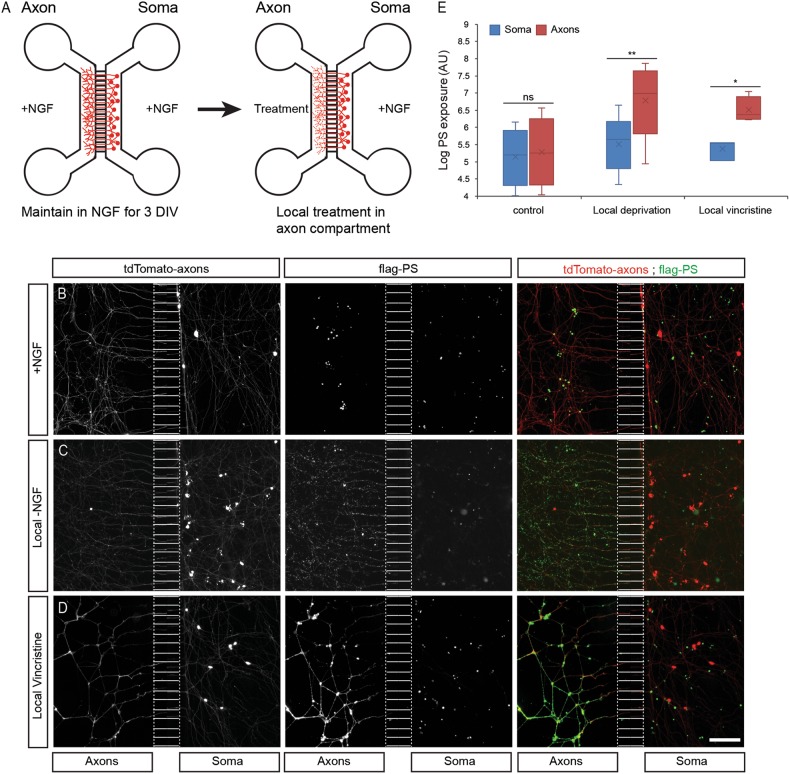
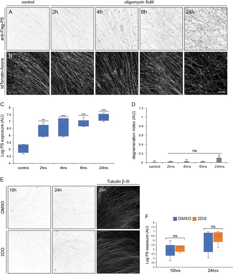
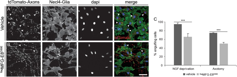
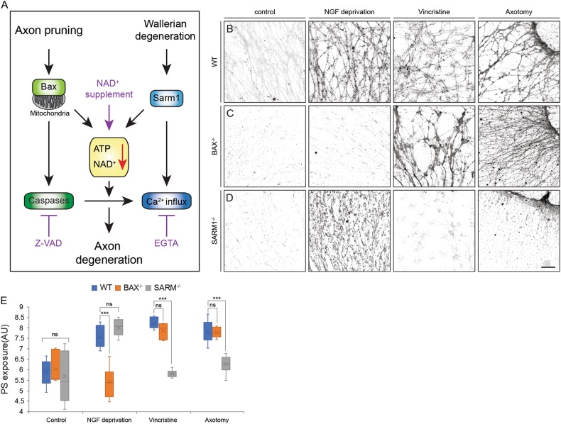
Apoptotic cells expose Phosphatidylserine (PS), that serves as an "eat me" signal for engulfing cells. Previous studies have shown that PS also marks degenerating axonsduring developmental pruning or in response to insults (Wallerian degeneration), but the pathways that control PS exposure on degenerating axons are largely unknown. Here, we used a series of in vitro assays to systematically explore the regulation of PS exposure during axonal degeneration. Our results show that PS exposure is regulated by the upstream activators of axonal pruning and Wallerian degeneration. However, our investigation of signaling further downstream revealed divergence between axon degeneration and PS exposure. Importantly, elevation of the axonal energetic status hindered PS exposure, while inhibition of mitochondrial activity caused PS exposure, without degeneration. Overall, our results suggest that the levels of PS on the outer axonal membrane can be dissociated from the degeneration process and that the axonal energetic status plays a key role in the regulation of PS exposure.

摘要

凋亡细胞暴露磷脂酰丝氨酸(PS),PS 作为吞噬细胞的“吃我”信号。先前的研究表明,PS 也标记发育性修剪过程中或受到损伤时(Wallerian 变性)退化轴突,但控制退化轴突上 PS 暴露的途径在很大程度上是未知的。在这里,我们使用一系列体外测定法系统地研究了轴突变性过程中 PS 暴露的调控。我们的结果表明,PS 暴露受轴突修剪和 Wallerian 变性的上游激活剂调控。然而,我们对信号的进一步下游研究表明,轴突变性和 PS 暴露之间存在分歧。重要的是,升高轴突能量状态会阻碍 PS 暴露,而抑制线粒体活性则会导致 PS 暴露而不导致变性。总的来说,我们的结果表明,外轴突膜上 PS 的水平可以与变性过程分离,并且轴突能量状态在 PS 暴露的调控中起着关键作用。

https://cdn.ncbi.nlm.nih.gov/pmc/blobs/6bc2/6214901/dc01d8bb67a1/41419_2018_1155_Fig7_HTML.jpg
https://cdn.ncbi.nlm.nih.gov/pmc/blobs/6bc2/6214901/33ceaa7ad386/41419_2018_1155_Fig1_HTML.jpg
https://cdn.ncbi.nlm.nih.gov/pmc/blobs/6bc2/6214901/e289505f17b6/41419_2018_1155_Fig2_HTML.jpg
https://cdn.ncbi.nlm.nih.gov/pmc/blobs/6bc2/6214901/75e924f236dc/41419_2018_1155_Fig3_HTML.jpg
https://cdn.ncbi.nlm.nih.gov/pmc/blobs/6bc2/6214901/197050b8f187/41419_2018_1155_Fig4_HTML.jpg
https://cdn.ncbi.nlm.nih.gov/pmc/blobs/6bc2/6214901/d32ac4a70585/41419_2018_1155_Fig5_HTML.jpg
https://cdn.ncbi.nlm.nih.gov/pmc/blobs/6bc2/6214901/009631569eac/41419_2018_1155_Fig6_HTML.jpg
https://cdn.ncbi.nlm.nih.gov/pmc/blobs/6bc2/6214901/dc01d8bb67a1/41419_2018_1155_Fig7_HTML.jpg

相似文献

1
Phosphatidylserine is a marker for axonal debris engulfment but its exposure can be decoupled from degeneration.磷脂酰丝氨酸是轴突碎片吞噬的标志物,但它的暴露可以与退化脱钩。
Cell Death Dis. 2018 Nov 2;9(11):1116. doi: 10.1038/s41419-018-1155-z.
2
Regulation of axon degeneration after injury and in development by the endogenous calpain inhibitor calpastatin.内源性钙蛋白酶抑制剂钙蛋白酶抑制素调控损伤和发育过程中的轴突变性。
Neuron. 2013 Dec 4;80(5):1175-89. doi: 10.1016/j.neuron.2013.08.034. Epub 2013 Nov 7.
3
Phagocytosis and self-destruction break down dendrites of sensory neurons at distinct steps of Wallerian degeneration.吞噬作用和自我破坏在 Wallerian 变性的不同步骤中断裂感觉神经元的树突。
Proc Natl Acad Sci U S A. 2022 Jan 25;119(4). doi: 10.1073/pnas.2111818119.
4
GSK3B-mediated phosphorylation of MCL1 regulates axonal autophagy to promote Wallerian degeneration.糖原合成酶激酶3β介导的MCL1磷酸化调节轴突自噬以促进沃勒变性。
J Cell Biol. 2017 Feb;216(2):477-493. doi: 10.1083/jcb.201606020. Epub 2017 Jan 4.
5
Toll/interleukin-1 receptor domain-containing adapter inducing interferon-β mediates microglial phagocytosis of degenerating axons.Toll/白细胞介素-1 受体结构域包含衔接子诱导干扰素-β 介导小胶质细胞吞噬退变轴突。
J Neurosci. 2012 May 30;32(22):7745-57. doi: 10.1523/JNEUROSCI.0203-12.2012.
6
The gene for slow Wallerian degeneration (Wld(s)) is also protective against vincristine neuropathy.慢沃勒氏变性基因(Wld(s))对长春新碱神经病变也具有保护作用。
Neurobiol Dis. 2001 Feb;8(1):155-61. doi: 10.1006/nbdi.2000.0334.
7
The dynein inhibitor Ciliobrevin D inhibits the bidirectional transport of organelles along sensory axons and impairs NGF-mediated regulation of growth cones and axon branches.动力蛋白抑制剂Ciliobrevin D可抑制细胞器沿感觉轴突的双向运输,并损害神经生长因子介导的生长锥和轴突分支调节。
Dev Neurobiol. 2015 Jul;75(7):757-77. doi: 10.1002/dneu.22246. Epub 2014 Nov 20.
8
Specific phospholipid scramblases are involved in exposure of phosphatidylserine, an "eat-me" signal for phagocytes, on degenerating axons.特定的磷脂翻转酶参与了磷脂酰丝氨酸(一种吞噬细胞的“吃我”信号)在退化轴突上的暴露过程。
Commun Integr Biol. 2017 Feb 17;10(2):e1296615. doi: 10.1080/19420889.2017.1296615. eCollection 2017.
9
Wallerian Degeneration Is Executed by an NMN-SARM1-Dependent Late Ca(2+) Influx but Only Modestly Influenced by Mitochondria.Wallerian 变性是由 NMN-SARM1 依赖的晚期 Ca(2+)内流执行的,但受线粒体的影响较小。
Cell Rep. 2015 Dec 22;13(11):2539-2552. doi: 10.1016/j.celrep.2015.11.032. Epub 2015 Dec 10.
10
The WldS protein protects against axonal degeneration: a model of gene therapy for peripheral neuropathy.WldS蛋白可预防轴突退化:一种用于治疗周围神经病变的基因疗法模型。
Ann Neurol. 2001 Dec;50(6):773-9. doi: 10.1002/ana.10039.

引用本文的文献

1
Sigma-1 receptor activation by PRE-084 attenuates sepsis-associated encephalopathy by targeting microglial p38 MAPK-mediated neuroinflammation and neuronal endoplasmic reticulum stress.PRE-084激活西格玛-1受体可通过靶向小胶质细胞p38丝裂原活化蛋白激酶介导的神经炎症和神经元内质网应激来减轻脓毒症相关脑病。
Inflamm Res. 2025 Sep 4;74(1):117. doi: 10.1007/s00011-025-02086-5.
2
Neuron-to-glia signaling drives critical period experience-dependent synapse pruning.神经元与神经胶质细胞间的信号传导驱动关键期内依赖经验的突触修剪。
Sci Rep. 2025 Jul 16;15(1):25744. doi: 10.1038/s41598-025-11528-3.
3
Suppressing phagocyte activation by overexpressing the phosphatidylserine lipase ABHD12 preserves sarmopathic nerves.

本文引用的文献

1
Phosphatidylserine Externalization Results from and Causes Neurite Degeneration in Drosophila.磷脂酰丝氨酸外化是果蝇轴突退化的结果和原因。
Cell Rep. 2018 Aug 28;24(9):2273-2286. doi: 10.1016/j.celrep.2018.07.095.
2
Specific phospholipid scramblases are involved in exposure of phosphatidylserine, an "eat-me" signal for phagocytes, on degenerating axons.特定的磷脂翻转酶参与了磷脂酰丝氨酸(一种吞噬细胞的“吃我”信号)在退化轴突上的暴露过程。
Commun Integr Biol. 2017 Feb 17;10(2):e1296615. doi: 10.1080/19420889.2017.1296615. eCollection 2017.
3
Axonal Degeneration in Retinal Ganglion Cells Is Associated with a Membrane Polarity-Sensitive Redox Process.
通过过表达磷脂酰丝氨酸脂肪酶ABHD12抑制吞噬细胞活化可保护脊髓性肌萎缩症神经。
iScience. 2025 May 9;28(6):112626. doi: 10.1016/j.isci.2025.112626. eCollection 2025 Jun 20.
4
Phagocytosis-driven neurodegeneration through opposing roles of an ABC transporter in neurons and phagocytes.通过ABC转运蛋白在神经元和吞噬细胞中的相反作用,吞噬作用驱动神经退行性变。
Sci Adv. 2025 Mar 14;11(11):eadr5448. doi: 10.1126/sciadv.adr5448. Epub 2025 Mar 12.
5
Axonal spheroids are regulated by Schwann cells after peripheral nerve injury.轴突球体在周围神经损伤后受施万细胞调控。
bioRxiv. 2024 Nov 8:2024.11.08.622649. doi: 10.1101/2024.11.08.622649.
6
Epigenetic and 3D genome reprogramming during the aging of human hippocampus.人类海马体衰老过程中的表观遗传和三维基因组重编程。
bioRxiv. 2024 Oct 17:2024.10.14.618338. doi: 10.1101/2024.10.14.618338.
7
Microglia Gravitate toward Amyloid Plaques Surrounded by Externalized Phosphatidylserine via TREM2.小胶质细胞通过 TREM2 向被磷脂酰丝氨酸外化包围的淀粉样斑块迁移。
Adv Sci (Weinh). 2024 Sep;11(34):e2400064. doi: 10.1002/advs.202400064. Epub 2024 Jul 9.
8
Mast cell-derived BH4 is a critical mediator of postoperative pain.肥大细胞衍生的四氢生物蝶呤是术后疼痛的关键介质。
bioRxiv. 2023 Jan 24:2023.01.24.525378. doi: 10.1101/2023.01.24.525378.
9
The chemokine-like Orion bridges phosphatidylserine and Draper in phagocytosis of neurons.趋化因子样 Orion 在神经元吞噬作用中连接磷脂酰丝氨酸和 Draper。
Proc Natl Acad Sci U S A. 2023 Jun 13;120(24):e2303392120. doi: 10.1073/pnas.2303392120. Epub 2023 Jun 5.
10
Phospholipid scramblase Xkr8 is required for developmental axon pruning via phosphatidylserine exposure.磷脂翻转酶 Xkr8 通过暴露磷脂酰丝氨酸参与发育性轴突修剪。
EMBO J. 2023 Jul 17;42(14):e111790. doi: 10.15252/embj.2022111790. Epub 2023 May 22.
视网膜神经节细胞中的轴突退变与膜极性敏感的氧化还原过程有关。
J Neurosci. 2017 Apr 5;37(14):3824-3839. doi: 10.1523/JNEUROSCI.3882-16.2017. Epub 2017 Mar 8.
4
NMN Deamidase Delays Wallerian Degeneration and Rescues Axonal Defects Caused by NMNAT2 Deficiency In Vivo.NMN 脱氨酶延迟 Wallerian 变性并挽救 NMNAT2 缺乏引起的体内轴突缺陷。
Curr Biol. 2017 Mar 20;27(6):784-794. doi: 10.1016/j.cub.2017.01.070. Epub 2017 Mar 2.
5
Axon degeneration induces glial responses through Draper-TRAF4-JNK signalling.轴突变性通过 Draper-TRAF4-JNK 信号诱导神经胶质反应。
Nat Commun. 2017 Feb 6;8:14355. doi: 10.1038/ncomms14355.
6
Phosphatidylserine Exposure Controls Viral Innate Immune Responses by Microglia.磷脂酰丝氨酸暴露通过小胶质细胞控制病毒先天性免疫反应。
Neuron. 2017 Feb 8;93(3):574-586.e8. doi: 10.1016/j.neuron.2016.12.021. Epub 2017 Jan 19.
7
GSK3B-mediated phosphorylation of MCL1 regulates axonal autophagy to promote Wallerian degeneration.糖原合成酶激酶3β介导的MCL1磷酸化调节轴突自噬以促进沃勒变性。
J Cell Biol. 2017 Feb;216(2):477-493. doi: 10.1083/jcb.201606020. Epub 2017 Jan 4.
8
NMNAT1 inhibits axon degeneration via blockade of SARM1-mediated NAD depletion.烟酰胺单核苷酸腺苷转移酶1通过阻断含 sterile α 和 armadillo 基序的蛋白1介导的烟酰胺腺嘌呤二核苷酸消耗来抑制轴突退化。
Elife. 2016 Oct 13;5:e19749. doi: 10.7554/eLife.19749.
9
Microglia: Phagocytosing to Clear, Sculpt, and Eliminate.小胶质细胞:吞噬以清除、塑造和消除。
Dev Cell. 2016 Jul 25;38(2):126-8. doi: 10.1016/j.devcel.2016.07.006.
10
Microglia contribute to circuit defects in Mecp2 null mice independent of microglia-specific loss of Mecp2 expression.小胶质细胞导致Mecp2基因敲除小鼠的神经回路缺陷,这与小胶质细胞特异性缺失Mecp2表达无关。
Elife. 2016 Jul 26;5:e15224. doi: 10.7554/eLife.15224.