Orthodontic Department, Stomatology Hospital of Chongqing Medical University, Chongqing Key Laboratory of Oral Disease and Biomedical Sciences, Chongqing Municipal Key Laboratory, Chongqing 401147, China; Division of Plastic and Reconstructive Surgery, Department of Surgery, Stanford School of Medicine, Stanford, CA 94305, USA.
Division of Plastic and Reconstructive Surgery, Department of Surgery, Stanford School of Medicine, Stanford, CA 94305, USA.
Bone. 2019 May;122:176-183. doi: 10.1016/j.bone.2018.10.023. Epub 2018 Oct 25.
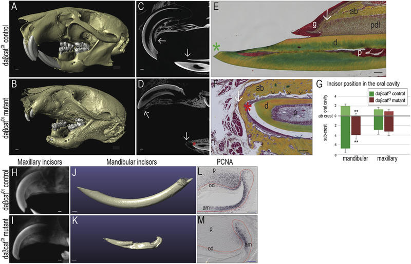
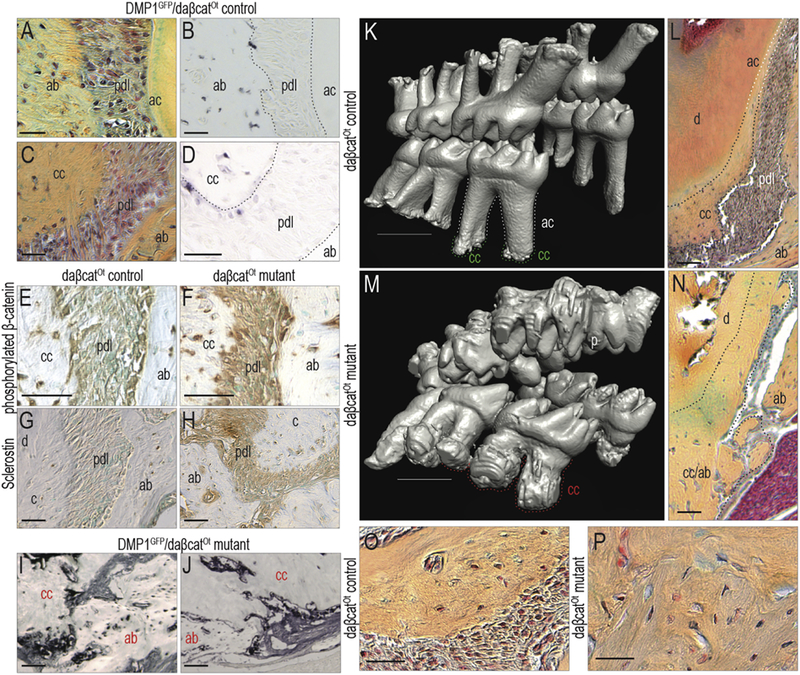
Vertebrate teeth are attached to the jawbones using a variety of methods but in mammals, a fibrous connection is the norm. This fibrous periodontal ligament (PDL) allows teeth to move in the jawbones in response to natural eruptive forces, mastication, and orthodontic tooth movement. In some disease states the PDL either calcifies or is replaced by a mineralized tissue and the result is ankylosis, where the tooth is fused to the alveolar bone. To understand how the PDL maintains this fibrous state, we examined a strain of mice in which tooth movement is arrested. Daβcat mice express a stabilized form of β-catenin in DMP1-positive alveolar bone osteocytes and cementocytes, which results in elevated Wnt signaling throughout the periodontium. As a consequence, there is an accrual of massive amounts of cellular cementum and alveolar bone, the PDL itself calcifies and teeth become ankylosed. These data suggest that to maintain its fibrous nature, Wnt signaling must normally be repressed in the PDL space.
脊椎动物的牙齿通过多种方式附着在颌骨上,但在哺乳动物中,纤维连接是常态。这种纤维牙周韧带(PDL)允许牙齿在颌骨中移动,以应对自然的出牙力、咀嚼和正畸牙齿移动。在某些疾病状态下,PDL 要么钙化,要么被矿化组织取代,结果是牙齿与牙槽骨融合,即牙骨质骨粘连。为了了解 PDL 如何保持这种纤维状态,我们研究了一种牙齿运动受阻的小鼠品系。在 Daβcat 小鼠中,β-连环蛋白的稳定形式在 DMP1 阳性牙槽骨成骨细胞和成牙骨质细胞中表达,导致整个牙周组织中 Wnt 信号升高。结果是大量细胞牙骨质和牙槽骨的积累,PDL 本身钙化,牙齿变得骨粘连。这些数据表明,为了保持其纤维特性,Wnt 信号在 PDL 空间中必须被正常抑制。