Department of Oral Biology, Yonsei University College of Dentistry, 134 Sinchon dong, Seodaemun-gu, Seoul, 120-752, Republic of Korea.
Department of Applied Life Science, The Graduate School, Yonsei University, Seoul, Republic of Korea.
J Transl Med. 2018 Nov 9;16(1):306. doi: 10.1186/s12967-018-1681-6.
Diabetes induces long bone loss and aggravation of periodontitis-induced alveolar bone loss. Simvastatin (SIM), which is a lipid-lowering agent is known to have an anabolic effect on bone. Therefore, we investigated effect of SIM on tibial and alveolar bone loss in type 1 diabetic rats with periodontitis.
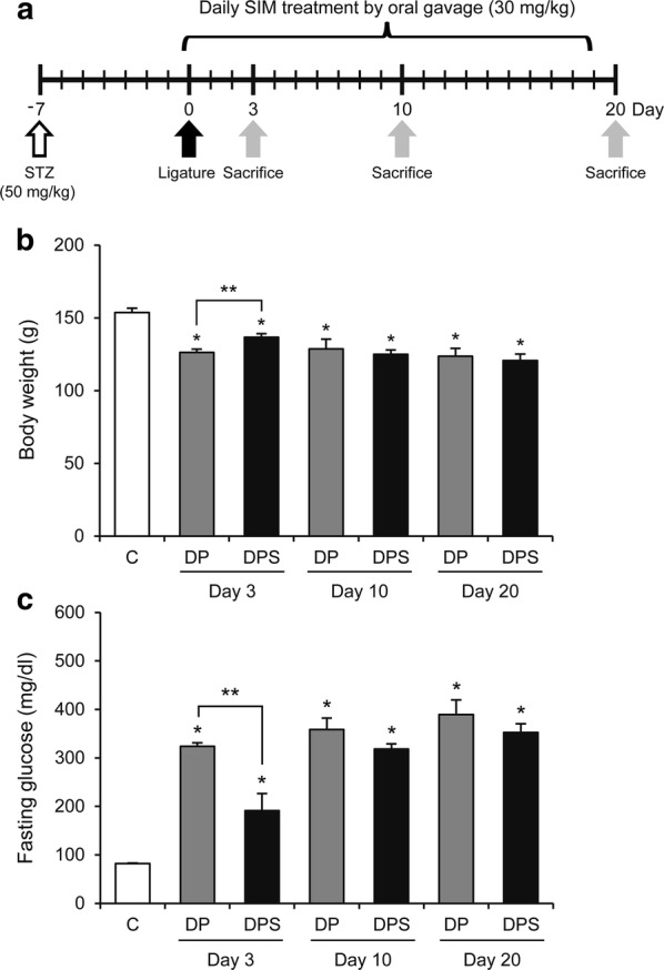
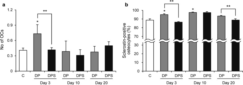
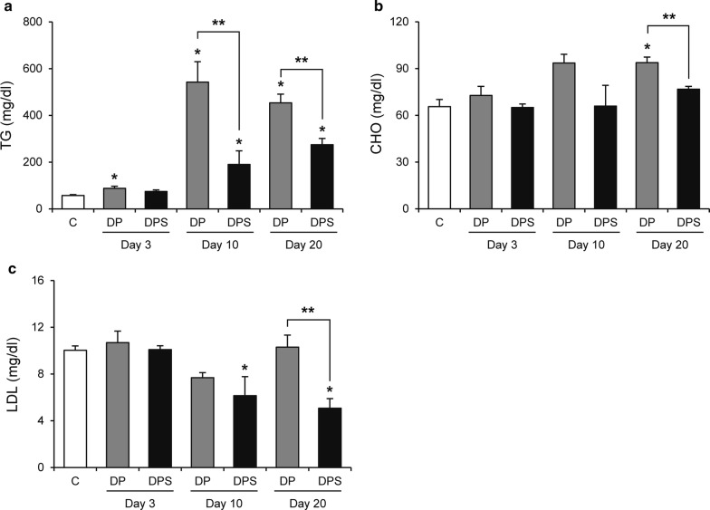
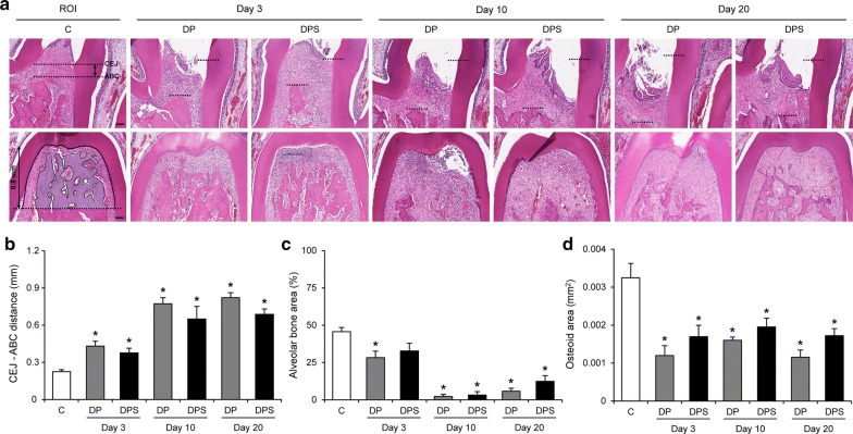
Rats were divided into control (C), diabetes with periodontitis (DP), and diabetes with periodontitis treated with SIM (DPS) groups. DP and DPS groups were intravenously injected with streptozotocin (50 mg/kg), and C group was injected with citrate buffer. Seven days later (day 0), periodontitis was induced by ligatures of mandibular first molars. DP and DPS groups were orally administered vehicle or SIM (30 mg/kg) from day 0 to days 3, 10, or 20. Alveolar and tibial bone loss was measured using histological and m-CT analysis alone or in combination. Osteoclast number and sclerostin-positive osteocytes in tibiae were evaluated by tartrate-resistant acid phosphatase and immunohistochemical staining, respectively. Glucose, triglyceride (TG), cholesterol (CHO), and low-density lipoprotein (LDL) were evaluated.
Consistent with diabetes induction, the DP group showed higher glucose and TG levels at all timepoints and higher CHO levels on day 20 than C group. Compared to the DP group, the DPS group exhibited reduced levels of glucose (day 3), TG (days 10 and 20), CHO, and LDL levels (day 20). Bone loss analysis revealed that the DP group had lower bone volume fraction, bone mineral density, bone surface density, and trabecular number in tibiae than C group at all timepoints. Interestingly, the DPS group exhibited elevation of these indices at early stages compared to the DP group. The DPS group showed reduction of osteoclasts (day 3) and sclerostin-positive osteocytes (days 3 and 20) compared with the DP group. There was no difference in alveolar bone loss between DP and DPS groups.
These results suggest that SIM attenuates tibial, but not alveolar bone loss in type 1 diabetic rats with periodontitis. Moreover, attenuation of tibial bone loss by SIM may be related to inhibition of osteoclast formation and reduction of sclerostin expression.
糖尿病可导致长骨丢失,并加重牙周炎引起的牙槽骨丢失。辛伐他汀(SIM)是一种降脂药物,已知其具有促进骨骼合成的作用。因此,我们研究了 SIM 对 1 型糖尿病伴牙周炎大鼠胫骨和牙槽骨丢失的影响。
将大鼠分为对照组(C)、糖尿病伴牙周炎组(DP)和糖尿病伴牙周炎加用 SIM 治疗组(DPS)。DP 和 DPS 组经尾静脉注射链脲佐菌素(50mg/kg),C 组注射柠檬酸盐缓冲液。7 天后(第 0 天),结扎下颌第一磨牙诱导牙周炎。DP 和 DPS 组从第 0 天开始连续 3、10 或 20 天经口给予载体或 SIM(30mg/kg)。通过组织学和 micro-CT 分析单独或联合检测牙槽骨和胫骨骨丢失。通过抗酒石酸酸性磷酸酶和免疫组织化学染色分别评估胫骨破骨细胞数量和骨硬化蛋白阳性成骨细胞。评估血糖、甘油三酯(TG)、胆固醇(CHO)和低密度脂蛋白(LDL)水平。
与糖尿病诱导一致,DP 组在所有时间点的血糖和 TG 水平均升高,CHO 水平在第 20 天高于 C 组。与 DP 组相比,DPS 组的血糖(第 3 天)、TG(第 10 和 20 天)、CHO 和 LDL 水平(第 20 天)降低。骨丢失分析显示,DP 组在所有时间点的胫骨骨体积分数、骨密度、骨表面密度和骨小梁数量均低于 C 组。有趣的是,DPS 组在早期与 DP 组相比,这些指标升高。与 DP 组相比,DPS 组的破骨细胞(第 3 天)和骨硬化蛋白阳性成骨细胞(第 3 和 20 天)减少。DP 和 DPS 组的牙槽骨丢失无差异。
这些结果表明,SIM 可减轻 1 型糖尿病伴牙周炎大鼠的胫骨骨丢失,但不能减轻牙槽骨丢失。此外,SIM 减轻胫骨骨丢失可能与抑制破骨细胞形成和降低骨硬化蛋白表达有关。