Department of Radiation Oncology, University of Miami Miller School of Medicine, Miami, FL, USA.
Department of Urology, University of Miami Miller School of Medicine, Miami, FL, USA.
Sci Rep. 2018 Nov 14;8(1):16801. doi: 10.1038/s41598-018-34916-4.
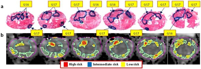
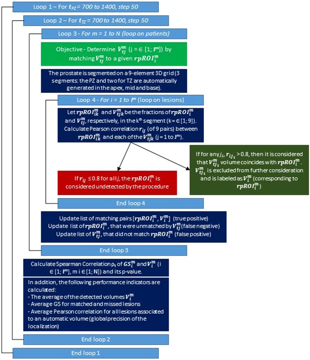
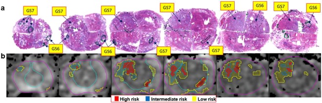
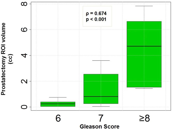
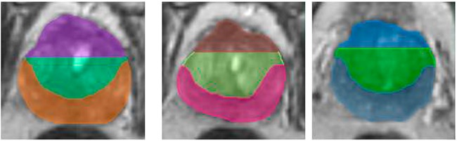
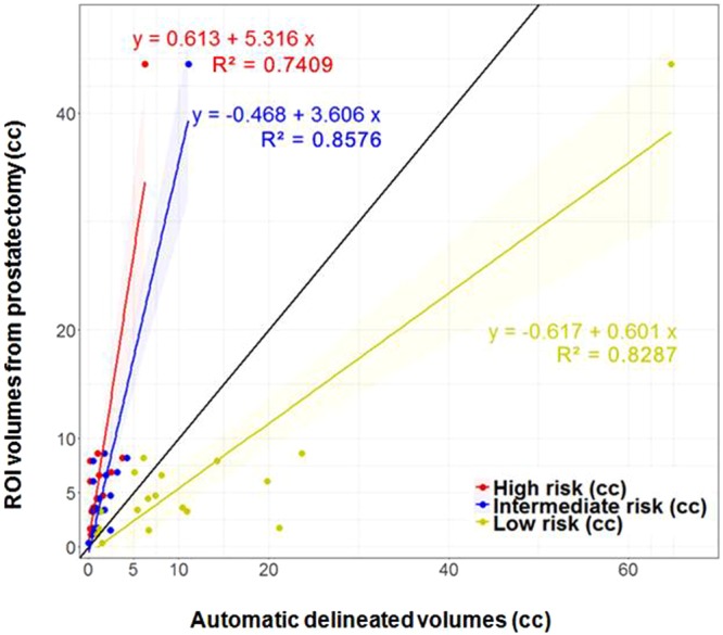
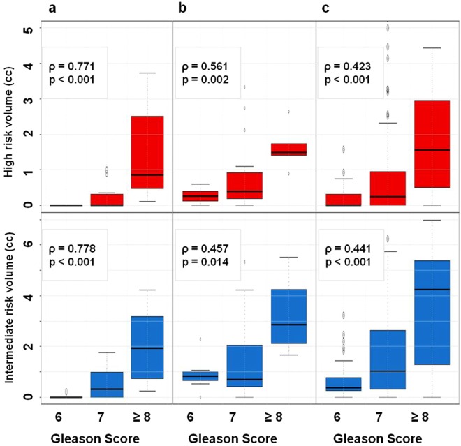
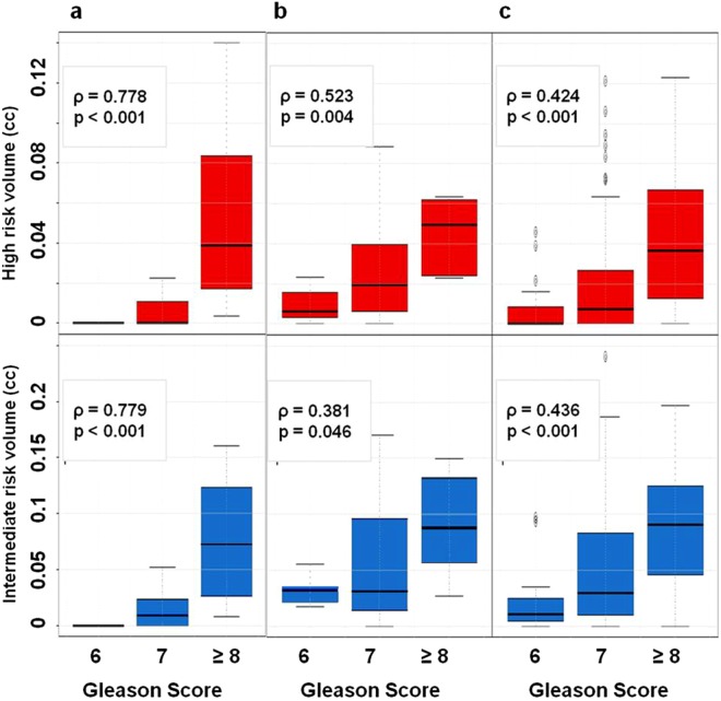
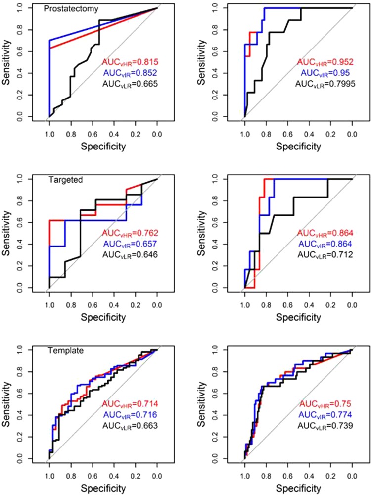
A procedure for identification of optimal Apparent Diffusion Coefficient (ADC) thresholds for automatic delineation of prostatic lesions with restricted diffusion at differing risk for cancer was developed. The relationship between the size of the identified Volumes of Interest (VOIs) and Gleason Score (GS) was evaluated. Patients with multiparametric (mp)MRI, acquired prior to radical prostatectomy (RP) (n = 18), mpMRI-ultrasound fused (MRI-US) (n = 21) or template biopsies (n = 139) were analyzed. A search algorithm, spanning ADC thresholds in 50 µm/s increments, determined VOIs that were matched to RP tumor nodules. Three ADC thresholds for both peripheral zone (PZ) and transition zone (TZ) were identified for estimation of VOIs at low, intermediate, and high risk of prostate cancer. The determined ADC thresholds for low, intermediate and high risk in PZ/TZ were: 900/800; 1100/850; and 1300/1050 µm/s. The correlation coefficients between the size of the high/intermediate/low risk VOIs and GS in the three cohorts were 0.771/0.778/0.369, 0.561/0.457/0.355 and 0.423/0.441/0.36 (p < 0.05). Low risk VOIs mapped all RP lesions; area under the curve (AUC) for intermediate risk VOIs to discriminate GS6 vs GS ≥ 7 was 0.852; for high risk VOIs to discriminate GS6,7 vs GS ≥ 8 was 0.952. In conclusion, the automatically delineated volumes in the prostate with restricted diffusion were found to strongly correlate with cancer aggressiveness.
开发了一种用于确定最佳表观扩散系数 (ADC) 阈值的方法,以便自动描绘具有受限扩散的前列腺病变的体积,这些病变的癌症风险不同。评估了确定的感兴趣区 (VOI) 大小与 Gleason 评分 (GS) 之间的关系。分析了接受多参数 MRI(在根治性前列腺切除术 (RP) 前获得,n=18)、MRI-超声融合 (MRI-US)(n=21)或模板活检 (n=139) 的患者。搜索算法跨越 50µm/s 增量的 ADC 阈值,确定与 RP 肿瘤结节匹配的 VOI。确定了用于估计前列腺癌低、中、高风险的外周带 (PZ) 和移行带 (TZ) 的三个 ADC 阈值。PZ/TZ 中低、中、高风险的 ADC 阈值分别为:900/800µm/s、1100/850µm/s 和 1300/1050µm/s。三个队列中,高/中/低风险 VOI 大小与 GS 的相关系数分别为 0.771/0.778/0.369、0.561/0.457/0.355 和 0.423/0.441/0.36(p<0.05)。低风险 VOI 映射了所有 RP 病变;中间风险 VOI 区分 GS6 与 GS≥7 的曲线下面积 (AUC) 为 0.852;高风险 VOI 区分 GS6、7 与 GS≥8 的 AUC 为 0.952。总之,发现具有受限扩散的前列腺中自动描绘的体积与癌症侵袭性密切相关。