Zhen Dan, Xia Wei, Yi Zhong Quan, Zhao Pan Wen, Zhong Jian Guo, Shi Hai Cun, Li Hua Liang, Dai Zhen Yu, Pan Ping Lei
1School of Nursing, Jiangsu Vocational College of Medicine, Yancheng, People's Republic of China.
2Department of Neurology, Affiliated Yancheng Hospital, School of Medicine, Southeast University, West Xindu Road 2#, Yancheng, Jiangsu Province, 224001 People's Republic of China.
Transl Neurodegener. 2018 Nov 7;7:26. doi: 10.1186/s40035-018-0134-8. eCollection 2018.
Resting-state functional magnetic resonance imaging studies using a regional homogeneity (ReHo) method have reported that amnestic mild cognitive impairment (aMCI) was associated with abnormalities in local functional connectivity. However, their results were not conclusive.
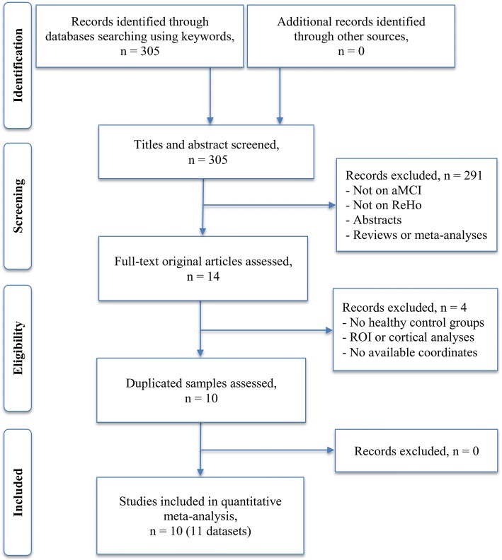
Seed-based Mapping was used to conduct a coordinate-based meta-analysis to identify consistent ReHo alterations in aMCI.
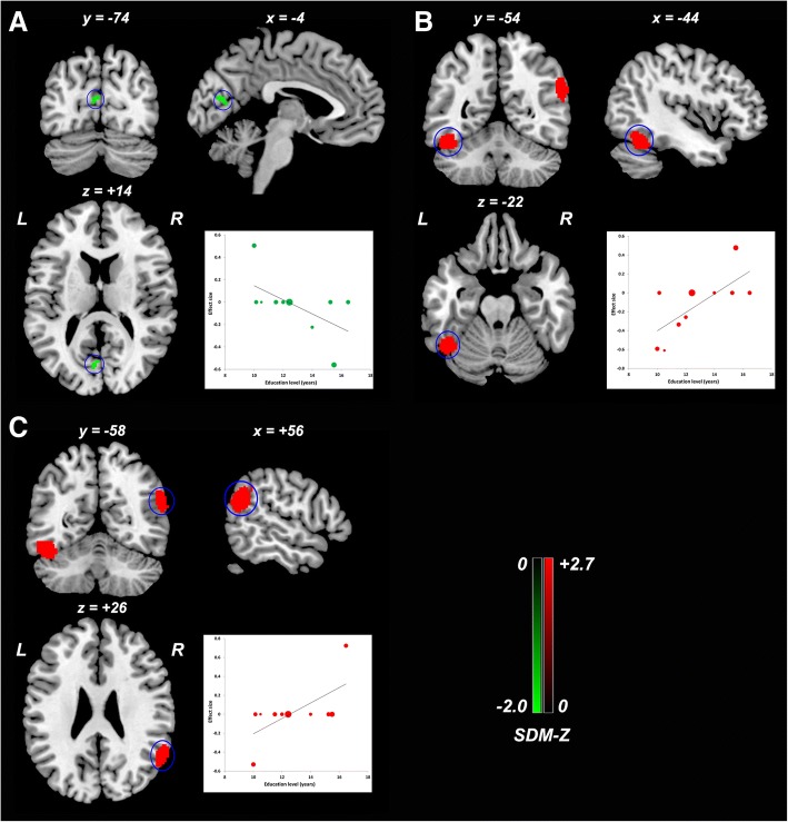
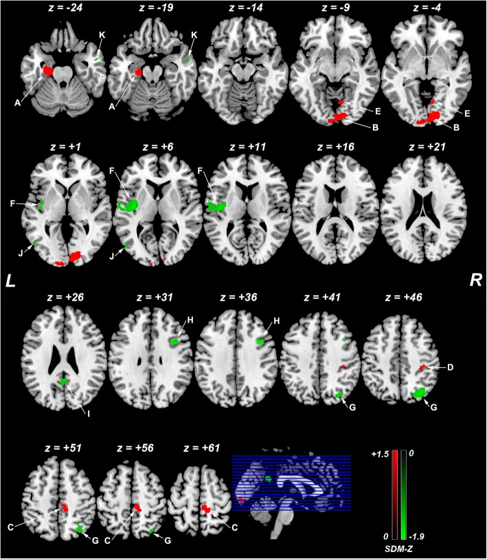
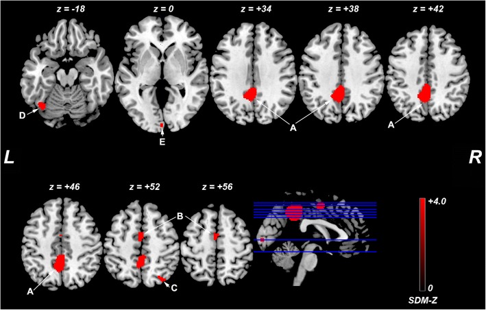
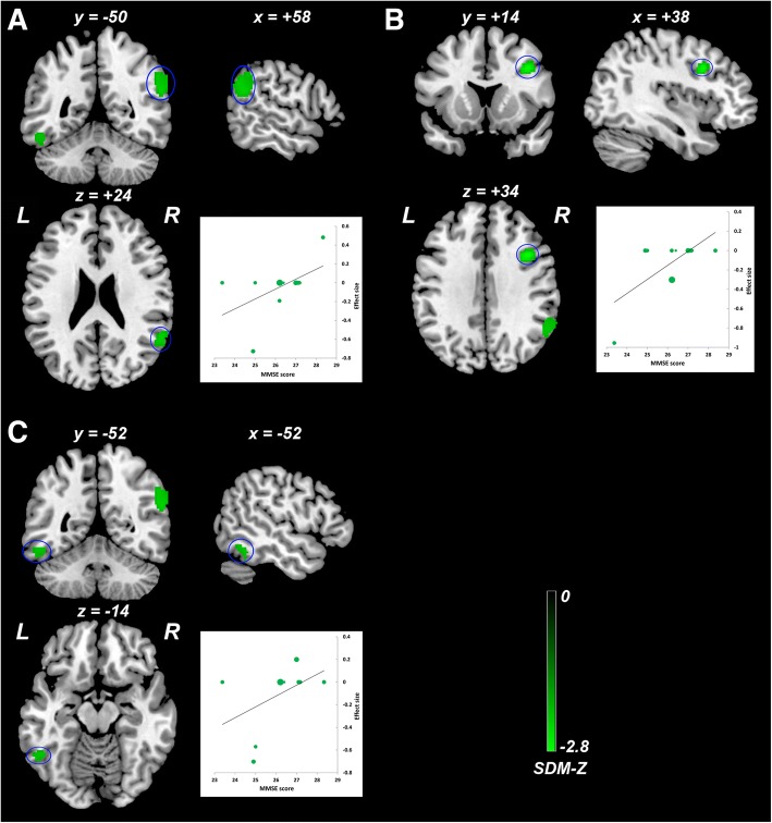
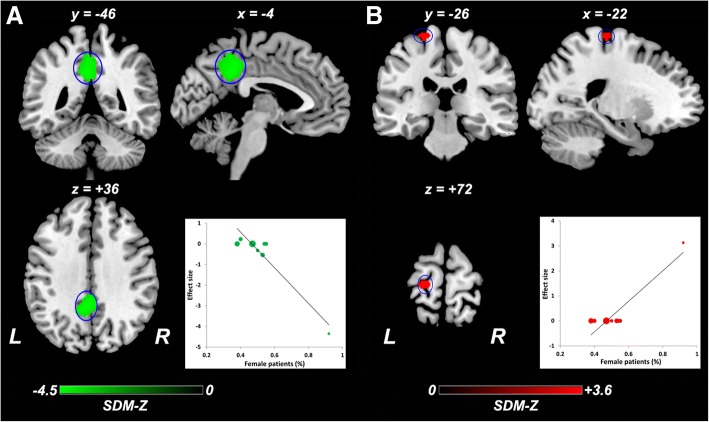
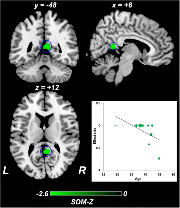
We identified 10 studies with 11 datasets suitable for inclusion, including 378 patients with aMCI and 435 healthy controls. This meta-analysis identified significant ReHo alterations in patients with aMCI relative to healthy controls, mainly within the default mode network (DMN) (bilateral posterior cingulate cortex [PCC], right angular gyrus, bilateral middle temporal gyri, and left parahippocampal gyrus/hippocampus), executive control network (right superior parietal lobule and dorsolateral prefrontal cortex), visual network (right lingual gyrus and left middle occipital gyrus), and sensorimotor network (right paracentral lobule/supplementary motor area, right postcentral gyrus and left posterior insula). Significant heterogeneity of ReHo alterations in the bilateral PCC, left parahippocampal gyrus/hippocampus, and right superior parietal lobule/angular gyrus was observed. Exploratory meta-regression analyses indicated that general cognitive function, gender distribution, age, and education level partially contributed to this heterogeneity.
This study provides provisional evidence that aMCI is associated with abnormal ReHo within the DMN, executive control network, visual network, and sensorimotor network. These local functional connectivity alterations suggest coexistence of functional deficits and compensation in these networks. These findings contribute to the modeling of brain functional connectomes and to a better understanding of the neural substrates of aMCI. Confounding factors merit much attention and warrant future investigations.
使用局部一致性(ReHo)方法的静息态功能磁共振成像研究报告称,遗忘型轻度认知障碍(aMCI)与局部功能连接异常有关。然而,他们的结果并不确凿。
基于种子点的映射用于进行基于坐标的荟萃分析,以确定aMCI中一致的ReHo改变。
我们确定了10项研究,其中11个数据集适合纳入,包括378例aMCI患者和435名健康对照。这项荟萃分析确定,与健康对照相比,aMCI患者存在显著的ReHo改变,主要在默认模式网络(DMN)(双侧后扣带回皮质[PCC]、右侧角回、双侧颞中回和左侧海马旁回/海马)、执行控制网络(右侧顶上小叶和背外侧前额叶皮质)、视觉网络(右侧舌回和左侧枕中回)以及感觉运动网络(右侧中央旁小叶/辅助运动区、右侧中央后回和左侧后岛叶)内。观察到双侧PCC、左侧海马旁回/海马和右侧顶上小叶/角回中ReHo改变存在显著异质性。探索性荟萃回归分析表明,一般认知功能、性别分布、年龄和教育水平部分导致了这种异质性。
本研究提供了初步证据,表明aMCI与DMN、执行控制网络、视觉网络和感觉运动网络内的ReHo异常有关。这些局部功能连接改变表明这些网络中存在功能缺陷和代偿共存。这些发现有助于脑功能连接组的建模,并有助于更好地理解aMCI的神经基础。混杂因素值得高度关注,并有待未来研究。