Jakobson Mo Susanna, Axelsson Jan, Jonasson Lars, Larsson Anne, Ögren Mattias J, Ögren Margareta, Varrone Andrea, Eriksson Linda, Bäckström David, Af Bjerkén Sara, Linder Jan, Riklund Katrine
Department of Radiation Sciences, Diagnostic Radiology, Umeå University, Umeå, Sweden.
Umeå Center for Functional Brain Imaging, Umeå University, Umeå, Sweden.
EJNMMI Res. 2018 Nov 15;8(1):100. doi: 10.1186/s13550-018-0450-0.
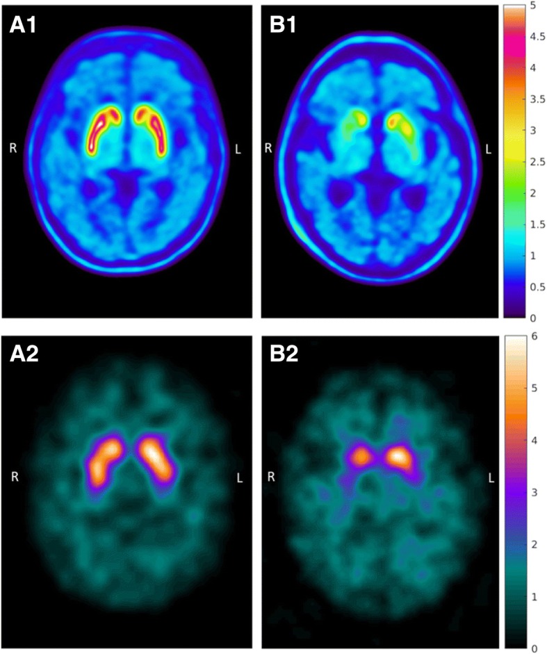
Dopamine transporter (DAT) imaging may be of diagnostic value in patients with clinically suspected parkinsonian disease. The purpose of this study was to compare the diagnostic performance of DAT imaging with positron emission computed tomography (PET), using the recently developed, highly DAT-selective radiopharmaceutical [F]FE-PE2I (FE-PE2I), to the commercially available and frequently used method with [I]FP-CIT (FP-CIT) single-photon emission computed tomography (SPECT) in early-stage idiopathic parkinsonian syndrome (PS).
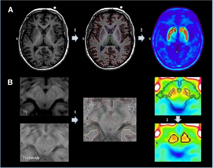
Twenty-two patients with a clinical de novo diagnosis of PS and 28 healthy controls (HC) participating in an on-going clinical trial of FE-PE2I were analyzed in this study. Within the trial protocol, participants are clinically reassessed 2 years after inclusion. A commercially available software was used for automatic calculation of FP-CIT-specific uptake ratio (SUR). MRI-based volumes of interest combined with threshold PET segmentation were used for FE-PE2I binding potential relative to non-displaceable binding (BP) quantification and specific uptake value ratios (SUVR).
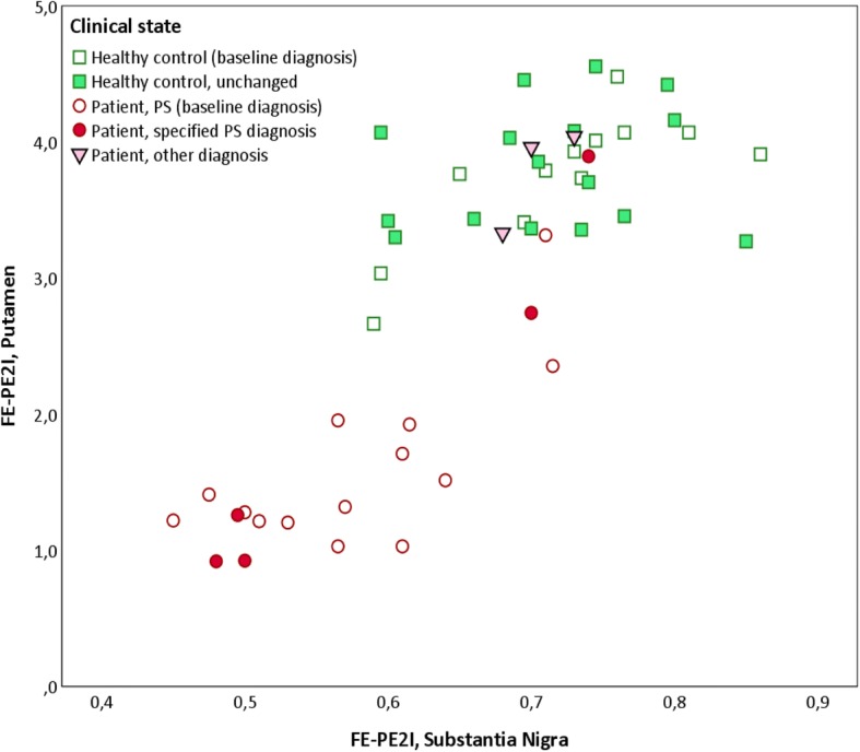
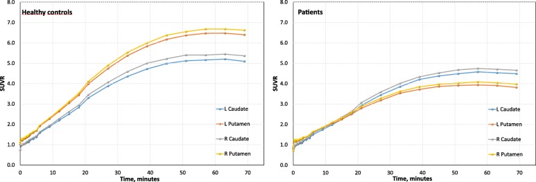
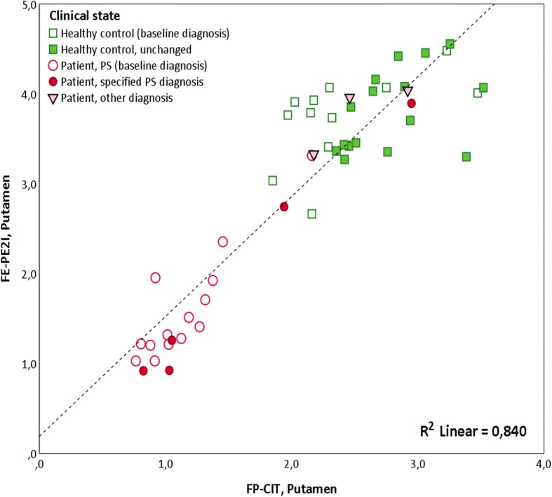
PET with FE-PE2I revealed significant differences between patients with a clinical de novo diagnosis of PS and healthy controls in striatal DAT availability (p < 0.001), with excellent accuracy of predicting dopaminergic deficit in early-stage PS. The effect sizes were calculated for FE-PE2I BP (Glass's Δ = 2.95), FE-PE2I SUVR (Glass's Δ = 2.57), and FP-CIT SUR (Glass's Δ = 2.29). The intraclass correlation (ICC) between FE-PE2I BP FP-CIT SUR was high in the caudate (ICC = 0.923), putamen (ICC = 0.922), and striatum (ICC = 0.946), p < 0.001. Five of the 22 patients displayed preserved striatal DAT availability in the striatum with both methods. At follow-up, a non-PS clinical diagnosis was confirmed in three of these, while one was clinically diagnosed with corticobasal syndrome. In these patients, FE-PE2I binding was also normal in the substantia nigra (SN), while significantly reduced in the remaining patients. FE-PE2I measurement of the mean DAT availability in the putamen was strongly correlated with BP in the SN (R = 0.816, p < 0.001). Olfaction and mean putamen DAT availability was correlated using both FE-PE2I BP and FP-CIT SUR (R ≥ 0.616, p < 0.001).
DAT imaging with FE-PE2I PET yields excellent basic diagnostic differentiation in early-stage PS, at least as good as FP-CIT SPECT.
多巴胺转运体(DAT)成像对于临床疑似帕金森病患者可能具有诊断价值。本研究的目的是比较使用最近开发的、高度DAT选择性放射性药物[F]FE-PE2I(FE-PE2I)的DAT成像与正电子发射计算机断层扫描(PET)的诊断性能,以及与市售且常用的[I]FP-CIT(FP-CIT)单光子发射计算机断层扫描(SPECT)在早期特发性帕金森综合征(PS)中的诊断性能。
本研究分析了22例临床初诊为PS的患者和28例健康对照(HC),这些患者参与了一项正在进行的FE-PE2I临床试验。在试验方案中,参与者在入组2年后进行临床重新评估。使用市售软件自动计算FP-CIT特异性摄取率(SUR)。基于MRI的感兴趣体积结合阈值PET分割用于FE-PE2I相对于不可置换结合(BP)的结合潜力定量和特异性摄取值比率(SUVR)。
使用FE-PE2I的PET显示,临床初诊为PS的患者与健康对照在纹状体DAT可用性方面存在显著差异(p < 0.001),在预测早期PS中的多巴胺能缺陷方面具有出色的准确性。计算了FE-PE2I BP(Glass's Δ = 2.95)、FE-PE2I SUVR(Glass's Δ = 2.57)和FP-CIT SUR(Glass's Δ = 2.29)的效应大小。FE-PE2I BP与FP-CIT SUR在尾状核(ICC = 0.923)、壳核(ICC = 0.922)和纹状体(ICC = 0.946)中的组内相关性较高,p < 0.001。22例患者中有5例使用两种方法均显示纹状体DAT可用性保留。在随访中,其中3例被确诊为非PS临床诊断,而1例临床诊断为皮质基底节综合征。在这些患者中,黑质(SN)中的FE-PE2I结合也正常,而其余患者中则显著降低。壳核中平均DAT可用性的FE-PE2I测量与SN中的BP密切相关(R = 0.816,p < 0.001)。使用FE-PE2I BP和FP-CIT SUR均显示嗅觉与壳核平均DAT可用性相关(R≥0.616,p < 0.001)。
使用FE-PE2I PET进行DAT成像在早期PS中产生了出色的基本诊断区分,至少与FP-CIT SPECT一样好。