• 文献检索
  • 文档翻译
  • 深度研究
  • 学术资讯
  • Suppr Zotero 插件Zotero 插件
  • 邀请有礼
  • 套餐&价格
  • 历史记录
应用&插件
Suppr Zotero 插件Zotero 插件浏览器插件Mac 客户端Windows 客户端微信小程序
定价
高级版会员购买积分包购买API积分包
服务
文献检索文档翻译深度研究API 文档MCP 服务
关于我们
关于 Suppr公司介绍联系我们用户协议隐私条款
关注我们

Suppr 超能文献

核心技术专利:CN118964589B侵权必究
粤ICP备2023148730 号-1Suppr @ 2026

文献检索

告别复杂PubMed语法,用中文像聊天一样搜索,搜遍4000万医学文献。AI智能推荐,让科研检索更轻松。

立即免费搜索

文件翻译

保留排版,准确专业,支持PDF/Word/PPT等文件格式,支持 12+语言互译。

免费翻译文档

深度研究

AI帮你快速写综述,25分钟生成高质量综述,智能提取关键信息,辅助科研写作。

立即免费体验

发育中婴儿连接组脑图谱的形状和生长脑网络图谱估计

ESTIMATION OF SHAPE AND GROWTH BRAIN NETWORK ATLASES FOR CONNECTOMIC BRAIN MAPPING IN DEVELOPING INFANTS.

作者信息

Rekik Islem, Li Gang, Lin Weili, Shen Dinggang

机构信息

BASIRA lab, CVIP group, School of Science and Engineering, Computing, University of Dundee, UK.

Department of Radiology and BRIC, University of North Carolina at Chapel Hill, NC, USA.

出版信息

Proc IEEE Int Symp Biomed Imaging. 2018 Apr;2018:985-989. doi: 10.1109/ISBI.2018.8363736. Epub 2018 May 24.

DOI:10.1109/ISBI.2018.8363736
PMID:30450154
原文链接:https://pmc.ncbi.nlm.nih.gov/articles/PMC6239168/
Abstract

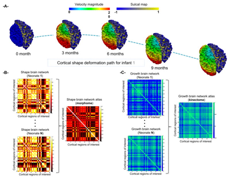
brain connectomics have heavily relied on using functional and diffusion Magnetic Resonance Imaging (MRI) modalities to examine functional and structural relationships between pairs of anatomical regions in the brain. However, research work on brain (i.e., shape-to-shape) connections, which can be derived from T1-w and T2-w MR images, in both typical and atypical development or ageing is very scarce. Furthermore, the brain cannot be only regarded as a static shape, since it is a dynamic complex system that changes at functional, structural and morphological levels. Hence, examining the 'connection' between brain and its changes with time (e.g., ) may help advance our understanding of connectomic brain dynamics as well as disorders that may affect it. To address these limitations, we unprecedentedly introduce two shape and growth connectivity analysis tools that further extend the field of connectomics to brain morphology and dynamics: . Specifically, for a population of anatomically labelled shapes, the identifies a network of anatomical shape regions that are connected when morphologically similar at a single timepoint, whereas the identifies anatomical shape regions that elicit similar evolution dynamics across successive timepoints. These proposed tools can be easily invested to examine how a baseline shape influences its deformation trajectory at later timepoints using longitudinal shape data. We evaluated these tools on 23 infants, with right and left cortical surfaces reconstructed at birth, 3, 6, 9 and 12 months of age. Investigating the relationship between the and the (from birth to 1 year of age) gave insights into brain connectivity at birth and how it develops over time.

摘要

脑连接组学在很大程度上依赖于使用功能磁共振成像和扩散磁共振成像(MRI)模态来检查大脑中各对解剖区域之间的功能和结构关系。然而,关于在典型和非典型发育或衰老过程中,可从T1加权和T2加权MR图像得出的脑(即形状与形状之间)连接的研究工作非常稀少。此外,大脑不能仅仅被视为一个静态形状,因为它是一个在功能、结构和形态层面都会发生变化的动态复杂系统。因此,研究大脑形状及其随时间的变化(例如)之间的“连接”,可能有助于推进我们对连接组脑动力学以及可能影响它的疾病的理解。为了解决这些局限性,我们前所未有的引入了两种形状和生长连通性分析工具,它们将连接组学领域进一步扩展到脑形态学和动力学:。具体来说,对于一组经解剖学标记的形状,在单个时间点形态相似时,识别出相互连接的解剖形状区域网络,而则识别出在连续时间点引发相似演化动力学的解剖形状区域。利用纵向形状数据,这些提出的工具可以很容易地用于研究基线形状如何影响其在后续时间点的变形轨迹。我们在23名婴儿身上评估了这些工具,这些婴儿在出生时、3个月、6个月、9个月和12个月大时重建了左右皮质表面。研究出生至1岁期间和之间的关系,有助于深入了解出生时的脑连接性及其随时间的发展情况。

https://cdn.ncbi.nlm.nih.gov/pmc/blobs/fb17/6239168/ab0f77a0756e/nihms969539f3.jpg
https://cdn.ncbi.nlm.nih.gov/pmc/blobs/fb17/6239168/cdea2ea44ad6/nihms969539f1.jpg
https://cdn.ncbi.nlm.nih.gov/pmc/blobs/fb17/6239168/269aa13752df/nihms969539f2.jpg
https://cdn.ncbi.nlm.nih.gov/pmc/blobs/fb17/6239168/ab0f77a0756e/nihms969539f3.jpg
https://cdn.ncbi.nlm.nih.gov/pmc/blobs/fb17/6239168/cdea2ea44ad6/nihms969539f1.jpg
https://cdn.ncbi.nlm.nih.gov/pmc/blobs/fb17/6239168/269aa13752df/nihms969539f2.jpg
https://cdn.ncbi.nlm.nih.gov/pmc/blobs/fb17/6239168/ab0f77a0756e/nihms969539f3.jpg

相似文献

1
ESTIMATION OF SHAPE AND GROWTH BRAIN NETWORK ATLASES FOR CONNECTOMIC BRAIN MAPPING IN DEVELOPING INFANTS.发育中婴儿连接组脑图谱的形状和生长脑网络图谱估计
Proc IEEE Int Symp Biomed Imaging. 2018 Apr;2018:985-989. doi: 10.1109/ISBI.2018.8363736. Epub 2018 May 24.
2
Unsupervised Manifold Learning Using High-Order Morphological Brain Networks Derived From T1-w MRI for Autism Diagnosis.使用源自T1加权磁共振成像的高阶形态学脑网络进行无监督流形学习以诊断自闭症
Front Neuroinform. 2018 Oct 26;12:70. doi: 10.3389/fninf.2018.00070. eCollection 2018.
3
Prediction of Longitudinal Development of Infant Cortical Surface Shape Using a 4D Current-Based Learning Framework.使用基于电流的4D学习框架预测婴儿皮质表面形状的纵向发展
Inf Process Med Imaging. 2015;24:576-87. doi: 10.1007/978-3-319-19992-4_45.
4
A comparative study of machine learning methods for predicting the evolution of brain connectivity from a baseline timepoint.一种用于从基线时间点预测脑连接性演变的机器学习方法的比较研究。
J Neurosci Methods. 2022 Feb 15;368:109475. doi: 10.1016/j.jneumeth.2022.109475. Epub 2022 Jan 4.
5
Toward Developmental Connectomics of the Human Brain.迈向人类大脑的发育连接组学
Front Neuroanat. 2016 Mar 31;10:25. doi: 10.3389/fnana.2016.00025. eCollection 2016.
6
The relation between structural and functional connectivity patterns in complex brain networks.复杂脑网络中结构与功能连接模式之间的关系。
Int J Psychophysiol. 2016 May;103:149-60. doi: 10.1016/j.ijpsycho.2015.02.011. Epub 2015 Feb 10.
7
Normalization of similarity-based individual brain networks from gray matter MRI and its association with neurodevelopment in infants with intrauterine growth restriction.基于相似性的正常化个体脑网络来自灰质 MRI 及其与宫内生长受限婴儿神经发育的关联。
Neuroimage. 2013 Dec;83:901-11. doi: 10.1016/j.neuroimage.2013.07.045. Epub 2013 Jul 22.
8
Altered small-world topology of structural brain networks in infants with intrauterine growth restriction and its association with later neurodevelopmental outcome.宫内生长受限婴儿的结构脑网络小世界拓扑结构改变及其与后期神经发育结局的关系。
Neuroimage. 2012 Apr 2;60(2):1352-66. doi: 10.1016/j.neuroimage.2012.01.059. Epub 2012 Jan 18.
9
It's All About the Networks.一切都与网络有关。
Epilepsy Curr. 2019 May-Jun;19(3):165-167. doi: 10.1177/1535759719843301. Epub 2019 Apr 29.
10
Function-specific and Enhanced Brain Structural Connectivity Mapping via Joint Modeling of Diffusion and Functional MRI.基于弥散张量成像和功能磁共振成像联合建模的功能特异性及增强脑结构连接图绘制
Sci Rep. 2018 Mar 16;8(1):4741. doi: 10.1038/s41598-018-23051-9.

本文引用的文献

1
Heritability of the shape of subcortical brain structures in the general population.一般人群亚皮质脑结构形状的遗传性。
Nat Commun. 2016 Dec 15;7:13738. doi: 10.1038/ncomms13738.
2
Multimodal population brain imaging in the UK Biobank prospective epidemiological study.英国生物银行前瞻性流行病学研究中的多模态人群脑成像
Nat Neurosci. 2016 Nov;19(11):1523-1536. doi: 10.1038/nn.4393. Epub 2016 Sep 19.
3
A multi-modal parcellation of human cerebral cortex.人类大脑皮层的多模态分区
Nature. 2016 Aug 11;536(7615):171-178. doi: 10.1038/nature18933. Epub 2016 Jul 20.
4
Multidirectional and Topography-based Dynamic-scale Varifold Representations with Application to Matching Developing Cortical Surfaces.基于多方向和地形的动态尺度变分曲面表示及其在匹配发育中的皮质表面中的应用
Neuroimage. 2016 Jul 15;135:152-62. doi: 10.1016/j.neuroimage.2016.04.037. Epub 2016 Apr 30.
5
Predicting infant cortical surface development using a 4D varifold-based learning framework and local topography-based shape morphing.使用基于4D变分曲面的学习框架和基于局部地形的形状变形预测婴儿皮质表面发育。
Med Image Anal. 2016 Feb;28:1-12. doi: 10.1016/j.media.2015.10.007. Epub 2015 Nov 10.
6
Age-Related Cortical Thickness Reduction in Non-Demented Down's Syndrome Subjects.非痴呆唐氏综合征患者与年龄相关的皮质厚度减少
J Neuroimaging. 2016 Jan-Feb;26(1):95-102. doi: 10.1111/jon.12259. Epub 2015 May 21.
7
Similarity network fusion for aggregating data types on a genomic scale.基于基因组尺度聚合数据类型的相似网络融合。
Nat Methods. 2014 Mar;11(3):333-7. doi: 10.1038/nmeth.2810. Epub 2014 Jan 26.
8
Measuring the dynamic longitudinal cortex development in infants by reconstruction of temporally consistent cortical surfaces.通过重建时间上一致的皮质表面来测量婴儿动态纵向皮质发育。
Neuroimage. 2014 Apr 15;90:266-79. doi: 10.1016/j.neuroimage.2013.12.038. Epub 2013 Dec 27.
9
Structural variations in prefrontal cortex mediate the relationship between early childhood stress and spatial working memory.前额皮质的结构变化介导了儿童早期压力与空间工作记忆之间的关系。
J Neurosci. 2012 Jun 6;32(23):7917-25. doi: 10.1523/JNEUROSCI.0307-12.2012.
10
The economy of brain network organization.大脑网络组织的经济学。
Nat Rev Neurosci. 2012 Apr 13;13(5):336-49. doi: 10.1038/nrn3214.