Bastiaens Alex J, Xie Sijia, Mustafa Dana A M, Frimat Jean-Philippe, den Toonder Jaap M J, Luttge Regina
Department of Mechanical Engineering, Microsystems Group and Institute for Complex Molecular Systems, Eindhoven University of Technology, Eindhoven, Netherlands.
MESA+ Institute for Nanotechnology, University of Twente, Enschede, Netherlands.
Front Cell Neurosci. 2018 Nov 6;12:415. doi: 10.3389/fncel.2018.00415. eCollection 2018.
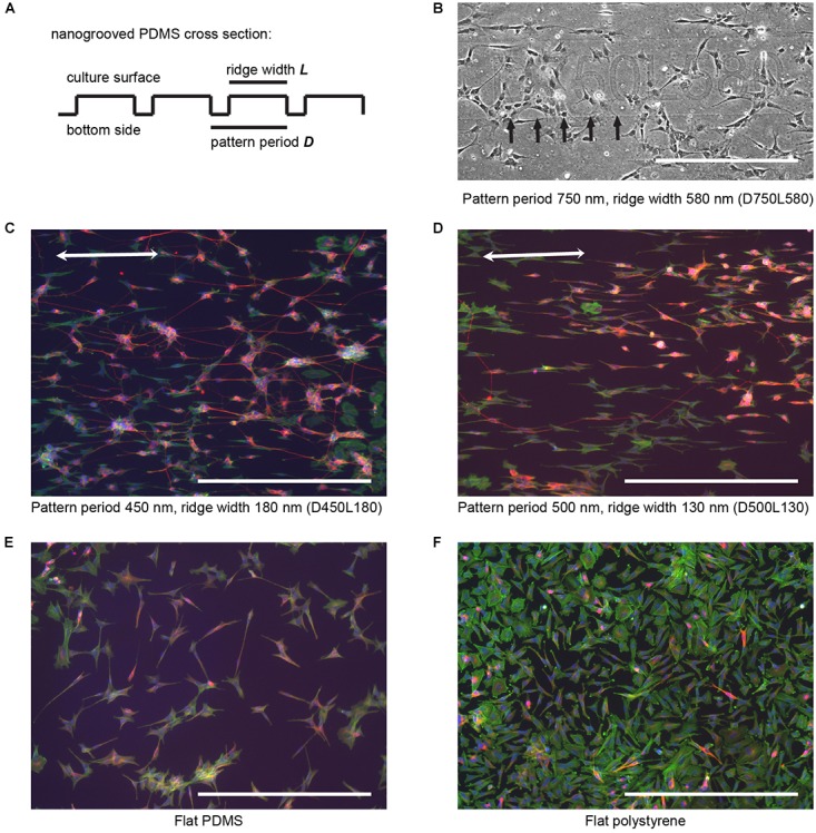
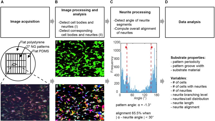
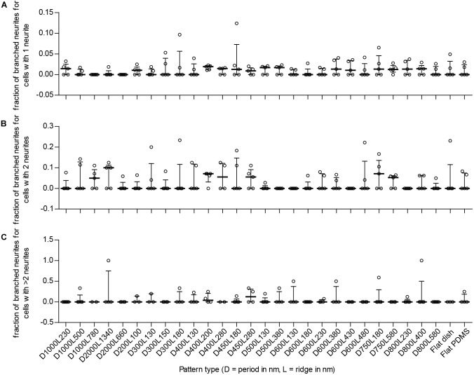
Research on neuronal differentiation and neuronal network guidance induced through nanotopographical cues generates large datasets, and therefore the analysis of such data can be aided by automatable, unbiased image screening tools. To link such tools, we present an image-based screening method to evaluate the influence of nanogroove pattern dimensions on neuronal differentiation. This new method consists of combining neuronal feature detection software, here HCA-Vision, and a Frangi vesselness algorithm to calculate neurite alignment values and quantify morphological aspects of neurons, which are measured via neurite length, neuronal polarity, and neurite branching, for differentiated SH-SY5Y cells cultured on nanogrooved polydimethylsiloxane (PDMS) patterns in the 200-2000 nm range. The applicability of this method is confirmed by our results, which find that the level of alignment is dependent on nanogroove dimensions. Furthermore, the screening method reveals that differentiation and alignment are correlated. In particular, patterns with groove widths >200 nm and with a low ridge width to pattern period ratio have a quantifiable influence on alignment, neurite length, and polarity. In summary, the novel combination of software that forms a base for this statistical analysis method demonstrates good potential for evaluating tissue microarchitecture, which depends on subtle design variation in substrate topography. Using the screening method, we obtained automated and sensitive quantified readouts from large datasets.
通过纳米拓扑线索诱导神经元分化和神经网络导向的研究产生了大量数据集,因此,此类数据的分析可借助可自动化、无偏差的图像筛选工具。为连接此类工具,我们提出一种基于图像的筛选方法,以评估纳米槽图案尺寸对神经元分化的影响。这种新方法包括将神经元特征检测软件(此处为HCA-Vision)与Frangi血管造影算法相结合,以计算神经突排列值并量化神经元的形态学方面,这些方面通过神经突长度、神经元极性和神经突分支来衡量,用于在200-2000 nm范围内的纳米槽聚二甲基硅氧烷(PDMS)图案上培养的分化型SH-SY5Y细胞。我们的结果证实了该方法的适用性,结果发现排列水平取决于纳米槽尺寸。此外,筛选方法表明分化与排列相关。特别是,槽宽>200 nm且脊宽与图案周期比低的图案对排列、神经突长度和极性有可量化的影响。总之,构成这种统计分析方法基础的软件新组合在评估组织微结构方面显示出良好潜力,组织微结构取决于底物拓扑的细微设计变化。使用筛选方法,我们从大型数据集中获得了自动化且灵敏的定量读数。