Yang Ying, Luo Hui, Zhou Can, Zhang Rongyi, Liu Si, Zhu Xiao, Ke Sha, Liu Hui, Lu Zhan, Chen Mao
1 Department of Clinical Medicine, North Sichuan Medical College, Nanchong, China; Department of Cardiology, Affiliated Hospital of North Sichuan Medical College, Nanchong, China.
2 Department of Cardiothoracic Surgery, Nanchong Central Hospital, Nanchong, China.
J Int Med Res. 2019 Jan;47(1):453-469. doi: 10.1177/0300060518809255. Epub 2018 Nov 26.
This study aimed to examine regulation of capillary tubules and lipid formation in vascular endothelial cells and macrophages via extracellular vesicle-mediated microRNA (miRNA)-4306 transfer.
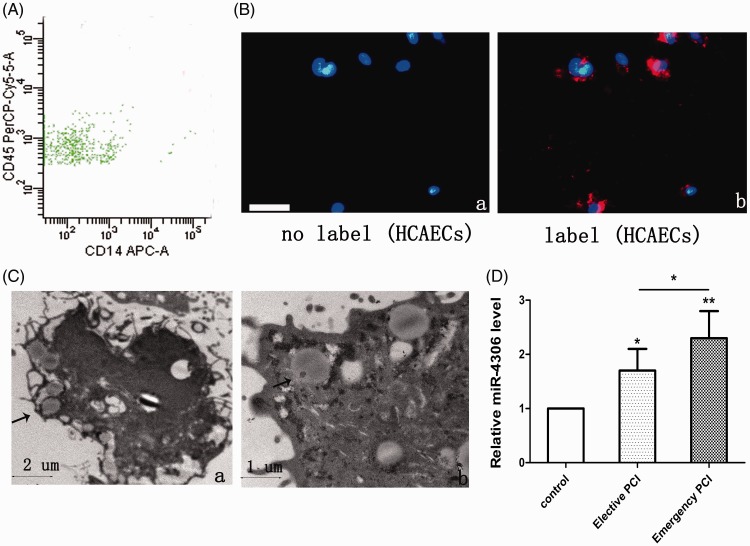
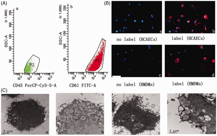
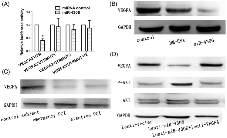
Whole blood samples (12 mL) were collected from 53 patients, and miR-4306 levels in extracellular vesicles (EVs) were analyzed by reverse transcription-polymerase chain reaction. Human coronary artery vascular endothelial cells (HCAECs) and human monocyte-derived macrophages (HMDMs) were transfected with a scrambled oligonucleotide, an miR-4306 mimic, or an anti-miR-4306 inhibitor. The direct effect of miR-4306 on the target gene was analyzed by a dual-luciferase reporter assay.
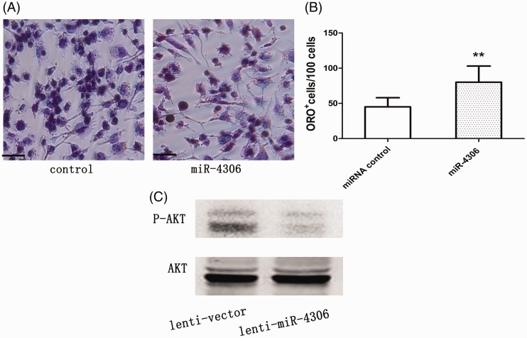
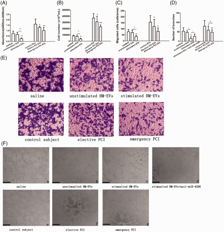
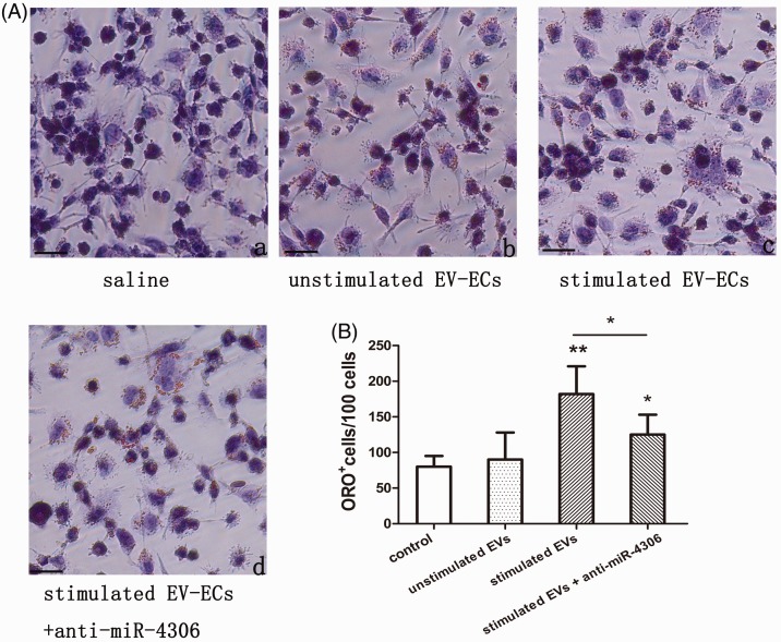
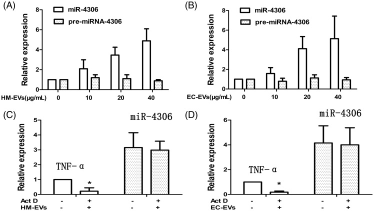
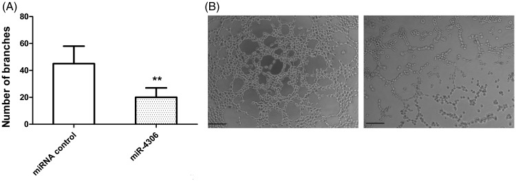
EV-contained miR-4306 released from HMDMs was significantly upregulated in coronary artery disease. Oxidized low-density lipoprotein (ox-LDL)-stimulated HMDM-derived EVs inhibited proliferation, migration, and angiogenesis abilities of HCAECs in vitro. However, ox-LDL-stimulated HCAEC-derived EVs enhanced lipid formation of HMDMs. The possible mechanism of these findings was partly due to EV-mediated miR-4306 upregulation of the Akt/nuclear factor kappa B signaling pathway.
Paracrine cellular crosstalk between HCAECs and HMDMs probably supports the pro-atherosclerotic effects of EVs under ox-LDL stress.
本研究旨在通过细胞外囊泡介导的微小RNA(miRNA)-4306转移,研究血管内皮细胞和巨噬细胞中毛细血管微管和脂质形成的调控机制。
收集53例患者的全血样本(12 mL),采用逆转录-聚合酶链反应分析细胞外囊泡(EVs)中miR-4306的水平。用人冠状动脉血管内皮细胞(HCAECs)和人单核细胞衍生的巨噬细胞(HMDMs)转染乱序寡核苷酸、miR-4306模拟物或抗miR-4306抑制剂。通过双荧光素酶报告基因检测分析miR-4306对靶基因的直接作用。
在冠状动脉疾病中,HMDMs释放的含miR-4306的EVs显著上调。氧化低密度脂蛋白(ox-LDL)刺激的HMDM来源的EVs在体外抑制HCAECs的增殖、迁移和血管生成能力。然而,ox-LDL刺激的HCAEC来源的EVs增强了HMDMs的脂质形成。这些发现的可能机制部分归因于EV介导的miR-4306上调Akt/核因子κB信号通路。
在ox-LDL应激下,HCAECs和HMDMs之间的旁分泌细胞间串扰可能支持了EVs的促动脉粥样硬化作用。