• 文献检索
  • 文档翻译
  • 深度研究
  • 学术资讯
  • Suppr Zotero 插件Zotero 插件
  • 邀请有礼
  • 套餐&价格
  • 历史记录
应用&插件
Suppr Zotero 插件Zotero 插件浏览器插件Mac 客户端Windows 客户端微信小程序
定价
高级版会员购买积分包购买API积分包
服务
文献检索文档翻译深度研究API 文档MCP 服务
关于我们
关于 Suppr公司介绍联系我们用户协议隐私条款
关注我们

Suppr 超能文献

核心技术专利:CN118964589B侵权必究
粤ICP备2023148730 号-1Suppr @ 2026

文献检索

告别复杂PubMed语法,用中文像聊天一样搜索,搜遍4000万医学文献。AI智能推荐,让科研检索更轻松。

立即免费搜索

文件翻译

保留排版,准确专业,支持PDF/Word/PPT等文件格式,支持 12+语言互译。

免费翻译文档

深度研究

AI帮你快速写综述,25分钟生成高质量综述,智能提取关键信息,辅助科研写作。

立即免费体验

蛋白酶激活受体-1 在慢性阻塞性肾病期间导致肾损伤和间质纤维化。

Protease-activated receptor-1 contributes to renal injury and interstitial fibrosis during chronic obstructive nephropathy.

机构信息

Center for Experimental and Molecular Medicine, Academic Medical Center Amsterdam, Amsterdam, The Netherlands.

Pathology, Academic Medical Center Amsterdam, Amsterdam, The Netherlands.

出版信息

J Cell Mol Med. 2019 Feb;23(2):1268-1279. doi: 10.1111/jcmm.14028. Epub 2018 Nov 28.

DOI:10.1111/jcmm.14028
PMID:30485646
原文链接:https://pmc.ncbi.nlm.nih.gov/articles/PMC6349177/
Abstract

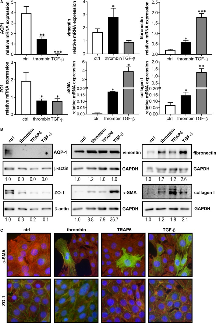
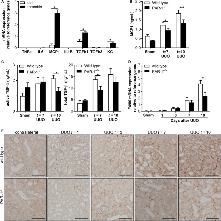
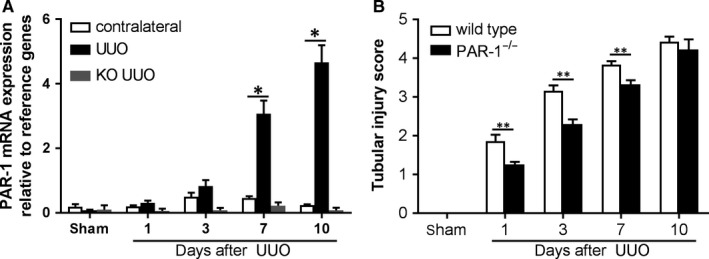
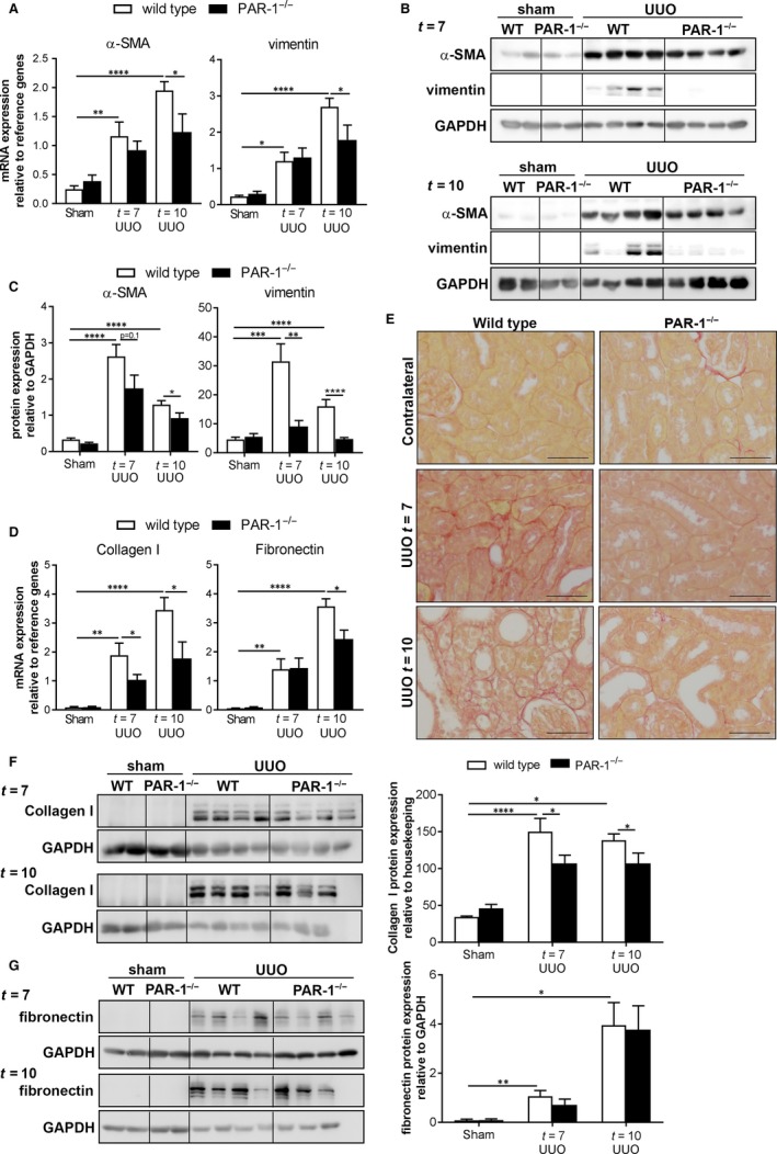
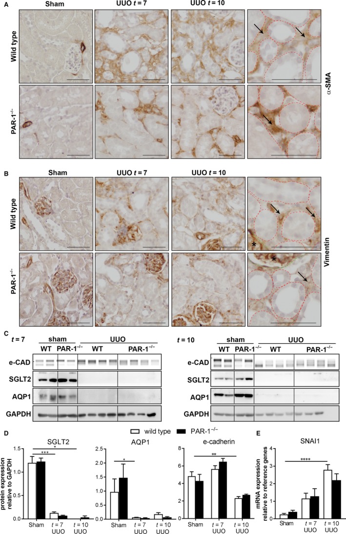
End-stage renal disease, the final stage of all chronic kidney disorders, is associated with renal fibrosis and inevitably leads to renal failure and death. Transition of tubular epithelial cells (TECs) into mesenchymal fibroblasts constitutes a proposed mechanism underlying the progression of renal fibrosis and here we assessed whether protease-activated receptor (PAR)-1, which recently emerged as an inducer of epithelial-to-mesenchymal transition (EMT), aggravates renal fibrosis. We show that PAR-1 activation on TECs reduces the expression of epithelial markers and simultaneously induces mesenchymal marker expression reminiscent of EMT. We next show that kidney damage was reduced in PAR-1-deficient mice during unilateral ureter obstruction (UUO) and that PAR-1-deficient mice develop a diminished fibrotic response. Importantly, however, we did hardly observe any signs of mesenchymal transition in both wild-type and PAR-1-deficient mice suggesting that diminished fibrosis in PAR-1-deficient mice is not due to reduced EMT. Instead, the accumulation of macrophages and fibroblasts was significantly reduced in PAR-1-deficient animals which were accompanied by diminished production of MCP-1 and TGF-β. Overall, we thus show that PAR-1 drives EMT of TECs in vitro and aggravates UUO-induced renal fibrosis although this is likely due to PAR-1-dependent pro-fibrotic cytokine production rather than EMT.

摘要

终末期肾病是所有慢性肾脏疾病的终末阶段,与肾纤维化有关,不可避免地导致肾衰竭和死亡。肾小管上皮细胞(TEC)向间充质成纤维细胞的转化构成了肾纤维化进展的一个潜在机制,在此我们评估了蛋白酶激活受体(PAR)-1是否会加重肾纤维化,PAR-1 最近被认为是上皮间质转化(EMT)的诱导剂。我们发现 PAR-1 在 TEC 上的激活会降低上皮标志物的表达,同时诱导间充质标志物的表达,类似于 EMT。接下来我们发现,在单侧输尿管梗阻(UUO)期间,PAR-1 缺陷小鼠的肾脏损伤减少,并且 PAR-1 缺陷小鼠的纤维化反应减弱。然而,重要的是,我们在野生型和 PAR-1 缺陷型小鼠中几乎没有观察到任何间质转化的迹象,这表明 PAR-1 缺陷型小鼠中减少的纤维化不是由于 EMT 减少所致。相反,PAR-1 缺陷型动物中巨噬细胞和成纤维细胞的积累明显减少,同时 MCP-1 和 TGF-β 的产生也减少。总体而言,我们表明 PAR-1 在体外驱动 TEC 的 EMT,并加重 UUO 诱导的肾纤维化,尽管这可能是由于 PAR-1 依赖性促纤维化细胞因子的产生而不是 EMT。

https://cdn.ncbi.nlm.nih.gov/pmc/blobs/0221/6349177/b10d06f1c219/JCMM-23-1268-g005.jpg
https://cdn.ncbi.nlm.nih.gov/pmc/blobs/0221/6349177/44037afc770d/JCMM-23-1268-g001.jpg
https://cdn.ncbi.nlm.nih.gov/pmc/blobs/0221/6349177/53a8ded3f024/JCMM-23-1268-g002.jpg
https://cdn.ncbi.nlm.nih.gov/pmc/blobs/0221/6349177/2f46ba1e6196/JCMM-23-1268-g003.jpg
https://cdn.ncbi.nlm.nih.gov/pmc/blobs/0221/6349177/7e2934875e93/JCMM-23-1268-g004.jpg
https://cdn.ncbi.nlm.nih.gov/pmc/blobs/0221/6349177/b10d06f1c219/JCMM-23-1268-g005.jpg

相似文献

1
Protease-activated receptor-1 contributes to renal injury and interstitial fibrosis during chronic obstructive nephropathy.蛋白酶激活受体-1 在慢性阻塞性肾病期间导致肾损伤和间质纤维化。
J Cell Mol Med. 2019 Feb;23(2):1268-1279. doi: 10.1111/jcmm.14028. Epub 2018 Nov 28.
2
Protease-activated receptor 2 does not contribute to renal inflammation or fibrosis in the obstructed kidney.蛋白酶激活受体 2 不参与梗阻肾脏的炎症或纤维化反应。
Nephrology (Carlton). 2019 Sep;24(9):983-991. doi: 10.1111/nep.13635. Epub 2019 Jul 31.
3
Targeted disruption of TGF-beta1/Smad3 signaling protects against renal tubulointerstitial fibrosis induced by unilateral ureteral obstruction.靶向破坏转化生长因子-β1/ Smad3信号通路可预防单侧输尿管梗阻诱导的肾小管间质纤维化。
J Clin Invest. 2003 Nov;112(10):1486-94. doi: 10.1172/JCI19270.
4
The PAR-1 antagonist vorapaxar ameliorates kidney injury and tubulointerstitial fibrosis.PAR-1 拮抗剂 vorapaxar 可改善肾损伤和肾小管间质纤维化。
Clin Sci (Lond). 2020 Nov 13;134(21):2873-2891. doi: 10.1042/CS20200923.
5
Nrf2 signaling attenuates epithelial-to-mesenchymal transition and renal interstitial fibrosis via PI3K/Akt signaling pathways.Nrf2 信号通路通过 PI3K/Akt 信号通路减弱上皮间质转化和肾间质纤维化。
Exp Mol Pathol. 2019 Dec;111:104296. doi: 10.1016/j.yexmp.2019.104296. Epub 2019 Aug 23.
6
A direct thrombin inhibitor, dabigatran etexilate protects from renal fibrosis by inhibiting protease activated receptor-1.直接凝血酶抑制剂达比加群酯通过抑制蛋白酶激活受体-1 来防止肾纤维化。
Eur J Pharmacol. 2021 Feb 15;893:173838. doi: 10.1016/j.ejphar.2020.173838. Epub 2020 Dec 24.
7
Plasmin(ogen) promotes renal interstitial fibrosis by promoting epithelial-to-mesenchymal transition: role of plasmin-activated signals.纤溶酶(原)通过促进上皮-间充质转化来促进肾间质纤维化:纤溶酶激活信号的作用。
J Am Soc Nephrol. 2007 Mar;18(3):846-59. doi: 10.1681/ASN.2006080886. Epub 2007 Jan 31.
8
Regulators of calcineurin 1 deficiency attenuates tubulointerstitial fibrosis through improving mitochondrial fitness.钙调磷酸酶 1 缺乏调节剂通过改善线粒体健康来减轻肾小管间质纤维化。
FASEB J. 2020 Nov;34(11). doi: 10.1096/fj.202000781RRR. Epub 2020 Sep 7.
9
Role of protease activated receptor-2 expression in renal interstitial fibrosis model in mice.蛋白酶激活受体-2表达在小鼠肾间质纤维化模型中的作用
J Huazhong Univ Sci Technolog Med Sci. 2005;25(5):523-6. doi: 10.1007/BF02896006.
10
Adenovirus-mediated P311 ameliorates renal fibrosis through inhibition of epithelial-mesenchymal transition via TGF-β1-Smad-ILK pathway in unilateral ureteral obstruction rats.腺病毒介导的 P311 通过 TGF-β1-Smad-ILK 通路抑制上皮间质转化改善单侧输尿管梗阻大鼠的肾纤维化。
Int J Mol Med. 2018 May;41(5):3015-3023. doi: 10.3892/ijmm.2018.3485. Epub 2018 Feb 12.

引用本文的文献

1
Serine proteases and protease-activated receptors signaling in the kidney.肾脏中的丝氨酸蛋白酶与蛋白酶激活受体信号传导
Am J Physiol Cell Physiol. 2025 Jul 1;329(1):C107-C117. doi: 10.1152/ajpcell.00143.2025. Epub 2025 Apr 17.
2
Matrix Metalloproteinases in Ureteropelvic Junction Obstruction: Their Role in Pathogenesis and Their Use as Clinical Markers.输尿管肾盂连接处梗阻中的基质金属蛋白酶:它们在发病机制中的作用及其作为临床标志物的应用
Cells. 2025 Mar 31;14(7):520. doi: 10.3390/cells14070520.
3
MicroRNA-204 may predict the renal function in patients with chronic kidney disease.

本文引用的文献

1
Renoprotective effects of a factor Xa inhibitor: fusion of basic research and a database analysis.Xa 因子抑制剂的肾脏保护作用:基础研究与数据库分析的融合。
Sci Rep. 2018 Jul 18;8(1):10858. doi: 10.1038/s41598-018-29008-2.
2
Plasmin reduces fibronectin deposition by mesangial cells in a protease-activated receptor-1 independent manner.纤溶酶以一种不依赖蛋白酶激活受体-1的方式减少系膜细胞中纤连蛋白的沉积。
Biochem Biophys Rep. 2017 Mar 29;10:152-156. doi: 10.1016/j.bbrep.2017.03.009. eCollection 2017 Jul.
3
NLRX1 dampens oxidative stress and apoptosis in tissue injury via control of mitochondrial activity.
微小RNA-204可能预测慢性肾脏病患者的肾功能。
Medicine (Baltimore). 2025 Jan 3;104(1):e41202. doi: 10.1097/MD.0000000000041202.
4
Unveiling Selected Influences on Chronic Kidney Disease Development and Progression.揭示慢性肾脏病发展和进展的部分影响因素。
Cells. 2024 Apr 26;13(9):751. doi: 10.3390/cells13090751.
5
Interaction of estradiol and renin-angiotensin system with microRNAs-21 and -29 in renal fibrosis: focus on TGF-β/smad signaling pathway.雌激素与肾素-血管紧张素系统与 microRNAs-21 和 -29 在肾纤维化中的相互作用:聚焦于 TGF-β/smad 信号通路。
Mol Biol Rep. 2024 Jan 18;51(1):137. doi: 10.1007/s11033-023-09127-4.
6
Role of protease-activated receptor 4 in mouse models of acute and chronic kidney injury.蛋白酶激活受体 4 在急性和慢性肾损伤小鼠模型中的作用。
Am J Physiol Renal Physiol. 2024 Feb 1;326(2):F219-F226. doi: 10.1152/ajprenal.00162.2023. Epub 2023 Nov 30.
7
Research progress on the mechanism of renal interstitial fibrosis in obstructive nephropathy.梗阻性肾病肾间质纤维化机制的研究进展
Heliyon. 2023 Jul 27;9(8):e18723. doi: 10.1016/j.heliyon.2023.e18723. eCollection 2023 Aug.
8
Application of nanoparticles in the diagnosis and treatment of chronic kidney disease.纳米颗粒在慢性肾脏病诊断与治疗中的应用。
Front Med (Lausanne). 2023 Apr 17;10:1132355. doi: 10.3389/fmed.2023.1132355. eCollection 2023.
9
Baicalin protects against renal interstitial fibrosis in mice by inhibiting the TGF-β/Smad signalling pathway.黄芩苷通过抑制 TGF-β/Smad 信号通路保护小鼠肾间质纤维化。
Pharm Biol. 2022 Dec;60(1):1407-1416. doi: 10.1080/13880209.2022.2097700.
10
Hirudin, a thrombin inhibitor, attenuates TGF-β-induced fibrosis in renal proximal tubular epithelial cells by inhibition of protease-activated receptor 1 expression via S1P/S1PR2/S1PR3 signaling.水蛭素是一种凝血酶抑制剂,它通过S1P/S1PR2/S1PR3信号传导抑制蛋白酶激活受体1的表达,从而减轻转化生长因子-β诱导的肾近端小管上皮细胞纤维化。
Exp Ther Med. 2022 Jan;23(1):3. doi: 10.3892/etm.2021.10924. Epub 2021 Oct 26.
NLRX1通过控制线粒体活性减轻组织损伤中的氧化应激和细胞凋亡。
J Exp Med. 2017 Aug 7;214(8):2405-2420. doi: 10.1084/jem.20161031. Epub 2017 Jun 16.
4
Protease-activated receptor-1 deficiency protects against streptozotocin-induced diabetic nephropathy in mice.蛋白酶激活受体-1 缺陷可预防链脲佐菌素诱导的小鼠糖尿病肾病。
Sci Rep. 2016 Sep 13;6:33030. doi: 10.1038/srep33030.
5
From the Cover: Coagulation-Driven Hepatic Fibrosis Requires Protease Activated Receptor-1 (PAR-1) in a Mouse Model of TCDD-Elicited Steatohepatitis.封面文章:在2,3,7,8-四氯二苯并对二恶英诱发的脂肪性肝炎小鼠模型中,凝血驱动的肝纤维化需要蛋白酶激活受体-1(PAR-1)
Toxicol Sci. 2016 Dec;154(2):381-391. doi: 10.1093/toxsci/kfw175. Epub 2016 Sep 9.
6
The emerging role of coagulation proteases in kidney disease.凝血蛋白酶在肾脏疾病中的新作用。
Nat Rev Nephrol. 2016 Feb;12(2):94-109. doi: 10.1038/nrneph.2015.177. Epub 2015 Nov 23.
7
Protease activated receptor-1 regulates macrophage-mediated cellular senescence: a risk for idiopathic pulmonary fibrosis.蛋白酶激活受体-1调节巨噬细胞介导的细胞衰老:特发性肺纤维化的一个风险因素。
Oncotarget. 2015 Nov 3;6(34):35304-14. doi: 10.18632/oncotarget.6095.
8
Epithelial-to-mesenchymal transition induces cell cycle arrest and parenchymal damage in renal fibrosis.上皮-间质转化诱导肾纤维化中的细胞周期停滞和实质损伤。
Nat Med. 2015 Sep;21(9):998-1009. doi: 10.1038/nm.3902. Epub 2015 Aug 3.
9
Protease activated receptor-1 deficiency diminishes bleomycin-induced skin fibrosis.蛋白酶激活受体-1缺乏可减轻博来霉素诱导的皮肤纤维化。
Mol Med. 2014 Sep 18;20(1):410-6. doi: 10.2119/molmed.2014.00027.
10
Protease-activated receptor-1 drives pancreatic cancer progression and chemoresistance.蛋白酶激活受体-1驱动胰腺癌进展和化疗耐药。
Int J Cancer. 2014 Nov 15;135(10):2294-304. doi: 10.1002/ijc.28726. Epub 2014 Jun 19.