Takkem Amer, Barakat Charif, Zakaraia Safa, Zaid Khaled, Najmeh Johnny, Ayoub Mahdi, Seirawan Mohammad Yaman
Department of Oral Histology and Pathology, University of Damascus, Damascus, Syria. Email:
Asian Pac J Cancer Prev. 2018 Nov 29;19(11):3279-3286. doi: 10.31557/APJCP.2018.19.11.3279.
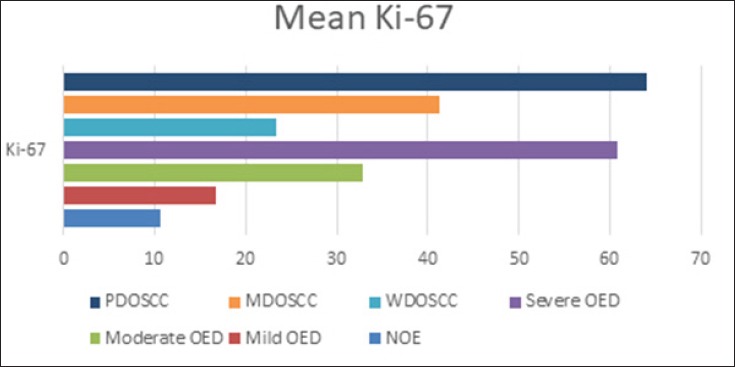
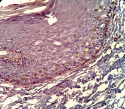
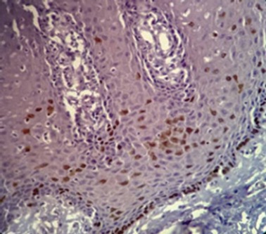
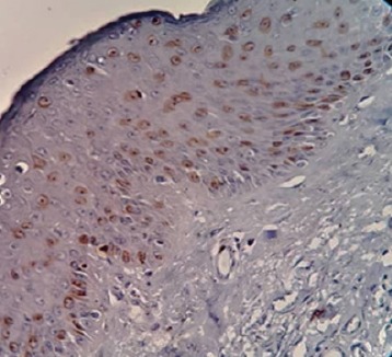
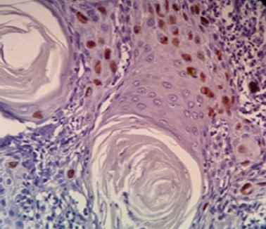
Introduction: Abnormal cell proliferation appears to be a possible predictor of tumorigenesis, Ki-67 protein expression is closely related to the cell proliferation and could be used as a biomarker for the growth in the most of human tumors. The aim of the study: Investigating of Ki-67 expression in the pathological grades of oral epithelial dysplasia and oral squamous cell carcinomas. Materials and Methods: The sample consisted of 30 formalin-fixed, paraffin-embedded specimens of oral epithelial dysplasia (OED), 30 other of oral squamous cell carcinomas (OSCC), and 10 normal oral epithelium (NOE) were conventionally stained with hematoxylin and eosin and immunohistochemically stained with Ki-67 monoclonal antibody. Results: Expression of Ki-67 was restricted to the basal layers in the normal oral epithelium whereas Ki-67 positive cells in oral epithelial dysplasia (OED) were located in the basal, suprabasal and spinous layers, Ki-67 expression was increased in high-risk cases. Ki-67 positive cells in well-differentiated (OSCC) were located mainly in the periphery of the tumor nests, in moderately-differentiated (OSCC) were located in both peripheral and part of a center of the tumor nests whereas it was diffused in most of the Poorly-differentiated (OSCC). Statistical analysis indicated a significant difference between the expression in (OED) and (NOE), (OSCC) and (NOE), and no differences between (OED) and (OSCC). Conclusion: This study has concluded that Ki-67 antigen could be used as a marker for the histological grading of OED and OSCC, Expression of Ki 67 increased according to the severity of oral epithelial dysplasia.
异常细胞增殖似乎是肿瘤发生的一个可能预测指标,Ki-67蛋白表达与细胞增殖密切相关,可作为大多数人类肿瘤生长的生物标志物。研究目的:研究Ki-67在口腔上皮发育异常和口腔鳞状细胞癌病理分级中的表达。材料与方法:样本包括30例福尔马林固定、石蜡包埋的口腔上皮发育异常(OED)标本、30例其他口腔鳞状细胞癌(OSCC)标本和10例正常口腔上皮(NOE)标本,常规苏木精-伊红染色,并采用Ki-67单克隆抗体进行免疫组化染色。结果:Ki-67在正常口腔上皮中仅局限于基底层表达,而在口腔上皮发育异常(OED)中,Ki-67阳性细胞位于基底层、基上层和棘层,高危病例中Ki-67表达增加。高分化(OSCC)中的Ki-67阳性细胞主要位于肿瘤巢周边,中分化(OSCC)中的Ki-67阳性细胞位于肿瘤巢周边及部分中央区域,而在大多数低分化(OSCC)中呈弥漫性分布。统计学分析表明,(OED)与(NOE)、(OSCC)与(NOE)之间的表达存在显著差异,而(OED)与(OSCC)之间无差异。结论:本研究得出结论,Ki-67抗原可作为OED和OSCC组织学分级的标志物,Ki-67的表达随口腔上皮发育异常的严重程度增加而升高。