Asiedu Kingsley O, Ferdousi Munira, Ton Phuongnga T, Adler Stephen S, Choyke Peter L, Sato Noriko
Molecular Imaging Program, Center for Cancer Research, National Cancer Institute, National Institutes of Health, NIH, Building 10, Room B3B406, Bethesda, MD, 20892-1002, USA.
Clinical Monitoring Research Program Directorate, Frederick National Laboratory for Cancer Research sponsored by the National Cancer Institute, Frederick, MD, 21702, USA.
EJNMMI Res. 2018 Dec 13;8(1):109. doi: 10.1186/s13550-018-0463-8.
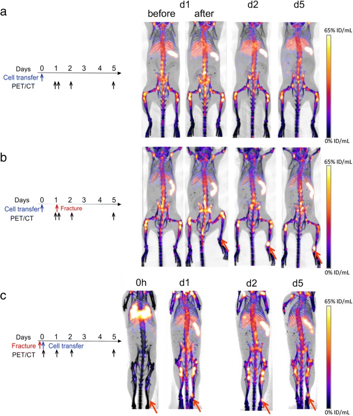
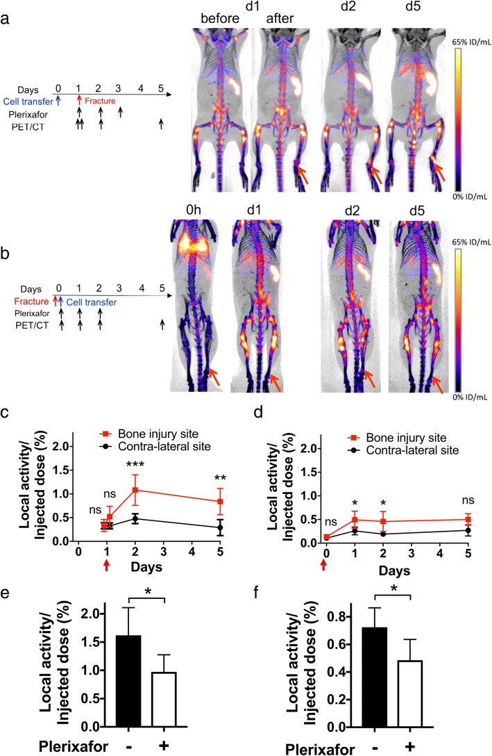
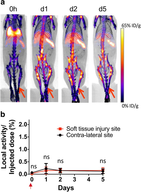
Bone fracture healing is dependent upon the rapid migration and engraftment of bone marrow (BM) progenitor and stem cells to the site of injury. Stromal cell-derived factor-1 plays a crucial role in recruiting BM cells expressing its receptor CXCR4. Recently, a CXCR4 antagonist, plerixafor, has been used to mobilize BM cells into the blood in efforts to enhance cell migration to sites of injury presumably improving healing. In this study, we employed zirconium-89 (Zr)-oxine-labeled BM cells imaged with positron emission tomography (PET)/computed tomography (CT) to visualize and quantitate BM cell trafficking following acute bone injury and to investigate the effect of plerixafor on BM cell homing. Unilateral 1-mm incisions were created in the distal tibia of mice either on the same day (d0) or 24 h (d1) after Zr-oxine-labeled BM cell transfer (n = 4-6, 2-2.3 × 10 cells at 9.65-15.7 kBq/10 cells). Serial microPET/CT imaging was performed and migration of Zr-labeled cells to the bone injury was quantified. The effects of three daily doses of plerixafor on cell trafficking were evaluated beginning on the day of fracture generation (n = 4-6). The labeled cells localizing to the fracture were analyzed by flow cytometry and immunohistochemistry.
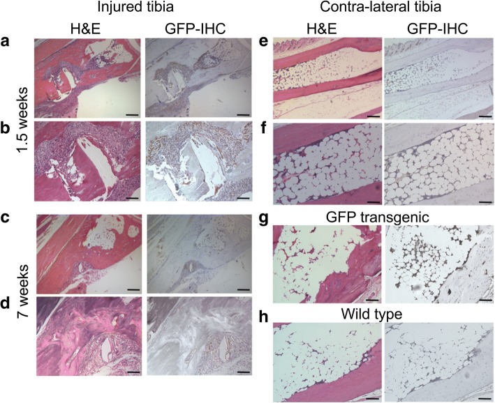
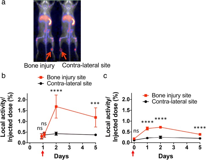
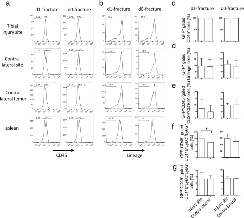
In d0- and d1-fracture groups, 0.7% and 1.7% of administered BM cells accumulated within the fracture, respectively. Plerixafor treatment reduced BM cell migration to the fracture by approximately one-third (p < 0.05 for both fracture groups). Flow cytometry analysis of donor cells collected from the injured site revealed a predominance of CD45 stem/progenitor cell populations and subsequent histological analysis demonstrated the presence of donor cells engrafted within sites of fracture repair.
Zr-oxine labeling enabled visualization and quantitation of BM cell recruitment to acute fractures and further demonstrated that plerixafor plays an inhibitory role in this recruitment.
骨折愈合依赖于骨髓(BM)祖细胞和干细胞快速迁移并植入损伤部位。基质细胞衍生因子-1在招募表达其受体CXCR4的BM细胞中起关键作用。最近,一种CXCR4拮抗剂普乐沙福已被用于将BM细胞动员到血液中,以增强细胞向损伤部位的迁移,推测这可能改善愈合。在本研究中,我们使用正电子发射断层扫描(PET)/计算机断层扫描(CT)成像的锆-89(Zr)-奥辛标记的BM细胞,以可视化和定量急性骨损伤后BM细胞的迁移,并研究普乐沙福对BM细胞归巢的影响。在Zr-奥辛标记的BM细胞转移后的同一天(d0)或24小时(d1),在小鼠胫骨远端制作单侧1毫米切口(n = 4 - 6,9.65 - 15.7 kBq/10个细胞时为2 - 2.3×10个细胞)。进行连续的微型PET/CT成像,并对Zr标记细胞向骨损伤部位的迁移进行定量。从骨折发生当天开始评估每日三次剂量的普乐沙福对细胞迁移的影响(n = 4 - 6)。通过流式细胞术和免疫组织化学分析定位于骨折部位的标记细胞。
在d0骨折组和d1骨折组中,分别有0.7%和1.7%的给药BM细胞积聚在骨折部位。普乐沙福治疗使BM细胞向骨折部位的迁移减少了约三分之一(两个骨折组均p < 0.05)。对从损伤部位收集的供体细胞进行流式细胞术分析,显示CD45干细胞/祖细胞群占优势,随后的组织学分析表明在骨折修复部位存在植入的供体细胞。
Zr-奥辛标记能够可视化和定量BM细胞向急性骨折部位的募集,并进一步证明普乐沙福在这种募集中起抑制作用。