• 文献检索
  • 文档翻译
  • 深度研究
  • 学术资讯
  • Suppr Zotero 插件Zotero 插件
  • 邀请有礼
  • 套餐&价格
  • 历史记录
应用&插件
Suppr Zotero 插件Zotero 插件浏览器插件Mac 客户端Windows 客户端微信小程序
定价
高级版会员购买积分包购买API积分包
服务
文献检索文档翻译深度研究API 文档MCP 服务
关于我们
关于 Suppr公司介绍联系我们用户协议隐私条款
关注我们

Suppr 超能文献

核心技术专利:CN118964589B侵权必究
粤ICP备2023148730 号-1Suppr @ 2026

文献检索

告别复杂PubMed语法,用中文像聊天一样搜索,搜遍4000万医学文献。AI智能推荐,让科研检索更轻松。

立即免费搜索

文件翻译

保留排版,准确专业,支持PDF/Word/PPT等文件格式,支持 12+语言互译。

免费翻译文档

深度研究

AI帮你快速写综述,25分钟生成高质量综述,智能提取关键信息,辅助科研写作。

立即免费体验

乳腺癌细胞质标志物免疫组织化学评估的计算机模拟方法

In Silico Approach for Immunohistochemical Evaluation of a Cytoplasmic Marker in Breast Cancer.

作者信息

Mazo Claudia, Orue-Etxebarria Estibaliz, Zabalza Ignacio, Vivanco Maria D M, Kypta Robert M, Beristain Andoni

机构信息

Vicomtech, eHealth and Biomedical Applications, 20009 San Sebastian-Donostia, Spain.

School of Computer Science, University College Dublin, D14 YH57 Dublin, Ireland.

出版信息

Cancers (Basel). 2018 Dec 15;10(12):517. doi: 10.3390/cancers10120517.

DOI:10.3390/cancers10120517
PMID:30558303
原文链接:https://pmc.ncbi.nlm.nih.gov/articles/PMC6316458/
Abstract

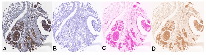
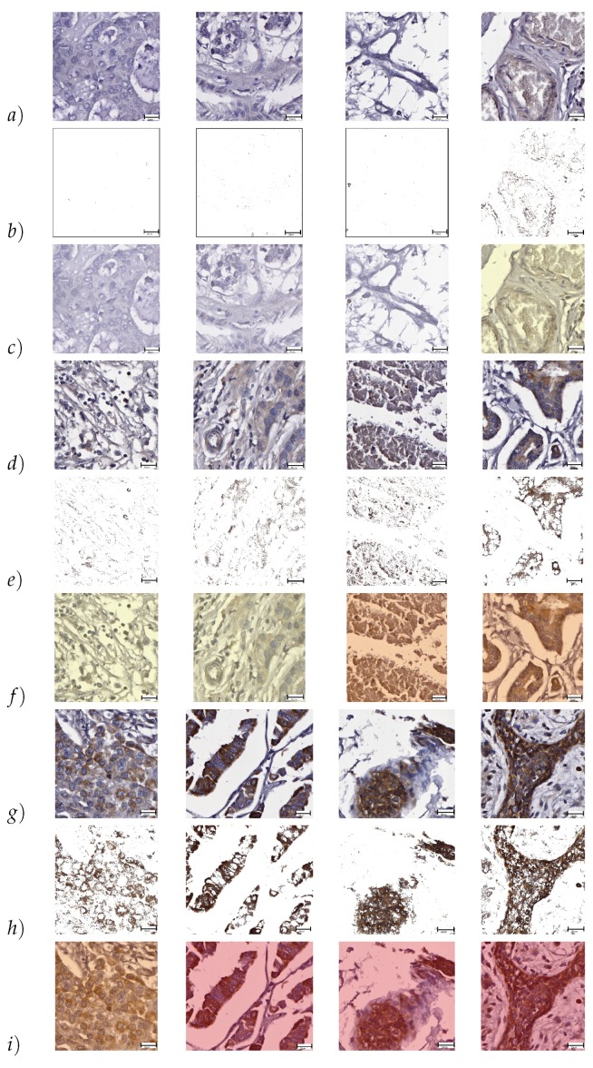
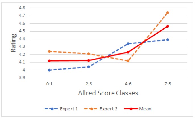
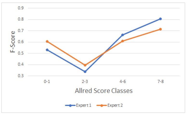
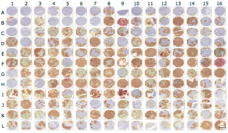
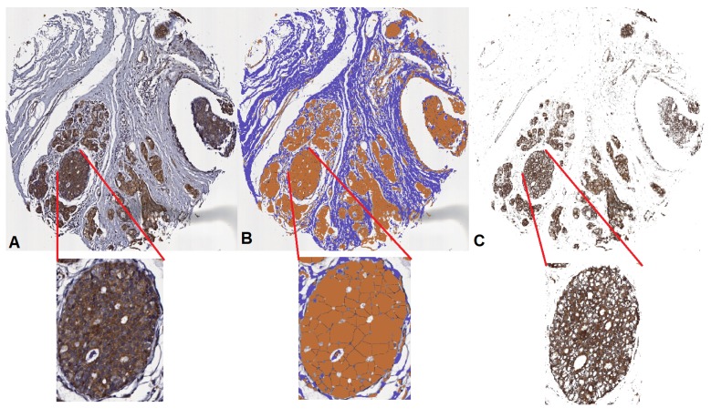
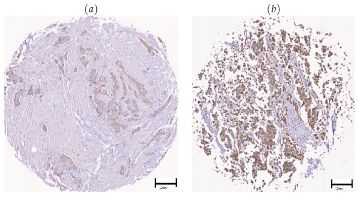
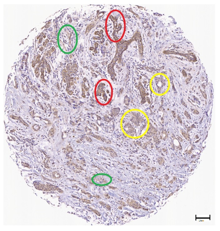
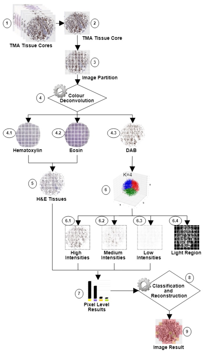
Breast cancer is the most frequently diagnosed cancer in women and the second most common cancer overall, with nearly 1.7 million new cases worldwide every year. Breast cancer patients need accurate tools for early diagnosis and to improve treatment. Biomarkers are increasingly used to describe and evaluate tumours for prognosis, to facilitate and predict response to therapy and to evaluate residual tumor, post-treatment. Here, we evaluate different methods to separate Diaminobenzidine (DAB) from Hematoxylin and Eosin (H&E) staining for , a potential cytoplasmic breast cancer biomarker. A method comprising clustering and Color deconvolution allowed us to recognize and quantify levels accurately at pixel levels. Experimental validation was conducted using a set of 12,288 blocks of m × n pixels without overlap, extracted from a Tissue Microarray (TMA) composed of 192 tissue cores. Intraclass Correlations (ICC) among evaluators of the data of 0.634 , 0.791 , 0.551 and 0.63 for each Allred class and an average ICC of 0.752 among evaluators and automatic classification were obtained. Furthermore, this method received an average rating of 4.26 out of 5 in the segmentation process from the evaluators.

摘要

乳腺癌是女性中最常被诊断出的癌症,也是总体上第二常见的癌症,全球每年有近170万新病例。乳腺癌患者需要准确的工具用于早期诊断和改善治疗。生物标志物越来越多地用于描述和评估肿瘤的预后,促进和预测对治疗的反应以及评估治疗后的残留肿瘤。在此,我们评估了从苏木精和伊红(H&E)染色中分离二氨基联苯胺(DAB)的不同方法,以用于一种潜在的细胞质乳腺癌生物标志物。一种包括聚类和颜色反卷积的方法使我们能够在像素水平上准确识别和量化 水平。使用从由192个组织芯组成的组织微阵列(TMA)中提取的一组12288个m×n像素的无重叠块进行了实验验证。对于每个艾尔雷德等级,评估者之间的数据类内相关性(ICC)分别为0.634、0.791、0.551和0.63,评估者与自动分类之间的平均ICC为0.752。此外,在评估者的 分割过程中,该方法在5分制中平均获得了4.26分的评分。

https://cdn.ncbi.nlm.nih.gov/pmc/blobs/7b84/6316458/910332c4a631/cancers-10-00517-g010.jpg
https://cdn.ncbi.nlm.nih.gov/pmc/blobs/7b84/6316458/62c5f8bd6ea4/cancers-10-00517-g001.jpg
https://cdn.ncbi.nlm.nih.gov/pmc/blobs/7b84/6316458/7e47b45a0550/cancers-10-00517-g002.jpg
https://cdn.ncbi.nlm.nih.gov/pmc/blobs/7b84/6316458/4c4d41351926/cancers-10-00517-g003.jpg
https://cdn.ncbi.nlm.nih.gov/pmc/blobs/7b84/6316458/05c8d3b14dea/cancers-10-00517-g004.jpg
https://cdn.ncbi.nlm.nih.gov/pmc/blobs/7b84/6316458/7e2b3cf5dfe0/cancers-10-00517-g005.jpg
https://cdn.ncbi.nlm.nih.gov/pmc/blobs/7b84/6316458/73da73adc42a/cancers-10-00517-g006.jpg
https://cdn.ncbi.nlm.nih.gov/pmc/blobs/7b84/6316458/5da2bb502e00/cancers-10-00517-g007.jpg
https://cdn.ncbi.nlm.nih.gov/pmc/blobs/7b84/6316458/e0a069e7fc98/cancers-10-00517-g008.jpg
https://cdn.ncbi.nlm.nih.gov/pmc/blobs/7b84/6316458/53d381040bdd/cancers-10-00517-g009.jpg
https://cdn.ncbi.nlm.nih.gov/pmc/blobs/7b84/6316458/910332c4a631/cancers-10-00517-g010.jpg

相似文献

1
In Silico Approach for Immunohistochemical Evaluation of a Cytoplasmic Marker in Breast Cancer.乳腺癌细胞质标志物免疫组织化学评估的计算机模拟方法
Cancers (Basel). 2018 Dec 15;10(12):517. doi: 10.3390/cancers10120517.
2
Fast unsupervised nuclear segmentation and classification scheme for automatic allred cancer scoring in immunohistochemical breast tissue images.快速无监督的核分割和分类方案,用于免疫组织化学乳腺组织图像中的自动 Allred 癌症评分。
Comput Methods Programs Biomed. 2018 Oct;165:37-51. doi: 10.1016/j.cmpb.2018.08.005. Epub 2018 Aug 10.
3
Automatic quantification of IHC stain in breast TMA using colour analysis.使用色彩分析对乳腺 TMA 的 IHC 染色进行自动定量。
Comput Med Imaging Graph. 2017 Nov;61:14-27. doi: 10.1016/j.compmedimag.2017.06.002. Epub 2017 Jun 13.
4
Digital separation of diaminobenzidine-stained tissues via an automatic color-filtering for immunohistochemical quantification.通过自动颜色过滤对二氨基联苯胺染色组织进行数字分离以进行免疫组织化学定量分析。
Biomed Opt Express. 2015 Jan 15;6(2):544-58. doi: 10.1364/BOE.6.000544. eCollection 2015 Feb 1.
5
Validation of tissue microarray biomarker expression of breast carcinomas in Saudi women.沙特女性乳腺癌组织芯片生物标志物表达的验证
Hematol Oncol Stem Cell Ther. 2009;2(3):394-8. doi: 10.1016/s1658-3876(09)50007-6.
6
Reinventing Nuclear Histo-score Utilizing Inherent Morphologic Cutoffs: Blue-brown Color H-score (BBC-HS).利用固有形态学截断值重新构建核组织学评分:蓝-棕颜色 H 评分(BBC-HS)。
Appl Immunohistochem Mol Morphol. 2023 Aug 1;31(7):500-506. doi: 10.1097/PAI.0000000000001095. Epub 2023 Jan 10.
7
Breast cancer dataset with biomarker Biglycan.带有生物标志物双糖链蛋白聚糖的乳腺癌数据集。
Data Brief. 2023 Feb 14;47:108978. doi: 10.1016/j.dib.2023.108978. eCollection 2023 Apr.
8
Impact of tissue sampling on accuracy of Ki67 immunohistochemistry evaluation in breast cancer.组织取样对乳腺癌中Ki67免疫组化评估准确性的影响。
Diagn Pathol. 2016 Aug 30;11(1):82. doi: 10.1186/s13000-016-0525-z.
9
An unsupervised automatic segmentation algorithm for breast tissue classification of dedicated breast computed tomography images.一种用于专用乳腺 CT 图像的乳腺组织分类的无监督自动分割算法。
Med Phys. 2018 Jun;45(6):2542-2559. doi: 10.1002/mp.12920. Epub 2018 May 9.
10
Segmentation of glandular epithelium in colorectal tumours to automatically compartmentalise IHC biomarker quantification: A deep learning approach.结直肠肿瘤中腺上皮的分割,以自动划分免疫组化生物标志物的定量:深度学习方法。
Med Image Anal. 2018 Oct;49:35-45. doi: 10.1016/j.media.2018.07.004. Epub 2018 Jul 12.

引用本文的文献

1
Advances in breast cancer diagnosis: a comprehensive review of imaging, biosensors, and emerging wearable technologies.乳腺癌诊断的进展:影像学、生物传感器及新兴可穿戴技术的全面综述
Front Oncol. 2025 Jun 18;15:1587517. doi: 10.3389/fonc.2025.1587517. eCollection 2025.
2
Correlation Between ImageJ and Conventional Manual Scoring Methods for Programmed Death-Ligand 1 Immuno-Histochemically Stained Sections.免疫组化染色的程序性死亡配体 1 图像分析与传统人工评分方法的相关性。
Technol Cancer Res Treat. 2024 Jan-Dec;23:15330338241242635. doi: 10.1177/15330338241242635.
3
Application of Artificial Intelligence Techniques to Predict Risk of Recurrence of Breast Cancer: A Systematic Review.

本文引用的文献

1
Transfer learning for classification of cardiovascular tissues in histological images.基于迁移学习的组织病理学图像心血管组织分类。
Comput Methods Programs Biomed. 2018 Oct;165:69-76. doi: 10.1016/j.cmpb.2018.08.006. Epub 2018 Aug 16.
2
QuPath: Open source software for digital pathology image analysis.QuPath:用于数字病理学图像分析的开源软件。
Sci Rep. 2017 Dec 4;7(1):16878. doi: 10.1038/s41598-017-17204-5.
3
Classification of cardiovascular tissues using LBP based descriptors and a cascade SVM.使用基于局部二值模式(LBP)的描述符和级联支持向量机(SVM)对心血管组织进行分类。
应用人工智能技术预测乳腺癌复发风险:一项系统综述。
J Pers Med. 2022 Sep 13;12(9):1496. doi: 10.3390/jpm12091496.
Comput Methods Programs Biomed. 2017 Aug;147:1-10. doi: 10.1016/j.cmpb.2017.06.003. Epub 2017 Jun 10.
4
Wnt signaling in cancer.癌症中的Wnt信号传导
Oncogene. 2017 Mar;36(11):1461-1473. doi: 10.1038/onc.2016.304. Epub 2016 Sep 12.
5
Automated Ki-67 Quantification of Immunohistochemical Staining Image of Human Nasopharyngeal Carcinoma Xenografts.人鼻咽癌异种移植组织免疫组化染色图像的 Ki-67 全自动定量分析。
Sci Rep. 2016 Aug 26;6:32127. doi: 10.1038/srep32127.
6
Automatic recognition of fundamental tissues on histology images of the human cardiovascular system.在人类心血管系统组织学图像上对基本组织的自动识别。
Micron. 2016 Oct;89:1-8. doi: 10.1016/j.micron.2016.07.001. Epub 2016 Jul 12.
7
Wnt1 and SFRP1 as potential prognostic factors and therapeutic targets in cutaneous squamous cell carcinoma.Wnt1和SFRP1作为皮肤鳞状细胞癌潜在的预后因素和治疗靶点。
Genet Mol Res. 2016 Jun 20;15(2):gmr8187. doi: 10.4238/gmr.15028187.
8
A Guideline of Selecting and Reporting Intraclass Correlation Coefficients for Reliability Research.可靠性研究中组内相关系数选择与报告指南
J Chiropr Med. 2016 Jun;15(2):155-63. doi: 10.1016/j.jcm.2016.02.012. Epub 2016 Mar 31.
9
Statistical colour models: an automated digital image analysis method for quantification of histological biomarkers.统计颜色模型:一种用于组织学生物标志物定量分析的自动数字图像分析方法。
Biomed Eng Online. 2016 Apr 27;15:46. doi: 10.1186/s12938-016-0161-6.
10
A Comparison of Visual Assessment and Automated Digital Image Analysis of Ki67 Labeling Index in Breast Cancer.乳腺癌中Ki67标记指数的视觉评估与自动数字图像分析比较
PLoS One. 2016 Feb 29;11(2):e0150505. doi: 10.1371/journal.pone.0150505. eCollection 2016.