Université Clermont Auvergne, INRA, UMR 1019, Unité de Nutrition Humaine, CRNH-Auvergne, F-63000, Clermont-Ferrand, France.
CHU Clermont-Ferrand, Centre Jean Perrin, Unité de Nutrition, CLARA, F-63000, Clermont-Ferrand, France.
BMC Cancer. 2018 Dec 18;18(1):1264. doi: 10.1186/s12885-018-5141-8.
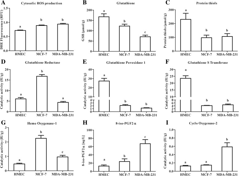
Obesity is associated with oxidative stress, a major factor in carcinogenesis, and with high leptin concentration. The aim of this study was to determine the effects of leptin on the antioxidant response in three human mammary epithelial cells each presenting a different neoplastic status: healthy human mammary epithelial cells (HMEC), oestrogen-receptor positive MCF-7 cells and triple-negative MDA-MB-231 cells.
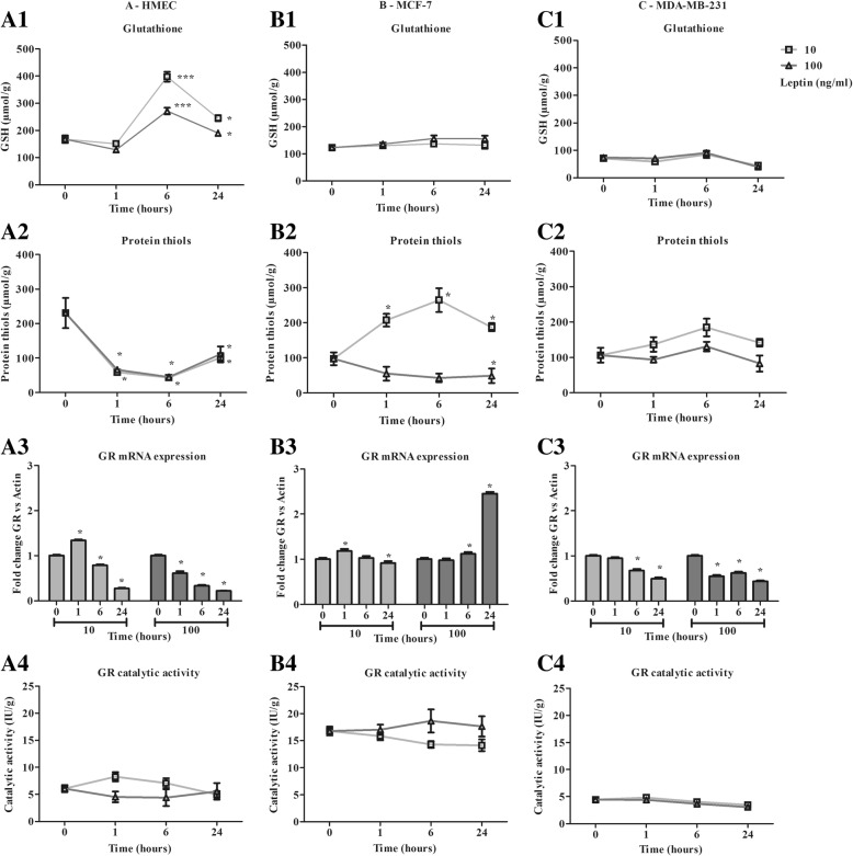
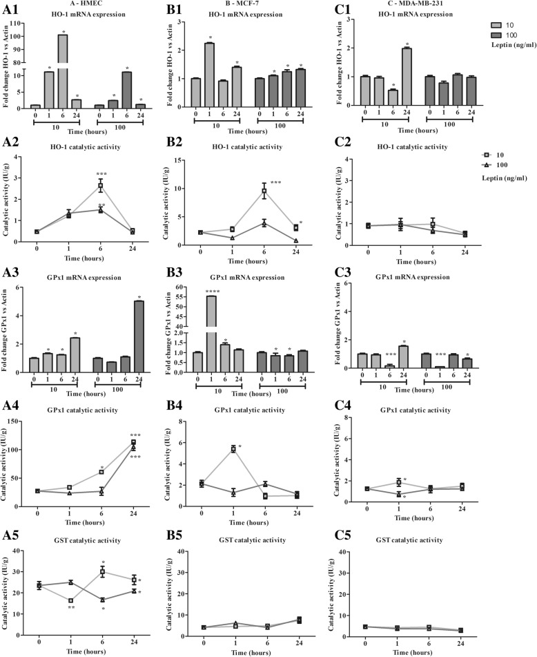
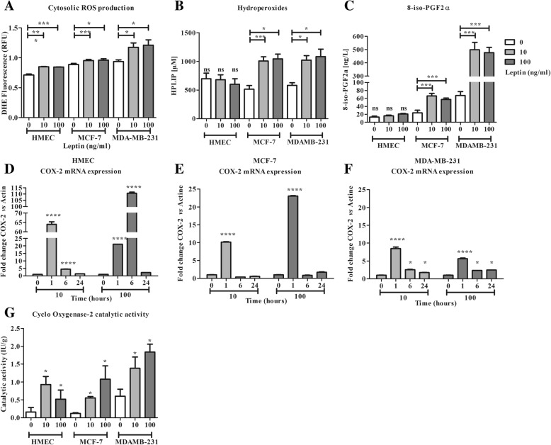
This in vitro kinetic study characterized the cell antioxidant response after 1, 6 and 24 h in the presence of leptin (10 or 100 ng/ml).The antioxidant response was defined in terms of cell glutathione content, gene expression and catalytic activity of antioxidant enzymes (i.e. glutathione peroxidase 1 (Gpx1), glutathione reductase (GR), glutathione S transferase (GST), heme-oxygenase 1 (HO-1) and cyclooxygenase-2 (COX-2)). Oxidative stress occurrence was assessed by lipid hydro peroxide (HPLIP) and isoprostane concentrations in culture media at 24 h.
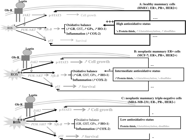
At both concentrations used, leptin induced ROS production in all cell models, contributing to various antioxidant responses linked to neoplastic cell status. HMEC developed a highly inducible antioxidant response based on antioxidant enzyme activation and an increase in cell GSH content at 10 ng/ml of leptin. However, at 100 ng/ml of leptin, activation of antioxidant response was lower. Conversely, in tumour cells, MCF-7 and MDA-MB-231, leptin did not induce an efficient antioxidant response, at either concentration, resulting in an increase of lipid peroxidation products.
Leptin can modulate the oxidative status of mammary epithelial cells differently according to their neoplastic state. These novel results shed light on oxidative status changes in mammary cells in the presence of leptin.
肥胖与氧化应激有关,氧化应激是致癌作用的一个主要因素,并且与瘦素浓度高有关。本研究旨在确定瘦素对三种人乳腺上皮细胞(分别具有不同的肿瘤状态:健康人乳腺上皮细胞(HMEC)、雌激素受体阳性 MCF-7 细胞和三阴性 MDA-MB-231 细胞)抗氧化反应的影响。
这项体外动力学研究在存在瘦素(10 或 100ng/ml)的情况下,分别在 1、6 和 24 小时后对细胞的抗氧化反应进行了特征描述。抗氧化反应的定义是根据细胞谷胱甘肽含量、抗氧化酶(即谷胱甘肽过氧化物酶 1(Gpx1)、谷胱甘肽还原酶(GR)、谷胱甘肽 S 转移酶(GST)、血红素加氧酶 1(HO-1)和环氧化酶-2(COX-2))的基因表达和催化活性来确定的。通过 24 小时时培养介质中的脂质氢过氧化物(HPLIP)和异前列腺素浓度来评估氧化应激的发生。
在两种使用浓度下,瘦素均诱导所有细胞模型中的 ROS 产生,从而导致与肿瘤细胞状态相关的各种抗氧化反应。HMEC 基于抗氧化酶激活和细胞 GSH 含量增加,在 10ng/ml 的瘦素下发展出高度可诱导的抗氧化反应。然而,在 100ng/ml 的瘦素下,抗氧化反应的激活较低。相反,在肿瘤细胞 MCF-7 和 MDA-MB-231 中,瘦素在两种浓度下均未诱导有效的抗氧化反应,导致脂质过氧化产物增加。
瘦素可以根据其肿瘤状态不同地调节乳腺上皮细胞的氧化状态。这些新的结果阐明了在存在瘦素的情况下乳腺细胞氧化状态的变化。