• 文献检索
  • 文档翻译
  • 深度研究
  • 学术资讯
  • Suppr Zotero 插件Zotero 插件
  • 邀请有礼
  • 套餐&价格
  • 历史记录
应用&插件
Suppr Zotero 插件Zotero 插件浏览器插件Mac 客户端Windows 客户端微信小程序
定价
高级版会员购买积分包购买API积分包
服务
文献检索文档翻译深度研究API 文档MCP 服务
关于我们
关于 Suppr公司介绍联系我们用户协议隐私条款
关注我们

Suppr 超能文献

核心技术专利:CN118964589B侵权必究
粤ICP备2023148730 号-1Suppr @ 2026

文献检索

告别复杂PubMed语法,用中文像聊天一样搜索,搜遍4000万医学文献。AI智能推荐,让科研检索更轻松。

立即免费搜索

文件翻译

保留排版,准确专业,支持PDF/Word/PPT等文件格式,支持 12+语言互译。

免费翻译文档

深度研究

AI帮你快速写综述,25分钟生成高质量综述,智能提取关键信息,辅助科研写作。

立即免费体验

肌萎缩侧索硬化症中的新兴磁共振成像技术与分析方法

Emerging Magnetic Resonance Imaging Techniques and Analysis Methods in Amyotrophic Lateral Sclerosis.

作者信息

Barritt Andrew W, Gabel Matt C, Cercignani Mara, Leigh P Nigel

机构信息

Clinical Imaging Sciences Centre, Brighton and Sussex Medical School, Falmer, United Kingdom.

Hurstwood Park Neurological Centre Haywards Heath, West Sussex, United Kingdom.

出版信息

Front Neurol. 2018 Dec 4;9:1065. doi: 10.3389/fneur.2018.01065. eCollection 2018.

DOI:10.3389/fneur.2018.01065
PMID:30564192
原文链接:https://pmc.ncbi.nlm.nih.gov/articles/PMC6288229/
Abstract

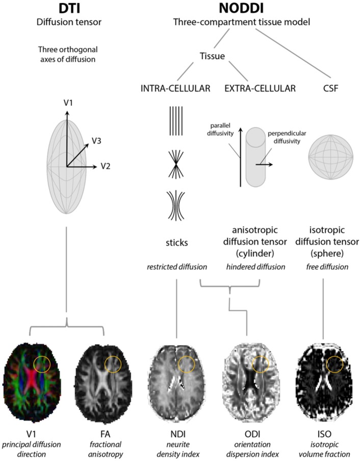
Objective markers of disease sensitive to the clinical activity, symptomatic progression, and underlying substrates of neurodegeneration are highly coveted in amyotrophic lateral sclerosis in order to more eloquently stratify the highly heterogeneous phenotype and facilitate the discovery of effective disease modifying treatments for patients. Magnetic resonance imaging (MRI) is a promising, non-invasive biomarker candidate whose acquisition techniques and analysis methods are undergoing constant evolution in the pursuit of parameters which more closely represent biologically-applicable tissue changes. Neurite Orientation Dispersion and Density Imaging (NODDI; a form of diffusion imaging), and quantitative Magnetization Transfer Imaging (qMTi) are two such emerging modalities which have each broadened the understanding of other neurological disorders and have the potential to provide new insights into structural alterations initiated by the disease process in ALS. Furthermore, novel neuroimaging data analysis approaches such as Event-Based Modeling (EBM) may be able to circumvent the requirement for longitudinal scanning as a means to comprehend the dynamic stages of neurodegeneration . Combining these and other innovative imaging protocols with more sophisticated techniques to analyse ever-increasing datasets holds the exciting prospect of transforming understanding of the biological processes and temporal evolution of the ALS syndrome, and can only benefit from multicentre collaboration across the entire ALS research community.

摘要

在肌萎缩侧索硬化症中,人们迫切渴望获得对疾病临床活动、症状进展和神经退行性变潜在底物敏感的客观标志物,以便更清晰地对高度异质性的表型进行分层,并促进为患者发现有效的疾病修饰治疗方法。磁共振成像(MRI)是一种很有前景的非侵入性生物标志物候选物,其采集技术和分析方法在不断发展,以寻求更能代表生物学上适用的组织变化的参数。神经突方向离散度和密度成像(NODDI;一种扩散成像形式)以及定量磁化传递成像(qMTi)是两种这样的新兴模式,它们各自拓宽了对其他神经系统疾病的理解,并有潜力为肌萎缩侧索硬化症疾病过程引发的结构改变提供新的见解。此外,诸如基于事件的建模(EBM)等新颖的神经影像数据分析方法可能能够规避纵向扫描的要求,以此作为理解神经退行性变动态阶段的一种手段。将这些以及其他创新成像方案与更复杂的技术相结合,以分析不断增加的数据集,有望改变对肌萎缩侧索硬化症综合征生物过程和时间演变的理解,并且只有通过整个肌萎缩侧索硬化症研究界的多中心合作才能从中受益。

https://cdn.ncbi.nlm.nih.gov/pmc/blobs/1aa8/6288229/bc4f5fd79e69/fneur-09-01065-g0002.jpg
https://cdn.ncbi.nlm.nih.gov/pmc/blobs/1aa8/6288229/edf5f2af9429/fneur-09-01065-g0001.jpg
https://cdn.ncbi.nlm.nih.gov/pmc/blobs/1aa8/6288229/bc4f5fd79e69/fneur-09-01065-g0002.jpg
https://cdn.ncbi.nlm.nih.gov/pmc/blobs/1aa8/6288229/edf5f2af9429/fneur-09-01065-g0001.jpg
https://cdn.ncbi.nlm.nih.gov/pmc/blobs/1aa8/6288229/bc4f5fd79e69/fneur-09-01065-g0002.jpg

相似文献

1
Emerging Magnetic Resonance Imaging Techniques and Analysis Methods in Amyotrophic Lateral Sclerosis.肌萎缩侧索硬化症中的新兴磁共振成像技术与分析方法
Front Neurol. 2018 Dec 4;9:1065. doi: 10.3389/fneur.2018.01065. eCollection 2018.
2
Assessment of Cerebral White Matter Involvement in Amyotrophic Lateral Sclerosis Patients With Disease Progression and Cognitive Impairment by Fixel-Based Analysis and Neurite Orientation Dispersion and Density Imaging.基于 Fixel 的分析和神经丝取向分散和密度成像评估疾病进展和认知障碍的肌萎缩侧索硬化症患者的脑白质受累情况。
J Magn Reson Imaging. 2024 Sep;60(3):900-908. doi: 10.1002/jmri.29171. Epub 2023 Dec 7.
3
Longitudinal microstructural MRI markers of demyelination and neurodegeneration in early relapsing-remitting multiple sclerosis: Magnetisation transfer, water diffusion and g-ratio.早期复发缓解型多发性硬化症的脱髓鞘和神经退行性变的纵向微观结构 MRI 标志物:磁化传递、水扩散和 g 比值。
Neuroimage Clin. 2022;36:103228. doi: 10.1016/j.nicl.2022.103228. Epub 2022 Oct 13.
4
Corticospinal fibers with different origins impair in amyotrophic lateral sclerosis: A neurite orientation dispersion and density imaging study.皮质脊髓束纤维起源不同在肌萎缩侧索硬化症中受损:一项神经丝取向弥散和密度成像研究。
CNS Neurosci Ther. 2023 Nov;29(11):3406-3415. doi: 10.1111/cns.14270. Epub 2023 May 19.
5
Neurite orientation dispersion and density imaging in myelin oligodendrocyte glycoprotein antibody-associated disease and neuromyelitis optica spectrum disorders.髓鞘少突胶质细胞糖蛋白抗体相关疾病和视神经脊髓炎谱系障碍中的神经突方向离散度与密度成像
Mult Scler Relat Disord. 2025 Mar;95:106324. doi: 10.1016/j.msard.2025.106324. Epub 2025 Feb 8.
6
Early and progressive dysfunction revealed by in vivo neurite imaging in the rNLS8 TDP-43 mouse model of ALS.TDP-43 基因突变 ALS 模型鼠体内神经突成像揭示的早期进行性功能障碍。
Neuroimage Clin. 2022;34:103016. doi: 10.1016/j.nicl.2022.103016. Epub 2022 Apr 22.
7
Cortico-efferent tract involvement in primary lateral sclerosis and amyotrophic lateral sclerosis: A two-centre tract of interest-based DTI analysis.皮质传出束在原发性侧索硬化症和肌萎缩侧索硬化症中的受累:基于两中心感兴趣区的 DTI 分析。
Neuroimage Clin. 2018;20:1062-1069. doi: 10.1016/j.nicl.2018.10.005. Epub 2018 Oct 9.
8
Neurite orientation and dispersion density imaging (NODDI) detects cortical and corticospinal tract degeneration in ALS.神经突方向分布密度成像(NODDI)可检测 ALS 中的皮质和皮质脊髓束变性。
J Neurol Neurosurg Psychiatry. 2019 Apr;90(4):404-411. doi: 10.1136/jnnp-2018-318830. Epub 2018 Oct 25.
9
Structural imaging differences and longitudinal changes in primary lateral sclerosis and amyotrophic lateral sclerosis.原发性侧索硬化症和肌萎缩性侧索硬化症的结构影像学差异和纵向变化。
Neuroimage Clin. 2012 Dec 24;2:151-60. doi: 10.1016/j.nicl.2012.12.003. eCollection 2012.
10
Neurite orientation dispersion and density imaging quantifies microstructural impairment in the thalamus and its connectivity in amyotrophic lateral sclerosis.神经突方向分散和密度成像定量测量肌萎缩侧索硬化症患者丘脑的结构损伤及其连接。
CNS Neurosci Ther. 2024 Feb;30(2):e14616. doi: 10.1111/cns.14616.

引用本文的文献

1
Role and Potential of Artificial Intelligence in Biomarker Discovery and Development of Treatment Strategies for Amyotrophic Lateral Sclerosis.人工智能在肌萎缩侧索硬化症生物标志物发现及治疗策略开发中的作用与潜力
Int J Mol Sci. 2025 May 2;26(9):4346. doi: 10.3390/ijms26094346.
2
New developments in imaging in ALS.肌萎缩侧索硬化症成像技术的新进展。
J Neurol. 2025 May 12;272(6):392. doi: 10.1007/s00415-025-13143-8.
3
T1-relaxation times along the corticospinal tract as a diagnostic marker in patients with amyotrophic lateral sclerosis.

本文引用的文献

1
Neurite orientation and dispersion density imaging (NODDI) detects cortical and corticospinal tract degeneration in ALS.神经突方向分布密度成像(NODDI)可检测 ALS 中的皮质和皮质脊髓束变性。
J Neurol Neurosurg Psychiatry. 2019 Apr;90(4):404-411. doi: 10.1136/jnnp-2018-318830. Epub 2018 Oct 25.
2
Neurite density is reduced in the presymptomatic phase of disease.神经突密度在疾病的无症状前期降低。
J Neurol Neurosurg Psychiatry. 2019 Apr;90(4):387-394. doi: 10.1136/jnnp-2018-318994. Epub 2018 Oct 24.
3
In vivo characterization of white matter pathology in premanifest huntington's disease.
作为肌萎缩侧索硬化症患者诊断标志物的沿皮质脊髓束的T1弛豫时间
Front Neuroimaging. 2025 Feb 13;4:1549727. doi: 10.3389/fnimg.2025.1549727. eCollection 2025.
4
Clinical, Cortical, Subcortical, and White Matter Features of Right Temporal Variant FTD.右侧颞叶变异型额颞叶痴呆的临床、皮质、皮质下及白质特征
Brain Sci. 2024 Aug 11;14(8):806. doi: 10.3390/brainsci14080806.
5
Limbic Network and Papez Circuit Involvement in ALS: Imaging and Clinical Profiles in GGGGCC Hexanucleotide Carriers in and -Negative Patients.边缘系统网络和帕佩兹环路与肌萎缩侧索硬化症的关系:C9orf72基因中GGGGCC六核苷酸重复序列携带者及阴性患者的影像学和临床特征
Biology (Basel). 2024 Jul 6;13(7):504. doi: 10.3390/biology13070504.
6
Reproducible Spinal Cord Quantitative MRI Analysis with the Spinal Cord Toolbox.可重现的脊髓定量 MRI 分析——脊髓工具箱。
Magn Reson Med Sci. 2024 Jul 1;23(3):307-315. doi: 10.2463/mrms.rev.2023-0159. Epub 2024 Mar 12.
7
MRI biomarkers and neuropsychological assessments of hippocampal and parahippocampal regions affected by ALS: A systematic review.磁共振成像生物标志物与 ALS 影响的海马和海马旁区域神经心理学评估:系统综述。
CNS Neurosci Ther. 2024 Feb;30(2):e14578. doi: 10.1111/cns.14578.
8
Neurite-based white matter alterations in MAPT mutation carriers: A multi-shell diffusion MRI study in the ALLFTD consortium.载有 MAPT 突变的个体的基于神经突的白质改变:ALLFTD 联盟的多壳层扩散 MRI 研究。
Neurobiol Aging. 2024 Feb;134:135-145. doi: 10.1016/j.neurobiolaging.2023.12.001. Epub 2023 Dec 10.
9
The involvement of language-associated networks, tracts, and cortical regions in frontotemporal dementia and amyotrophic lateral sclerosis: Structural and functional alterations.语言相关网络、束和皮质区域在前额颞叶痴呆和肌萎缩侧索硬化症中的作用:结构和功能改变。
Brain Behav. 2023 Nov;13(11):e3250. doi: 10.1002/brb3.3250. Epub 2023 Sep 11.
10
Emotional Regulation and Adolescent Concussion: Overview and Role of Neuroimaging.情绪调节与青少年脑震荡:概述及神经影像学作用。
Int J Environ Res Public Health. 2023 Jul 1;20(13):6274. doi: 10.3390/ijerph20136274.
在症状前亨廷顿病中白质病理学的体内特征描述。
Ann Neurol. 2018 Oct;84(4):497-504. doi: 10.1002/ana.25309. Epub 2018 Sep 15.
4
Microstructural imaging in the spinal cord and validation strategies.脊髓微观结构成像及验证策略。
Neuroimage. 2018 Nov 15;182:169-183. doi: 10.1016/j.neuroimage.2018.04.009. Epub 2018 Apr 7.
5
Data-driven models of dominantly-inherited Alzheimer's disease progression.基于数据驱动的显性遗传性阿尔茨海默病进展模型。
Brain. 2018 May 1;141(5):1529-1544. doi: 10.1093/brain/awy050.
6
Cortical microstructure in young onset Alzheimer's disease using neurite orientation dispersion and density imaging.利用神经丝取向分散和密度成像研究早发性阿尔茨海默病的皮质微观结构。
Hum Brain Mapp. 2018 Jul;39(7):3005-3017. doi: 10.1002/hbm.24056. Epub 2018 Mar 25.
7
Multimodal spinal cord MRI offers accurate diagnostic classification in ALS.多模态脊髓磁共振成像可为肌萎缩侧索硬化症提供准确的诊断分类。
J Neurol Neurosurg Psychiatry. 2018 Nov;89(11):1220-1221. doi: 10.1136/jnnp-2017-317214. Epub 2018 Jan 20.
8
A study of neurite orientation dispersion and density imaging in wilson's disease.脑神经元纤维取向分散度与密度成像在威尔逊病中的研究。
J Magn Reson Imaging. 2018 Aug;48(2):423-430. doi: 10.1002/jmri.25930. Epub 2017 Dec 18.
9
Gray Matter Surface based Spatial Statistics (GS-BSS) in Diffusion Microstructure.基于灰质表面的扩散微结构空间统计学(GS-BSS)
Med Image Comput Comput Assist Interv. 2017 Sep;10433:638-646. doi: 10.1007/978-3-319-66182-7_73. Epub 2017 Sep 4.
10
Histological validation of diffusion MRI fiber orientation distributions and dispersion.弥散磁共振纤维方向分布和分散的组织学验证。
Neuroimage. 2018 Jan 15;165:200-221. doi: 10.1016/j.neuroimage.2017.10.046. Epub 2017 Oct 23.