• 文献检索
  • 文档翻译
  • 深度研究
  • 学术资讯
  • Suppr Zotero 插件Zotero 插件
  • 邀请有礼
  • 套餐&价格
  • 历史记录
应用&插件
Suppr Zotero 插件Zotero 插件浏览器插件Mac 客户端Windows 客户端微信小程序
定价
高级版会员购买积分包购买API积分包
服务
文献检索文档翻译深度研究API 文档MCP 服务
关于我们
关于 Suppr公司介绍联系我们用户协议隐私条款
关注我们

Suppr 超能文献

核心技术专利:CN118964589B侵权必究
粤ICP备2023148730 号-1Suppr @ 2026

文献检索

告别复杂PubMed语法,用中文像聊天一样搜索,搜遍4000万医学文献。AI智能推荐,让科研检索更轻松。

立即免费搜索

文件翻译

保留排版,准确专业,支持PDF/Word/PPT等文件格式,支持 12+语言互译。

免费翻译文档

深度研究

AI帮你快速写综述,25分钟生成高质量综述,智能提取关键信息,辅助科研写作。

立即免费体验

通过测量视网膜中的血管和神经源性变化来研究认知障碍的多模态诊断眼部生物标志物。

Investigating Multimodal Diagnostic Eye Biomarkers of Cognitive Impairment by Measuring Vascular and Neurogenic Changes in the Retina.

作者信息

Cabrera DeBuc Delia, Somfai Gabor Mark, Arthur Edmund, Kostic Maja, Oropesa Susel, Mendoza Santiesteban Carlos

机构信息

Department of Ophthalmology, Bascom Palmer Eye Institute, University of Miami, Miami, FL, United States.

Retinology Unit, Pallas Kliniken, Olten, Switzerland.

出版信息

Front Physiol. 2018 Dec 6;9:1721. doi: 10.3389/fphys.2018.01721. eCollection 2018.

DOI:10.3389/fphys.2018.01721
PMID:30574092
原文链接:https://pmc.ncbi.nlm.nih.gov/articles/PMC6291749/
Abstract

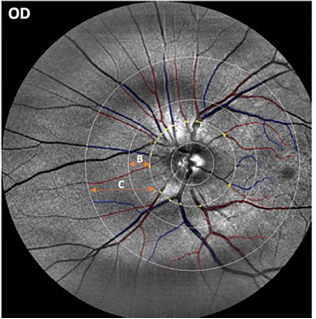
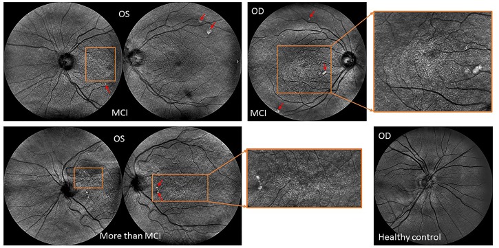
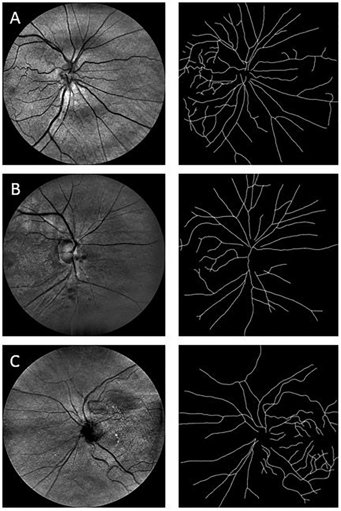
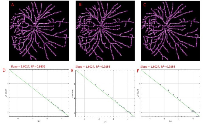
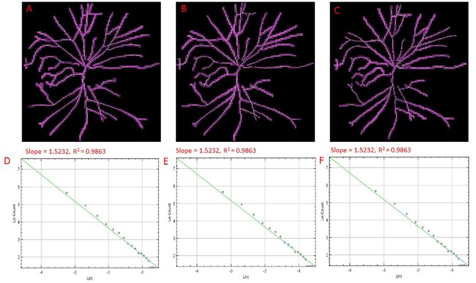
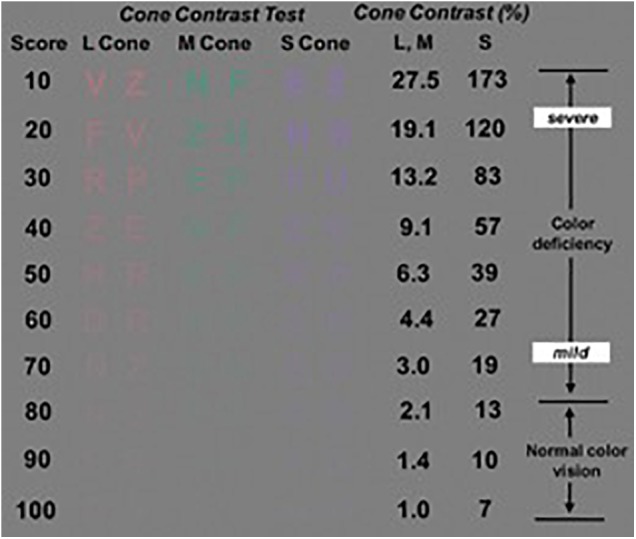
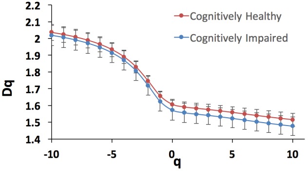
Previous studies have demonstrated that cognitive impairment (CI) is not limited to the brain but also affects the retina. In this pilot study, we investigated the correlation between the retinal vascular complexity and neurodegenerative changes in patients with CI using a low-cost multimodal approach. Quantification of the retinal structure and function were conducted for every subject ( = 69) using advanced retinal imaging, full-field electroretinogram (ERG) and visual performance exams. The retinal vascular parameters were calculated using the Singapore Institute Vessel Assessment software. The Montreal Cognitive Assessment was used to measure CI. Pearson product moment correlation was performed between variables. Of the 69 participants, 32 had CI (46%). We found significantly altered microvascular network in individuals with CI (larger venular-asymmetry factor: 0.7 ± 0.2) compared with controls (0.6 ± 0.2). The vascular fractal dimension was lower in individuals with CI (capacity, information and correlation dimensions: D, D and D (mean ± SD): 1.57 ± 0.06; 1.56 ± 0.06; 1.55 ± 0.06; age 81 ± 6years) vs. controls (1.61 ± 0.03; 1.59 ± 0.03; 1.58 ± 0.03; age: 80 ± 7 years). Also, drusen-like regions in the peripheral retina along with pigment dispersion were noted in subjects with mild CI. Functional loss in color vision as well as smaller ERG amplitudes and larger peak times were observed in the subjects with CI. Pearson product moment correlation showed significant associations between the vascular parameters (artery-vein ratio, total length-diameter ratio, D, D, D and the implicit time (IT) of the flicker response but these associations were not significant in the partial correlations. This study illustrates that there are multimodal retinal markers that may be sensitive to CI decline, and adds to the evidence that there is a statistical trend pointing to the correlation between retinal neuronal dysfunction and microvasculature changes suggesting that retinal geometric vascular and functional parameters might be associated with physiological changes in the retina due to CI. We suspect our analysis of combined structural-functional parameters, instead of individual biomarkers, may provide a useful clinical marker of CI that could also provide increased sensitivity and specificity for the differential diagnosis of CI. However, because of our study sample was small, the full extent of clinical applicability of our approach is provocative and still to be determined.

摘要

以往研究表明,认知障碍(CI)并不局限于大脑,还会影响视网膜。在这项初步研究中,我们采用低成本多模态方法,调查了CI患者视网膜血管复杂性与神经退行性变之间的相关性。使用先进的视网膜成像、全视野视网膜电图(ERG)和视觉性能检查,对每位受试者(n = 69)的视网膜结构和功能进行了量化。使用新加坡国立眼科中心血管评估软件计算视网膜血管参数。采用蒙特利尔认知评估量表来测量CI。对各变量进行Pearson积矩相关分析。69名参与者中,32人患有CI(46%)。我们发现,与对照组(0.6±0.2)相比,CI患者的微血管网络有显著改变(较大的静脉不对称因子:0.7±0.2)。CI患者的血管分形维数较低(容量维、信息维和关联维:D0、D1和D2(均值±标准差):1.57±0.06;1.56±0.06;1.55±0.06;年龄81±6岁),而对照组为(1.61±0.03;1.59±0.03;1.58±0.03;年龄:80±7岁)。此外,轻度CI患者的周边视网膜出现了类玻璃膜疣区域以及色素分散。CI患者出现了色觉功能丧失,以及较小的ERG振幅和较长的峰值时间。Pearson积矩相关分析显示,血管参数(动静脉比、总长度直径比、D0、D1、D2和闪烁反应的隐含时间(IT))之间存在显著关联,但这些关联在偏相关分析中并不显著。本研究表明,存在多种可能对CI衰退敏感的视网膜标志物,并进一步证明存在一种统计趋势,表明视网膜神经元功能障碍与微血管变化之间存在相关性,提示视网膜几何血管和功能参数可能与CI导致的视网膜生理变化有关。我们推测,对结构 - 功能参数进行综合分析,而非单个生物标志物,可能会提供一种有用的CI临床标志物,也可能提高CI鉴别诊断的敏感性和特异性。然而,由于我们的研究样本量较小,我们方法的临床应用的全面程度仍有待探讨和确定。

https://cdn.ncbi.nlm.nih.gov/pmc/blobs/ff33/6291749/cbc46f9fc7d7/fphys-09-01721-g007.jpg
https://cdn.ncbi.nlm.nih.gov/pmc/blobs/ff33/6291749/f3550b09c5df/fphys-09-01721-g001.jpg
https://cdn.ncbi.nlm.nih.gov/pmc/blobs/ff33/6291749/4f97849f3cf4/fphys-09-01721-g002.jpg
https://cdn.ncbi.nlm.nih.gov/pmc/blobs/ff33/6291749/17975151d617/fphys-09-01721-g003.jpg
https://cdn.ncbi.nlm.nih.gov/pmc/blobs/ff33/6291749/cff89dde3231/fphys-09-01721-g004.jpg
https://cdn.ncbi.nlm.nih.gov/pmc/blobs/ff33/6291749/3ce74cdbc409/fphys-09-01721-g005.jpg
https://cdn.ncbi.nlm.nih.gov/pmc/blobs/ff33/6291749/8ca0a82b94ac/fphys-09-01721-g006.jpg
https://cdn.ncbi.nlm.nih.gov/pmc/blobs/ff33/6291749/cbc46f9fc7d7/fphys-09-01721-g007.jpg

相似文献

1
Investigating Multimodal Diagnostic Eye Biomarkers of Cognitive Impairment by Measuring Vascular and Neurogenic Changes in the Retina.通过测量视网膜中的血管和神经源性变化来研究认知障碍的多模态诊断眼部生物标志物。
Front Physiol. 2018 Dec 6;9:1721. doi: 10.3389/fphys.2018.01721. eCollection 2018.
2
Investigating Vascular Complexity and Neurogenic Alterations in Sectoral Regions of the Retina in Patients With Cognitive Impairment.研究认知障碍患者视网膜扇形区域的血管复杂性和神经源性改变。
Front Physiol. 2020 Nov 9;11:570412. doi: 10.3389/fphys.2020.570412. eCollection 2020.
3
Distinguishing cognitive impairment by using singularity spectrum and lacunarity analysis of the retinal vascular network.利用视网膜血管网络的奇异谱和孔隙率分析来区分认知障碍。
Neurophotonics. 2019 Oct;6(4):041109. doi: 10.1117/1.NPh.6.4.041109. Epub 2019 Sep 23.
4
Ocular biomarkers of cognitive decline based on deep-learning retinal vessel segmentation.基于深度学习视网膜血管分割的认知能力下降的眼部生物标志物。
BMC Geriatr. 2024 Jan 6;24(1):28. doi: 10.1186/s12877-023-04593-8.
5
Retinal microvasculature changes in amyloid-negative subcortical vascular cognitive impairment compared to amyloid-positive Alzheimer's disease.与淀粉样蛋白阳性阿尔茨海默病相比,无淀粉样蛋白阴性皮质下血管性认知障碍的视网膜微血管变化。
J Neurol Sci. 2019 Jan 15;396:94-101. doi: 10.1016/j.jns.2018.10.025. Epub 2018 Oct 31.
6
Retinal vascular fractals and cognitive impairment.视网膜血管分形与认知障碍。
Dement Geriatr Cogn Dis Extra. 2014 Aug 27;4(2):305-13. doi: 10.1159/000363286. eCollection 2014 May.
7
Correlation between retinal oscillatory potentials and retinal vascular caliber in type 2 diabetes.2 型糖尿病患者视网膜振荡电位与视网膜血管口径的相关性。
Invest Ophthalmol Vis Sci. 2010 Jan;51(1):482-6. doi: 10.1167/iovs.09-4069. Epub 2009 Aug 26.
8
Retinal dysfunction in a presymptomatic patient with Huntington's disease.亨廷顿病症状前患者的视网膜功能障碍
Doc Ophthalmol. 2018 Jun;136(3):213-221. doi: 10.1007/s10633-018-9632-3. Epub 2018 Apr 24.
9
Evaluation of retinal function and flicker light-induced retinal vascular response in normotensive patients with diabetes without retinopathy.评估无糖尿病视网膜病变的正常血压糖尿病患者的视网膜功能和闪烁光诱导的视网膜血管反应。
Invest Ophthalmol Vis Sci. 2011 May 2;52(6):2861-7. doi: 10.1167/iovs.10-5960.
10
Relationships between quantitative retinal microvascular characteristics and cognitive function based on automated artificial intelligence measurements.基于人工智能自动测量的视网膜微血管定量特征与认知功能之间的关系。
Front Cell Dev Biol. 2023 Jun 21;11:1174984. doi: 10.3389/fcell.2023.1174984. eCollection 2023.

引用本文的文献

1
Ocular changes as potential biomarkers for early diagnosis of Alzheimer's disease.眼部变化作为阿尔茨海默病早期诊断的潜在生物标志物。
Alzheimers Dement. 2025 Aug;21(8):e70476. doi: 10.1002/alz.70476.
2
Clinical Applications of the Cone Contrast Test in Ophthalmology and Neurology.圆锥对比度测试在眼科和神经学中的临床应用。
J Clin Med. 2025 Apr 29;14(9):3079. doi: 10.3390/jcm14093079.
3
Comparative analysis of retinal vascular structural parameters in populations with different glucose metabolism status based on color fundus photography and artificial intelligence.

本文引用的文献

1
Peripheral Retinal Imaging Biomarkers for Alzheimer's Disease: A Pilot Study.用于阿尔茨海默病的周边视网膜成像生物标志物:一项初步研究。
Ophthalmic Res. 2018;59(4):182-192. doi: 10.1159/000487053. Epub 2018 Apr 5.
2
Revolution of Alzheimer Precision Neurology. Passageway of Systems Biology and Neurophysiology.阿尔茨海默病精准神经医学的革命。系统生物学和神经生理学的通道。
J Alzheimers Dis. 2018;64(s1):S47-S105. doi: 10.3233/JAD-179932.
3
Retinal amyloid pathology and proof-of-concept imaging trial in Alzheimer's disease.视网膜淀粉样病变与阿尔茨海默病的概念验证成像试验
基于彩色眼底摄影和人工智能对不同糖代谢状态人群视网膜血管结构参数的比较分析
Front Cell Dev Biol. 2025 Feb 3;13:1550176. doi: 10.3389/fcell.2025.1550176. eCollection 2025.
4
Association and multimodal model of retinal and blood-based biomarkers for detection of preclinical Alzheimer's disease.用于检测临床前阿尔茨海默病的视网膜和血液生物标志物的关联及多模态模型
Alzheimers Res Ther. 2025 Jan 10;17(1):19. doi: 10.1186/s13195-024-01668-5.
5
Analysis and comparison of retinal vascular parameters under different glucose metabolic status based on deep learning.基于深度学习的不同葡萄糖代谢状态下视网膜血管参数的分析与比较
Int J Ophthalmol. 2024 Sep 18;17(9):1581-1591. doi: 10.18240/ijo.2024.09.02. eCollection 2024.
6
Retinal electrophysiological alterations are associated with cognition in early course psychosis.视网膜电生理改变与早期精神病性病程中的认知有关。
Schizophr Res. 2024 Dec;274:137-141. doi: 10.1016/j.schres.2024.09.013. Epub 2024 Sep 17.
7
Retinal Vascular and Structural Changes in the Murine Alzheimer's Model from 6 to 20 Months.从 6 个月到 20 个月的小鼠阿尔茨海默病模型中的视网膜血管和结构变化。
Biomolecules. 2024 Jul 10;14(7):828. doi: 10.3390/biom14070828.
8
Retinal peri-arteriolar versus peri-venular amyloidosis, hippocampal atrophy, and cognitive impairment: exploratory trial.视网膜动静脉周围淀粉样变性、海马萎缩与认知障碍:探索性试验。
Acta Neuropathol Commun. 2024 Jun 28;12(1):109. doi: 10.1186/s40478-024-01810-2.
9
Alzheimer's disease pathophysiology in the Retina.视网膜中的阿尔茨海默病病理生理学。
Prog Retin Eye Res. 2024 Jul;101:101273. doi: 10.1016/j.preteyeres.2024.101273. Epub 2024 May 15.
10
Progress in clinical research and applications of retinal vessel quantification technology based on fundus imaging.基于眼底成像的视网膜血管量化技术的临床研究与应用进展
Front Bioeng Biotechnol. 2024 Feb 22;12:1329263. doi: 10.3389/fbioe.2024.1329263. eCollection 2024.
JCI Insight. 2017 Aug 17;2(16). doi: 10.1172/jci.insight.93621.
4
Evaluation of clinical validity of the Rabin cone contrast test in normal phakic or pseudophakic eyes and severely dichromatic eyes.评估 Rabin 锥体对比试验在正常眼或人工晶状体眼及严重双色眼中的临床有效性。
Acta Ophthalmol. 2018 Mar;96(2):e164-e167. doi: 10.1111/aos.13495. Epub 2017 May 29.
5
Retinal microvascular network alterations: potential biomarkers of cerebrovascular and neural diseases.视网膜微血管网络改变:脑血管和神经疾病的潜在生物标志物。
Am J Physiol Heart Circ Physiol. 2017 Feb 1;312(2):H201-H212. doi: 10.1152/ajpheart.00201.2016. Epub 2016 Dec 6.
6
Predicting the transition from normal aging to Alzheimer's disease: A statistical mechanistic evaluation of FDG-PET data.预测从正常衰老到阿尔茨海默病的转变:对氟代脱氧葡萄糖正电子发射断层扫描(FDG-PET)数据的统计力学评估
Neuroimage. 2016 Nov 1;141:282-290. doi: 10.1016/j.neuroimage.2016.07.043. Epub 2016 Jul 22.
7
Acquired color vision deficiency.获得性色觉障碍。
Surv Ophthalmol. 2016 Mar-Apr;61(2):132-55. doi: 10.1016/j.survophthal.2015.11.004. Epub 2015 Nov 30.
8
Retinal microvascular network attenuation in Alzheimer's disease.阿尔茨海默病中的视网膜微血管网络衰减
Alzheimers Dement (Amst). 2015 May 16;1(2):229-235. doi: 10.1016/j.dadm.2015.04.001. eCollection 2015 Jun.
9
Effect of Pupil Size on Flicker ERGs Recorded With RETeval System: New Mydriasis-Free Full-Field ERG System.瞳孔大小对使用RETeval系统记录的闪光视网膜电图的影响:新型免散瞳全视野视网膜电图系统
Invest Ophthalmol Vis Sci. 2015 Jun;56(6):3684-90. doi: 10.1167/iovs.14-16349.
10
ISCEV Standard for full-field clinical electroretinography (2015 update).国际临床视觉电生理学会全视野临床视网膜电图标准(2015年更新版)
Doc Ophthalmol. 2015 Feb;130(1):1-12. doi: 10.1007/s10633-014-9473-7. Epub 2014 Dec 14.