Nuffield Department of Women's and Reproductive Health, University of Oxford, Women's Centre, Level 3, John Radcliffe Hospital, Oxford, United Kingdom.
IVF Centre, Hong Kong Sanatorium and Hospital, Happy Valley, Hong Kong.
Reproduction. 2019 Feb 1;157(2):135-148. doi: 10.1530/REP-18-0115.
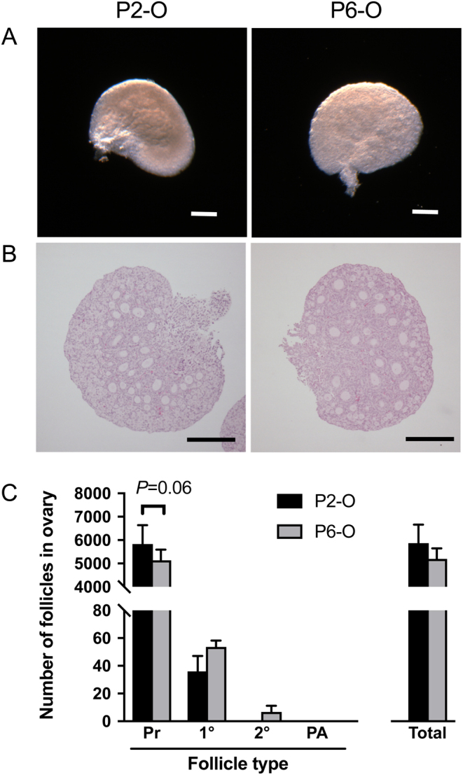
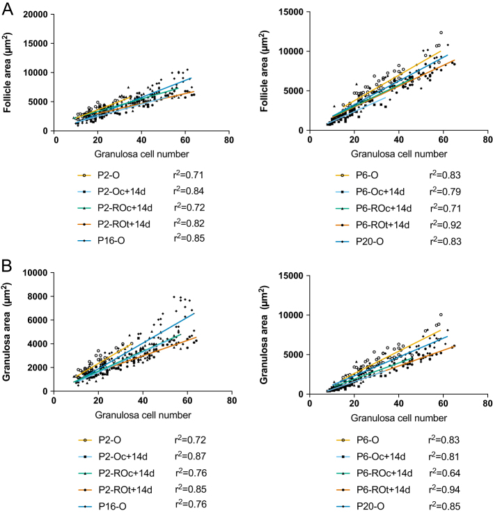
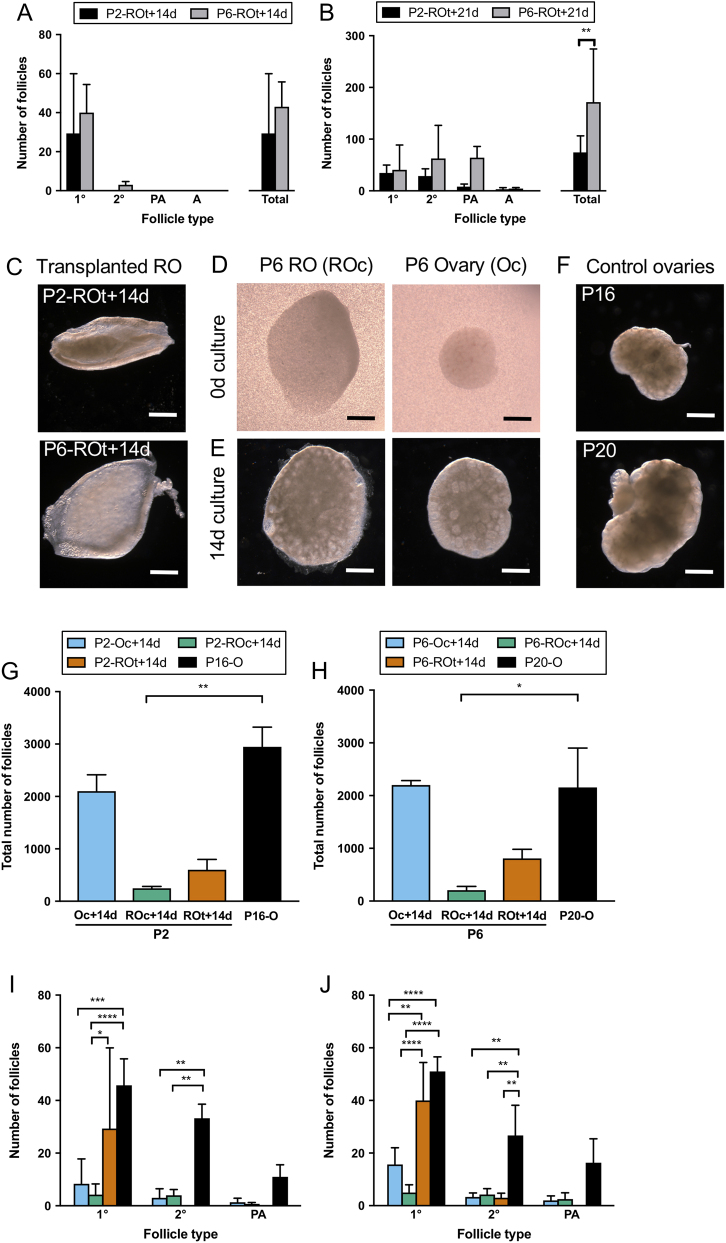
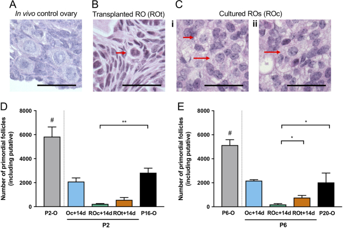
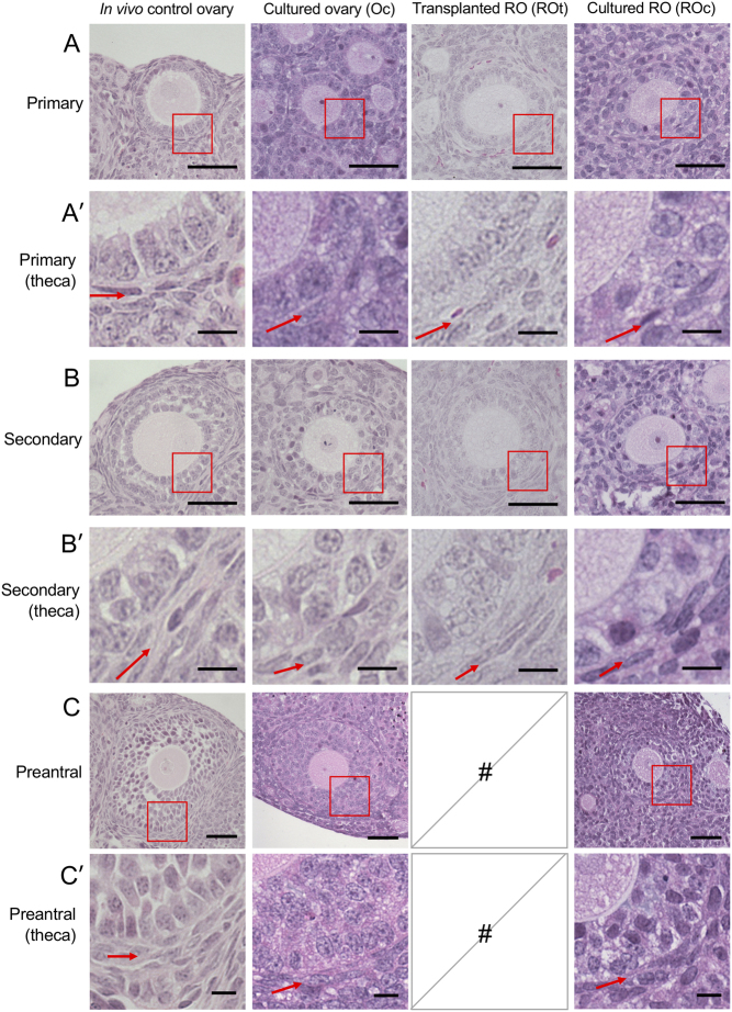
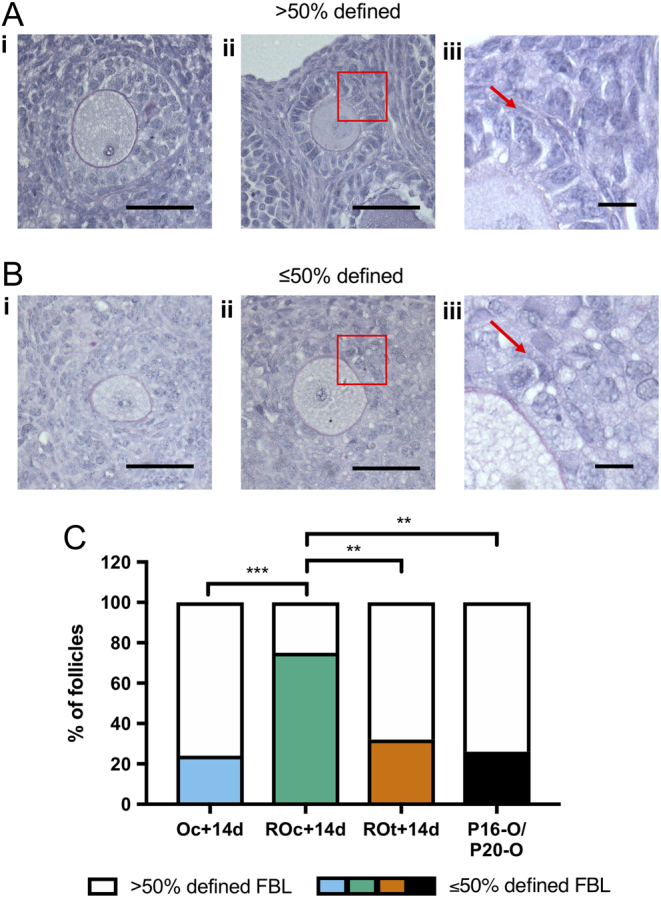
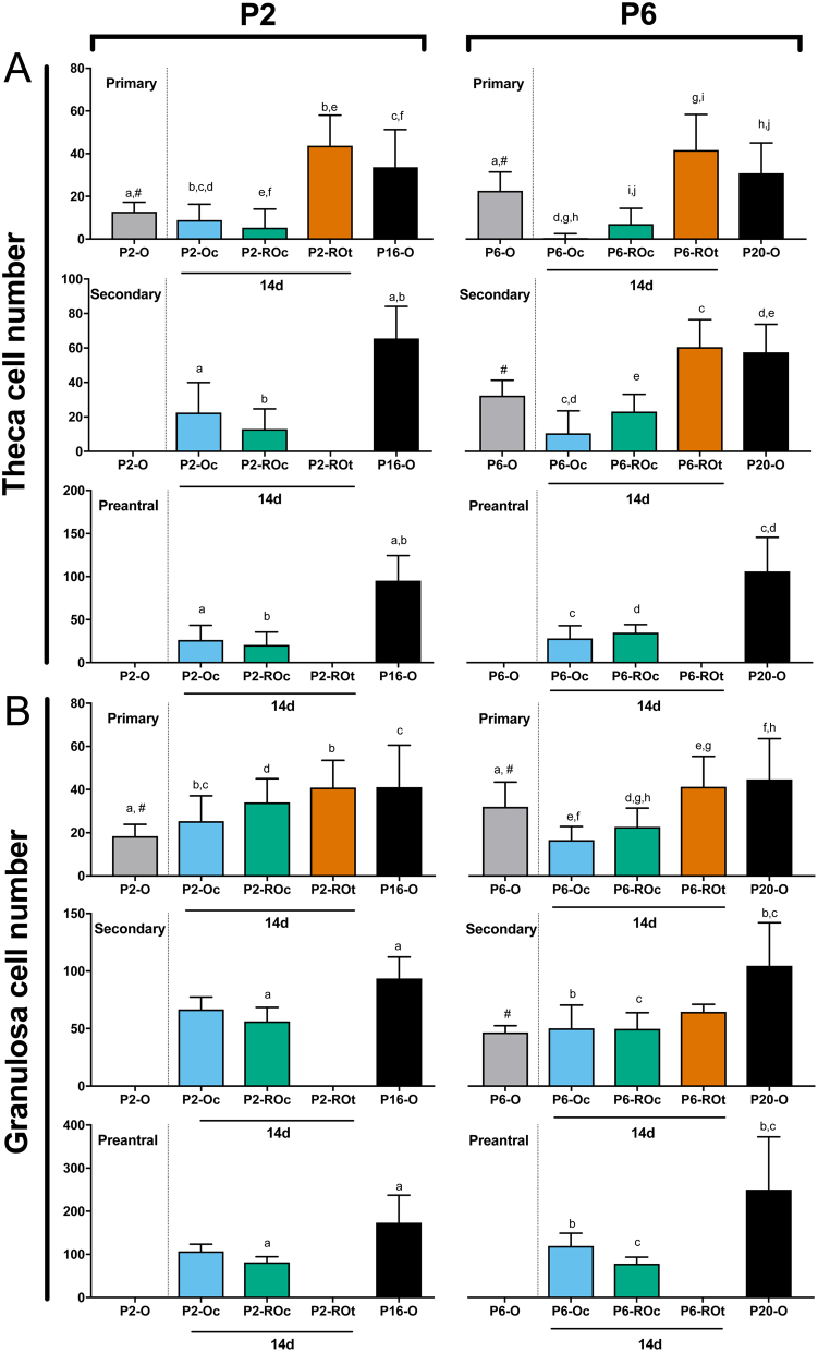
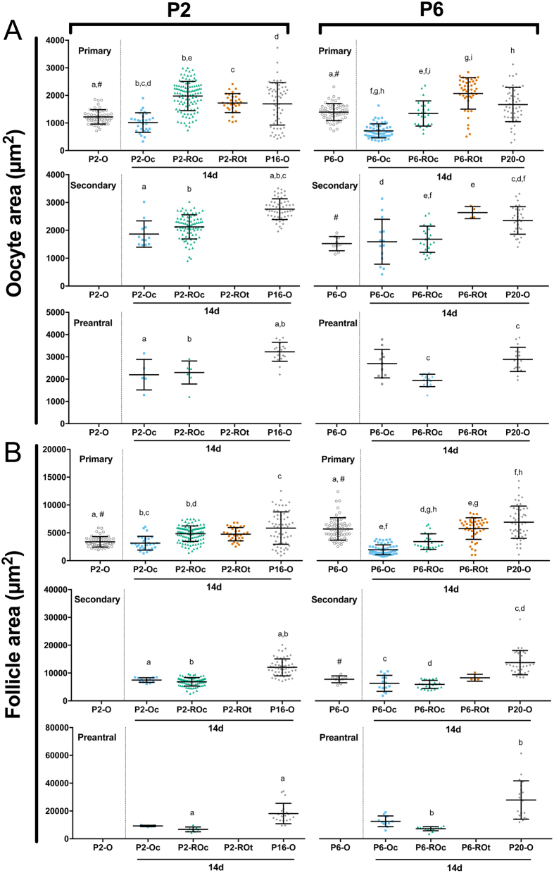
Follicle development requires complex and coordinated interactions between both the oocyte and its associated somatic cells. In ovarian dysfunction, follicle development may be abnormal due to defective somatic cell function; for example, premature ovarian insufficiency or malignancies. Replacing defective somatic cells, using the reaggregated ovary (RO) technique, may 'rescue' follicle development. ROs containing mature follicles have been generated when transplanted to a host mouse to develop. We have developed a RO culture technique and the aims were to determine how follicle development differed between transplanted and cultured ROs, and the influence of ovarian age (P2 vs P6). Mouse ROs were cultured for 14 days; P2 and P6 ovaries cultured as Controls. Follicle development was compared to ROs transplanted for 14 days and ovaries from P16 and P20 mice. ROs generated from either P2 or P6 exhibited similar follicle development in culture whereas in vivo follicle development was more advanced in P6 ROs. Follicles were more developed in cultured ROs than transplanted ROs. However, follicles in cultured ROs and ovaries had smaller oocytes with fewer theca and granulosa cells than in vivo counterparts. Our results demonstrate the fluidity of follicle development despite ovary dissociation and that environment is more important to basal lamina formation and theca cell development. Furthermore, follicle development within cultured ROs appears to be independent of oocyte nest breakdown and primordial follicle formation in source ovaries. Our results highlight the need for understanding follicle development in vitro, particularly in the development of the RO technique as a potential fertility treatment.
卵泡发育需要卵母细胞与其相关的体细胞之间复杂而协调的相互作用。在卵巢功能障碍中,由于体细胞功能缺陷,卵泡发育可能异常;例如,卵巢早衰或恶性肿瘤。使用聚集卵巢(RO)技术替换有缺陷的体细胞,可能会“挽救”卵泡发育。当将 RO 移植到宿主小鼠中以发育时,已经生成了含有成熟卵泡的 RO。我们已经开发了一种 RO 培养技术,其目的是确定移植和培养的 RO 之间的卵泡发育有何不同,以及卵巢年龄(P2 与 P6)的影响。将小鼠 RO 培养 14 天;将 P2 和 P6 卵巢作为对照进行培养。将卵泡发育与移植 14 天的 RO 和来自 P16 和 P20 小鼠的卵巢进行比较。从 P2 或 P6 产生的 RO 在培养中显示出相似的卵泡发育,而体内 P6 RO 的卵泡发育更为先进。与移植 RO 相比,培养 RO 中的卵泡发育更为成熟。然而,与体内对应物相比,培养 RO 中的卵泡具有更小的卵母细胞,并且其卵泡膜细胞和颗粒细胞更少。我们的结果表明,尽管卵巢分离,但卵泡发育具有流动性,并且环境对基底膜形成和卵泡膜细胞发育更为重要。此外,培养 RO 中的卵泡发育似乎独立于来源卵巢中的卵母细胞巢破裂和原始卵泡形成。我们的结果强调了在体外理解卵泡发育的必要性,特别是在 RO 技术作为潜在的生育治疗方法的发展方面。