Saleh Thekra Fadel, Sultan Ghada Abdulrhman, Altaey Omar Younis
Department of Anatomy, College of Veterinary Medicine, University of Mosul, Mosul, Iraq.
Open Vet J. 2025 May;15(5):2171-2181. doi: 10.5455/OVJ.2025.v15.i5.35. Epub 2025 May 31.
Dwarf hamsters are extensively used as models in reproductive disorders studies, reproductive endocrinology, embryo transplantation, and and egg fertilization.
This study aimed to explore the morphological and morphometric development of the ovary with T lymphocyte distribution at different ages using immunohistochemical cluster of differentiation 40 (CD40) expression.
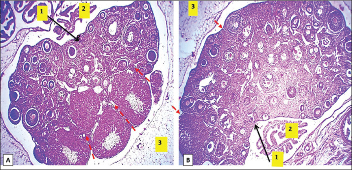
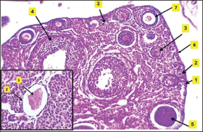
Fifteen dwarf hamster () females were used in this study and divided into three groups at three different ages (2, 4, and 8 weeks) after birth. Samples were collected, and the length, width, and weight were measured. Standard histological processing was performed, and the slides were stained with hematoxylin and eosin. Histomorphometrical analysis was performed using a 3.0 USB microscopic camera for determining the numbers and diameters of the primordial, primary, secondary, and Graafian follicles and the diameter of the mature oocyte and corpus luteum. Immunohistochemical analysis using CD40 anti-Mouse ligand antibody expression to evaluate T lymphocyte distribution within ovarian parenchyma.
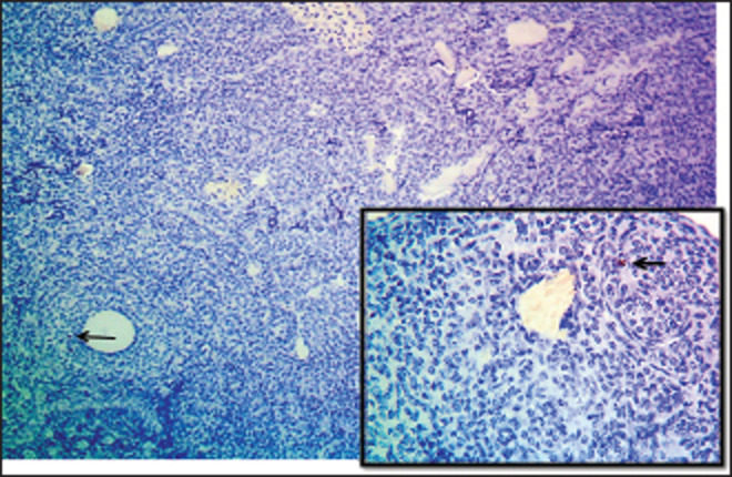
At 2 weeks of age, the observations showed symmetric development of the ovaries, with no significant differences in dimensions and weight. Histologically, the ovaries displayed early follicle development with sparse T lymphocytes. At 4 weeks, follicular size asymmetry emerged with the presence of corpus luteum and increased T lymphocyte counts. By 8 weeks, the ovaries exhibited more developed ovarian follicles, larger corpus luteum, and a further increase in T-lymphocyte density.
This investigation sheds light on the development of the ovary in dwarf hamsters, an understudied model with a short maturity age. These findings provide a promising understanding of ovarian morphological development and the diagnosis of various infertility developmental disorders.
矮仓鼠被广泛用作生殖障碍研究、生殖内分泌学、胚胎移植和卵子受精的模型。
本研究旨在利用免疫组织化学分化簇40(CD40)表达,探讨不同年龄卵巢的形态和形态计量学发育以及T淋巴细胞分布。
本研究使用了15只雌性矮仓鼠,在出生后的三个不同年龄(2周、4周和8周)分为三组。采集样本,测量其长度、宽度和重量。进行标准组织学处理,玻片用苏木精和伊红染色。使用3.0 USB显微镜相机进行组织形态计量学分析,以确定原始卵泡、初级卵泡、次级卵泡和格拉夫卵泡的数量和直径,以及成熟卵母细胞和黄体的直径。使用CD40抗小鼠配体抗体表达进行免疫组织化学分析,以评估卵巢实质内的T淋巴细胞分布。
2周龄时,观察到卵巢对称发育,尺寸和重量无显著差异。组织学上,卵巢显示早期卵泡发育,T淋巴细胞稀少。4周时,卵泡大小出现不对称,有黄体存在,T淋巴细胞计数增加。到8周时,卵巢显示出更发达的卵泡、更大的黄体,T淋巴细胞密度进一步增加。
本研究揭示了矮仓鼠卵巢的发育情况,矮仓鼠是一种成熟年龄短但研究较少的模型。这些发现为卵巢形态发育及各种不孕发育障碍的诊断提供了有前景的认识。