Tsonis Ioannis, Karamani Lydia, Xaplanteri Panagiota, Kolonitsiou Fevronia, Zampakis Petros, Gatzounis Georgios, Marangos Markos, Assimakopoulos Stelios F
Department of Neurosurgery, University of Patras Medical School, Patras 26504, Greece.
Department of Microbiology, University of Patras Medical School, Patras 26504, Greece.
World J Clin Cases. 2018 Dec 26;6(16):1169-1174. doi: 10.12998/wjcc.v6.i16.1169.
() is considered a non-pathogenic microorganism of the genus and a common laboratory contaminant. Only scarce reports of central nervous system infection have been reported, mainly in the form of pyogenic meningitis, usually in cases of direct inoculation by trauma or iatrogenically.
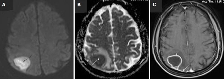
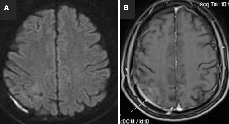
A 51-year-old man, with a free previous medical history, presented to the Emergency Department of our hospital complaining of recurrent episodes of left upper limb weakness, during the last month, which had been worsened the last 48 h. During his presentation in Emergency Department he experienced a generalized tonic-clonic grand mal seizure. Brain magnetic resonance imaging (MRI) scan with intravenous Gadolinium revealed a 3.3 cm × 2.7 cm lesion at the right parietal lobe surrounded by mild vasogenic edema, which included the posterior central gyrus. The core of the lesion showed relatively homogenous restricted diffusion. Post Gadolinium T1W1 image, revealed a ring-shaped enhancement. Due to the imaging findings, brain abscess was our primary consideration. Detailed examination for clinical signs of infectious foci revealed only poor oral hygiene with severe tooth decay and periodontal disease, but without detection of dental abscess. The patient underwent surgical treatment with right parietal craniotomy and total excision of the lesion. Pus and capsule tissue grew and according to antibiogram intravenous ceftriaxone 2 g bids was administered for 4 wk. The patient remained asymptomatic and follow-up MRI scan two months after operation showed complete removal of the abscess.
This case highlights the ultimate importance of appropriate oral hygiene and dental care to avoid potentially serious infectious complications and second, should not be considered merely as laboratory contaminant especially when cultivated by appropriate central nervous system specimen.
()被认为是该属的一种非致病性微生物,是常见的实验室污染物。仅有关于其引起中枢神经系统感染的罕见报道,主要表现为化脓性脑膜炎,通常发生在创伤直接接种或医源性感染的病例中。
一名51岁男性,既往无病史,因近一个月反复出现左上肢无力症状就诊于我院急诊科,近48小时症状加重。在急诊科就诊期间,他发生了一次全身性强直阵挛性癫痫大发作。静脉注射钆对比剂后的脑部磁共振成像(MRI)扫描显示,右侧顶叶有一个3.3 cm×2.7 cm的病灶,周围有轻度血管源性水肿,累及中央后回。病灶核心显示相对均匀的扩散受限。钆增强T1加权像显示环形强化。根据影像学表现,脑脓肿是首要考虑的诊断。对感染灶的临床体征进行详细检查,仅发现口腔卫生差,有严重龋齿和牙周病,但未发现牙脓肿。患者接受了右侧顶叶开颅手术并完整切除病灶。脓液和包膜组织培养出(),根据药敏结果,静脉注射头孢曲松2 g,每日两次,持续4周。患者术后无症状,术后两个月的MRI随访扫描显示脓肿已完全清除。
该病例强调了保持适当口腔卫生和牙齿护理以避免潜在严重感染并发症的极端重要性,其次,()不应仅仅被视为实验室污染物,特别是当它从合适的中枢神经系统标本中培养出来时。