• 文献检索
  • 文档翻译
  • 深度研究
  • 学术资讯
  • Suppr Zotero 插件Zotero 插件
  • 邀请有礼
  • 套餐&价格
  • 历史记录
应用&插件
Suppr Zotero 插件Zotero 插件浏览器插件Mac 客户端Windows 客户端微信小程序
定价
高级版会员购买积分包购买API积分包
服务
文献检索文档翻译深度研究API 文档MCP 服务
关于我们
关于 Suppr公司介绍联系我们用户协议隐私条款
关注我们

Suppr 超能文献

核心技术专利:CN118964589B侵权必究
粤ICP备2023148730 号-1Suppr @ 2026

文献检索

告别复杂PubMed语法,用中文像聊天一样搜索,搜遍4000万医学文献。AI智能推荐,让科研检索更轻松。

立即免费搜索

文件翻译

保留排版,准确专业,支持PDF/Word/PPT等文件格式,支持 12+语言互译。

免费翻译文档

深度研究

AI帮你快速写综述,25分钟生成高质量综述,智能提取关键信息,辅助科研写作。

立即免费体验

心肌细胞中的二元可塑性。

Dyadic Plasticity in Cardiomyocytes.

作者信息

Jones Peter P, MacQuaide Niall, Louch William E

机构信息

Department of Physiology, School of Biomedical Sciences, University of Otago, Dunedin, New Zealand.

HeartOtago, University of Otago, Dunedin, New Zealand.

出版信息

Front Physiol. 2018 Dec 11;9:1773. doi: 10.3389/fphys.2018.01773. eCollection 2018.

DOI:10.3389/fphys.2018.01773
PMID:30618792
原文链接:https://pmc.ncbi.nlm.nih.gov/articles/PMC6298195/
Abstract

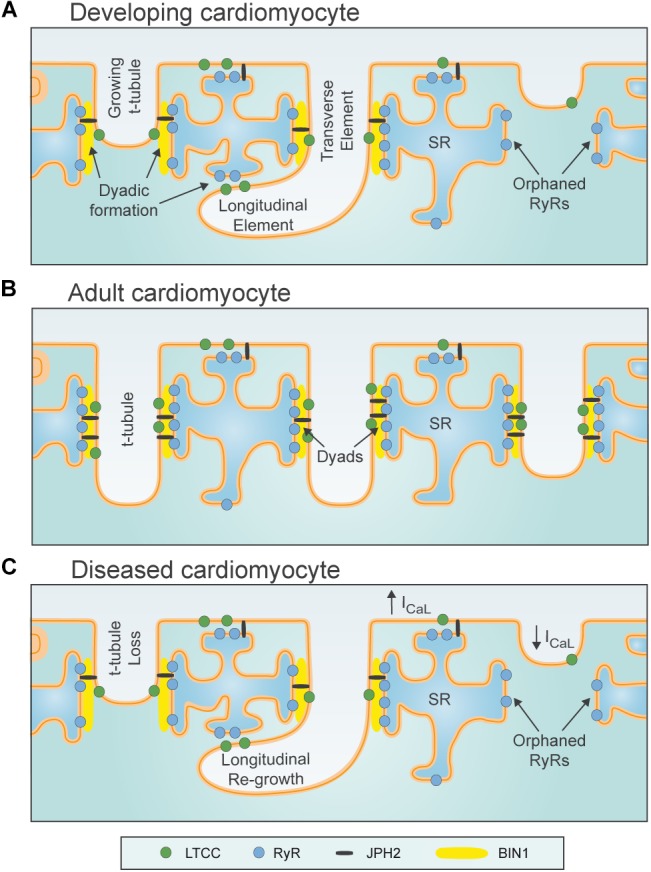
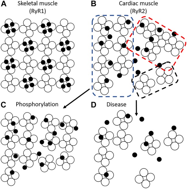
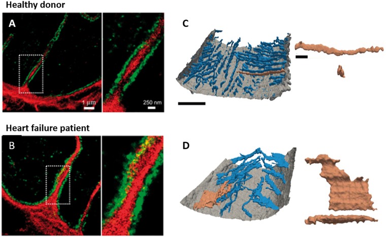
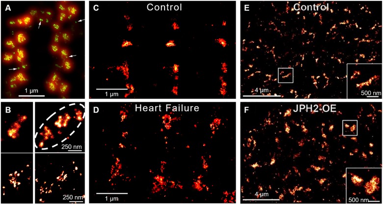
Contraction of cardiomyocytes is dependent on sub-cellular structures called dyads, where invaginations of the surface membrane (t-tubules) form functional junctions with the sarcoplasmic reticulum (SR). Within each dyad, Ca entry through t-tubular L-type Ca channels (LTCCs) elicits Ca release from closely apposed Ryanodine Receptors (RyRs) in the SR membrane. The efficiency of this process is dependent on the density and macroscale arrangement of dyads, but also on the nanoscale organization of LTCCs and RyRs within them. We presently review accumulating data demonstrating the remarkable plasticity of these structures. Dyads are known to form gradually during development, with progressive assembly of both t-tubules and junctional SR terminals, and precise trafficking of LTCCs and RyRs. While dyads can exhibit compensatory remodeling when required, dyadic degradation is believed to promote impaired contractility and arrythmogenesis in cardiac disease. Recent data indicate that this plasticity of dyadic structure/function is dependent on the regulatory proteins junctophilin-2, amphiphysin-2 (BIN1), and caveolin-3, which critically arrange dyadic membranes while stabilizing the position and activity of LTCCs and RyRs. Indeed, emerging evidence indicates that clustering of both channels enables "coupled gating", implying that nanoscale localization and function are intimately linked, and may allow fine-tuning of LTCC-RyR crosstalk. We anticipate that improved understanding of dyadic plasticity will provide greater insight into the processes of cardiac compensation and decompensation, and new opportunities to target the basic mechanisms underlying heart disease.

摘要

心肌细胞的收缩依赖于一种称为二联体的亚细胞结构,在这种结构中,表面膜(横小管)的内陷与肌浆网(SR)形成功能性连接。在每个二联体中,通过横小管L型钙通道(LTCCs)进入的钙离子会引发肌浆网膜上紧密相邻的兰尼碱受体(RyRs)释放钙离子。这一过程的效率不仅取决于二联体的密度和宏观排列,还取决于其中LTCCs和RyRs的纳米级组织。我们目前回顾了越来越多的数据,这些数据表明了这些结构具有显著的可塑性。已知二联体在发育过程中逐渐形成,伴随着横小管和连接性肌浆网终末的逐步组装,以及LTCCs和RyRs的精确运输。虽然二联体在需要时可以表现出代偿性重塑,但二联体的退化被认为会促进心脏病中收缩功能受损和心律失常的发生。最近的数据表明,二联体结构/功能的这种可塑性依赖于调节蛋白连接蛋白-2、发动蛋白-2(BIN1)和小窝蛋白-3,它们在稳定LTCCs和RyRs的位置和活性的同时,关键地排列二联体膜。事实上,新出现的证据表明,两种通道的聚集能够实现“耦合门控”,这意味着纳米级定位和功能密切相关,并且可能允许对LTCC-RyR相互作用进行微调。我们预计,对二联体可塑性的更好理解将为心脏代偿和失代偿过程提供更深入的见解,并为针对心脏病潜在基本机制提供新的机会。

https://cdn.ncbi.nlm.nih.gov/pmc/blobs/bf3d/6298195/752e73863a7b/fphys-09-01773-g006.jpg
https://cdn.ncbi.nlm.nih.gov/pmc/blobs/bf3d/6298195/7bf02c82eb6c/fphys-09-01773-g001.jpg
https://cdn.ncbi.nlm.nih.gov/pmc/blobs/bf3d/6298195/72b51be8a5bd/fphys-09-01773-g002.jpg
https://cdn.ncbi.nlm.nih.gov/pmc/blobs/bf3d/6298195/0fac6877e299/fphys-09-01773-g003.jpg
https://cdn.ncbi.nlm.nih.gov/pmc/blobs/bf3d/6298195/42a08c12f39f/fphys-09-01773-g004.jpg
https://cdn.ncbi.nlm.nih.gov/pmc/blobs/bf3d/6298195/e53efac9887e/fphys-09-01773-g005.jpg
https://cdn.ncbi.nlm.nih.gov/pmc/blobs/bf3d/6298195/752e73863a7b/fphys-09-01773-g006.jpg

相似文献

1
Dyadic Plasticity in Cardiomyocytes.心肌细胞中的二元可塑性。
Front Physiol. 2018 Dec 11;9:1773. doi: 10.3389/fphys.2018.01773. eCollection 2018.
2
Interaction of the Joining Region in Junctophilin-2 With the L-Type Ca Channel Is Pivotal for Cardiac Dyad Assembly and Intracellular Ca Dynamics.衔接蛋白 Junctophilin-2 的连接区与 L 型钙通道的相互作用对于心肌二联体的组装和细胞内钙动力学至关重要。
Circ Res. 2021 Jan 8;128(1):92-114. doi: 10.1161/CIRCRESAHA.119.315715. Epub 2020 Oct 23.
3
The Physiology and Pathophysiology of T-Tubules in the Heart.心脏中横管的生理学与病理生理学
Front Physiol. 2021 Sep 9;12:718404. doi: 10.3389/fphys.2021.718404. eCollection 2021.
4
Augmenting workload drives T-tubule assembly in developing cardiomyocytes.工作负荷增加可促进心肌细胞分化过程中的 T 小管组装。
J Physiol. 2024 Sep;602(18):4461-4486. doi: 10.1113/JP284538. Epub 2023 May 21.
5
Junctophilin-2 tethers T-tubules and recruits functional L-type calcium channels to lipid rafts in adult cardiomyocytes.连接蛋白-2 将 T 小管固定在成年心肌细胞的脂筏上,并募集功能性 L 型钙通道。
Cardiovasc Res. 2021 Jan 1;117(1):149-161. doi: 10.1093/cvr/cvaa033.
6
The architecture and function of cardiac dyads.心肌二联体的结构与功能。
Biophys Rev. 2020 Aug;12(4):1007-1017. doi: 10.1007/s12551-020-00729-x. Epub 2020 Jul 13.
7
Cardiomyocyte substructure reverts to an immature phenotype during heart failure.在心力衰竭过程中心肌细胞亚结构恢复为未成熟表型。
J Physiol. 2019 Apr;597(7):1833-1853. doi: 10.1113/JP277273. Epub 2019 Feb 27.
8
Calcium/calmodulin-dependent kinase II and nitric oxide synthase 1-dependent modulation of ryanodine receptors during β-adrenergic stimulation is restricted to the dyadic cleft.在β-肾上腺素能刺激过程中,钙/钙调蛋白依赖性激酶II和一氧化氮合酶1对兰尼碱受体的调节作用局限于二联体裂隙。
J Physiol. 2016 Oct 15;594(20):5923-5939. doi: 10.1113/JP271965. Epub 2016 Jul 3.
9
Cardiac BIN1 (cBIN1) is a regulator of cardiac contractile function and an emerging biomarker of heart muscle health.心脏 BIN1(cBIN1)是心脏收缩功能的调节剂,也是心肌健康的新兴生物标志物。
Sci China Life Sci. 2017 Mar;60(3):257-263. doi: 10.1007/s11427-016-0249-x. Epub 2016 Nov 23.
10
Regulation of Cardiomyocyte T-Tubular Structure: Opportunities for Therapy.心肌细胞T小管结构的调控:治疗机遇
Curr Heart Fail Rep. 2017 Jun;14(3):167-178. doi: 10.1007/s11897-017-0329-9.

引用本文的文献

1
A Better Understanding of Atrial-like and Ventricular-like Action Potentials in Stem Cell-Derived Cardiomyocytes: The Underestimated Role of the L-Type Ca Current.更好地理解干细胞衍生心肌细胞中的心房样和心室样动作电位:L型钙电流被低估的作用
Cells. 2025 Aug 8;14(16):1226. doi: 10.3390/cells14161226.
2
Electrodiffusion dynamics in the cardiomyocyte dyad at nano-scale resolution using the Poisson-Nernst-Planck (PNP) equations.使用泊松-能斯特-普朗克(PNP)方程在纳米尺度分辨率下研究心肌细胞二联体中的电扩散动力学。
PLoS Comput Biol. 2025 Jun 12;21(6):e1013149. doi: 10.1371/journal.pcbi.1013149. eCollection 2025 Jun.
3
Ryanodine Receptors in Islet Cell Function: Calcium Signaling, Hormone Secretion, and Diabetes.

本文引用的文献

1
Ryanodine receptor dispersion disrupts Ca release in failing cardiac myocytes.兰尼碱受体弥散破坏衰竭心肌细胞的钙释放。
Elife. 2018 Oct 30;7:e39427. doi: 10.7554/eLife.39427.
2
Dynamic L-type Ca1.2 channel trafficking facilitates Ca1.2 clustering and cooperative gating.动态 L 型 Ca1.2 通道运输有助于 Ca1.2 簇集和协同门控。
Biochim Biophys Acta Mol Cell Res. 2018 Sep;1865(9):1341-1355. doi: 10.1016/j.bbamcr.2018.06.013. Epub 2018 Jun 28.
3
Hyperactive ryanodine receptors in human heart failure and ischaemic cardiomyopathy reside outside of couplons.
胰岛细胞功能中的兰尼碱受体:钙信号传导、激素分泌与糖尿病
Cells. 2025 May 10;14(10):690. doi: 10.3390/cells14100690.
4
Virally delivered CMYA5 enhances the assembly of cardiac dyads.病毒递送的CMYA5增强了心肌二联体的组装。
Nat Biomed Eng. 2025 May;9(5):730-741. doi: 10.1038/s41551-024-01253-z. Epub 2024 Sep 5.
5
T cell Ca microdomains through the lens of computational modeling.T 细胞钙微区的计算建模视角。
Front Immunol. 2023 Oct 4;14:1235737. doi: 10.3389/fimmu.2023.1235737. eCollection 2023.
6
Endoplasmic Reticulum-Plasma Membrane Junctions as Sites of Depolarization-Induced Ca Signaling in Excitable Cells.内质网-质膜连接作为可兴奋细胞中去极化诱导钙信号的位点。
Annu Rev Physiol. 2023 Feb 10;85:217-243. doi: 10.1146/annurev-physiol-032122-104610. Epub 2022 Oct 6.
7
Ontogeny of cardiomyocytes: ultrastructure optimization to meet the demand for tight communication in excitation-contraction coupling and energy transfer.心肌细胞的个体发生:超微结构的优化以满足兴奋-收缩偶联和能量传递中紧密通讯的需求。
Philos Trans R Soc Lond B Biol Sci. 2022 Nov 21;377(1864):20210321. doi: 10.1098/rstb.2021.0321. Epub 2022 Oct 3.
8
Three-dimensional visualization of the cardiac ryanodine receptor clusters and the molecular-scale fraying of dyads.心脏兰尼碱受体簇的三维可视化和二联体的分子级磨损。
Philos Trans R Soc Lond B Biol Sci. 2022 Nov 21;377(1864):20210316. doi: 10.1098/rstb.2021.0316. Epub 2022 Oct 3.
9
Nanoscale organization of ryanodine receptor distribution and phosphorylation pattern determines the dynamics of calcium sparks.兰尼碱受体分布和磷酸化模式的纳米级组织决定钙火花的动力学。
PLoS Comput Biol. 2022 Jun 6;18(6):e1010126. doi: 10.1371/journal.pcbi.1010126. eCollection 2022 Jun.
10
Biological noise is a key determinant of the reproducibility and adaptability of cardiac pacemaking and EC coupling.生物噪声是心脏起搏和 EC 偶联的可重复性和适应性的关键决定因素。
J Gen Physiol. 2022 Sep 5;154(9). doi: 10.1085/jgp.202012613. Epub 2022 Apr 28.
在人类心力衰竭和缺血性心肌病中,活性过高的兰尼碱受体位于偶联点之外。
Cardiovasc Res. 2018 Sep 1;114(11):1512-1524. doi: 10.1093/cvr/cvy088.
4
Dynamic and Irregular Distribution of RyR2 Clusters in the Periphery of Live Ventricular Myocytes.活心室心肌细胞周边 RyR2 簇的动态和不规则分布。
Biophys J. 2018 Jan 23;114(2):343-354. doi: 10.1016/j.bpj.2017.11.026.
5
True Molecular Scale Visualization of Variable Clustering Properties of Ryanodine Receptors.真分子水平可视化兰尼碱受体的变构簇性质。
Cell Rep. 2018 Jan 9;22(2):557-567. doi: 10.1016/j.celrep.2017.12.045.
6
Transient activation of PKC results in long-lasting detrimental effects on systolic [Ca] in cardiomyocytes by altering actin cytoskeletal dynamics and T-tubule integrity.短暂激活蛋白激酶 C 会通过改变肌动蛋白细胞骨架动力学和 T 管完整性对心肌细胞的收缩期 [Ca] 产生持久的有害影响。
J Mol Cell Cardiol. 2018 Feb;115:104-114. doi: 10.1016/j.yjmcc.2018.01.003. Epub 2018 Jan 4.
7
T-tubule remodeling and increased heterogeneity of calcium release during the progression to heart failure in intact rat ventricle.在完整大鼠心室向心力衰竭进展过程中T小管重塑及钙释放异质性增加
Physiol Rep. 2017 Dec;5(24). doi: 10.14814/phy2.13540. Epub 2017 Dec 26.
8
Loss of caveolin-3-dependent regulation of I in rat ventricular myocytes in heart failure.心力衰竭大鼠心室肌细胞中窖蛋白-3依赖的I调节丧失。
Am J Physiol Heart Circ Physiol. 2018 Mar 1;314(3):H521-H529. doi: 10.1152/ajpheart.00458.2017. Epub 2017 Nov 3.
9
Calcium release-dependent inactivation precedes formation of the tubular system in developing rat cardiac myocytes.钙释放依赖性失活先于发育中的大鼠心肌细胞中管状系统的形成。
Eur Biophys J. 2017 Dec;46(8):691-703. doi: 10.1007/s00249-017-1249-z. Epub 2017 Sep 14.
10
Remodeling of the transverse tubular system after myocardial infarction in rabbit correlates with local fibrosis: A potential role of biomechanics.兔心肌梗死后横管系统重塑与局部纤维化相关:生物力学的潜在作用
Prog Biophys Mol Biol. 2017 Nov;130(Pt B):302-314. doi: 10.1016/j.pbiomolbio.2017.07.006. Epub 2017 Jul 11.