• 文献检索
  • 文档翻译
  • 深度研究
  • 学术资讯
  • Suppr Zotero 插件Zotero 插件
  • 邀请有礼
  • 套餐&价格
  • 历史记录
应用&插件
Suppr Zotero 插件Zotero 插件浏览器插件Mac 客户端Windows 客户端微信小程序
定价
高级版会员购买积分包购买API积分包
服务
文献检索文档翻译深度研究API 文档MCP 服务
关于我们
关于 Suppr公司介绍联系我们用户协议隐私条款
关注我们

Suppr 超能文献

核心技术专利:CN118964589B侵权必究
粤ICP备2023148730 号-1Suppr @ 2026

文献检索

告别复杂PubMed语法,用中文像聊天一样搜索,搜遍4000万医学文献。AI智能推荐,让科研检索更轻松。

立即免费搜索

文件翻译

保留排版,准确专业,支持PDF/Word/PPT等文件格式,支持 12+语言互译。

免费翻译文档

深度研究

AI帮你快速写综述,25分钟生成高质量综述,智能提取关键信息,辅助科研写作。

立即免费体验

使用光学相干断层扫描技术检测视神经通路胶质瘤患儿的视网膜神经纤维损失。

Use of Optical Coherence Tomography to Detect Retinal Nerve Fiber Loss in Children With Optic Pathway Glioma.

作者信息

Zahavi Alon, Toledano Helen, Cohen Rony, Sella Sara, Luckman Judith, Michowiz Shalom, Goldenberg-Cohen Nitza

机构信息

Department of Ophthalmology, Rabin Medical Center-Beilinson Hospital, Petah Tikva, Israel.

Sackler Faculty of Medicine, Tel Aviv University, Tel Aviv, Israel.

出版信息

Front Neurol. 2018 Dec 20;9:1102. doi: 10.3389/fneur.2018.01102. eCollection 2018.

DOI:10.3389/fneur.2018.01102
PMID:30619059
原文链接:https://pmc.ncbi.nlm.nih.gov/articles/PMC6306407/
Abstract

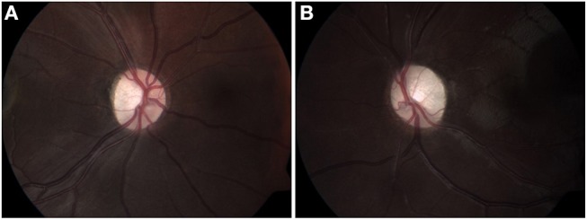
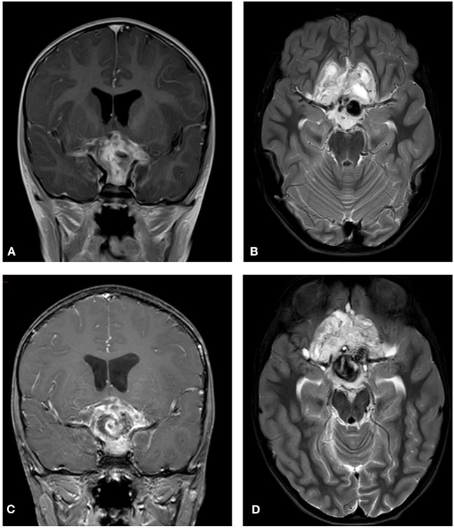
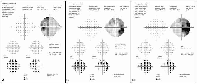
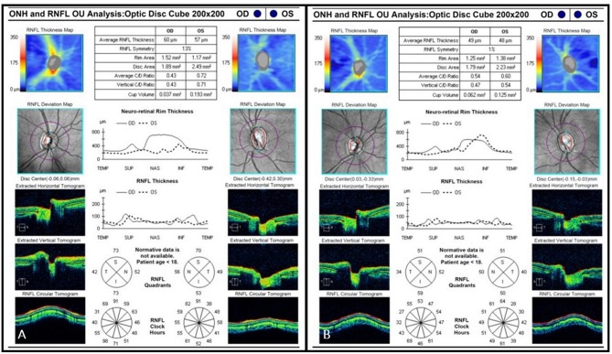
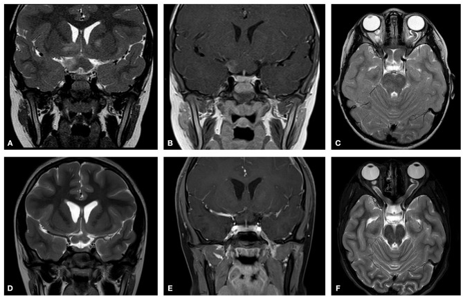
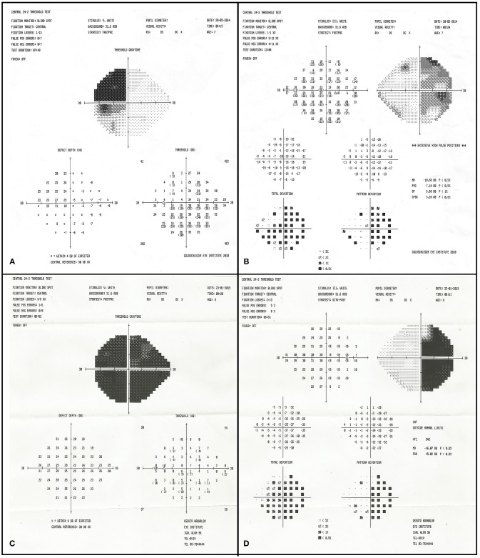
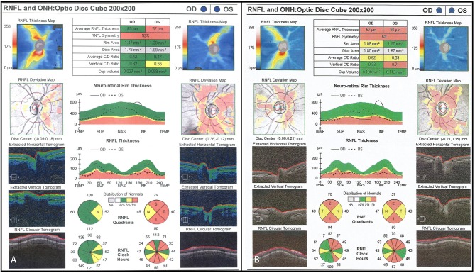
Optic pathway glioma (OPG) presents in childhood and can cause significant morbidity and visual loss. Magnetic resonance imaging (MRI) is the current imaging modality of choice for evaluation of OPG progression, but it is a relatively limited resource often requiring sedation in the pediatric age group. Additionally, OPG progression on MRI does not always correlate with clinical progression. As a result, several other modalities for evaluating OPG are being investigated, including optical coherence tomography (OCT), a readily available imaging technique in ophthalmic practice. The purpose of the present study was to examine the association between retinal nerve fiber layer (RNFL) thickness measured using OCT and optic nerve function in children with OPG with and without neurofibromatosis-1 (NF-1). A retrospective chart review was conducted to identify children diagnosed with OPG from 2001 to 2015 at a tertiary pediatric medical center. The correlation between OCT measurements and clinical visual parameters was statistically analyzed. Included were 23 children with imaging-confirmed OPG and spectral domain OCT: 10 with NF-1 (mean age at diagnosis 5.8 years) and 13 without (mean age at diagnosis 5.9 years). The glioma involved the chiasma-hypothalamus in 19 patients, optic nerve in 11, and optic tract in 7; more than one anatomic site was affected in 15. Symptoms were reported in 2 patients with NF-1 and most patients without NF-1. Visual field defects included monocular, bitemporal, nasal, and homonymous hemianopia. Initial mean RNFL was 85.4 μm in the NF-1 group and 65 μm in the non-NF-1 group. Visual acuity deteriorated in 1/10 patients and 5/13 patients, respectively. Repeated OCT showed continued RNFL thinning in 3 patients (5 eyes) in the NF-1 group and in 8 patients (11 eyes) in the non-NF-1 group, often associated with a decrease in optic nerve function. In conclusion, visual function in children with OPG is correlated with repeated OCT measurements and weakly with neuroimaging. Children without NF-1 are usually symptomatic and have a worse clinical outcome. These findings may have important implications when considering initiating, continuing or stopping chemotherapy for OPG. The application of OCT in the assessment of OPG and the correlation of the findings to clinical progression can have a significant impact on OPG patient management.

摘要

视路胶质瘤(OPG)多在儿童期发病,可导致严重的发病情况及视力丧失。磁共振成像(MRI)是目前评估OPG进展的首选影像学检查方法,但它是一种相对有限的资源,在儿科年龄组中通常需要镇静。此外,MRI上OPG的进展并不总是与临床进展相关。因此,正在研究其他几种评估OPG的方法,包括光学相干断层扫描(OCT),这是眼科实践中一种易于获得的成像技术。本研究的目的是检查在患有和未患有1型神经纤维瘤病(NF-1)的OPG儿童中,使用OCT测量的视网膜神经纤维层(RNFL)厚度与视神经功能之间的关联。进行了一项回顾性病历审查,以确定2001年至2015年在一家三级儿科医疗中心被诊断为OPG的儿童。对OCT测量值与临床视觉参数之间的相关性进行了统计分析。纳入了23例经影像学证实患有OPG并接受了光谱域OCT检查的儿童:10例患有NF-1(诊断时平均年龄5.8岁),13例未患有NF-1(诊断时平均年龄5.9岁)。胶质瘤累及视交叉-下丘脑的有19例患者,累及视神经的有11例,累及视束的有7例;15例患者有一个以上的解剖部位受累。10例患有NF-1的患者中有2例报告有症状,大多数未患有NF-1的患者有症状。视野缺损包括单眼、双颞侧、鼻侧和同向性偏盲。NF-1组初始平均RNFL为85.4μm,非NF-1组为65μm。视力分别在1/10例和5/13例患者中恶化。重复OCT检查显示NF-1组3例患者(5只眼)和非NF-1组8例患者(11只眼)的RNFL持续变薄,这通常与视神经功能下降有关。总之,OPG儿童的视觉功能与重复的OCT测量值相关,与神经影像学的相关性较弱。未患有NF-1的儿童通常有症状,临床结局较差。在考虑开始、继续或停止OPG化疗时,这些发现可能具有重要意义。OCT在OPG评估中的应用以及检查结果与临床进展的相关性可能会对OPG患者的管理产生重大影响。

https://cdn.ncbi.nlm.nih.gov/pmc/blobs/2ae4/6306407/353192b60637/fneur-09-01102-g0007.jpg
https://cdn.ncbi.nlm.nih.gov/pmc/blobs/2ae4/6306407/d1fbaab1ecf9/fneur-09-01102-g0001.jpg
https://cdn.ncbi.nlm.nih.gov/pmc/blobs/2ae4/6306407/a5984d294cac/fneur-09-01102-g0002.jpg
https://cdn.ncbi.nlm.nih.gov/pmc/blobs/2ae4/6306407/9e3014b43e75/fneur-09-01102-g0003.jpg
https://cdn.ncbi.nlm.nih.gov/pmc/blobs/2ae4/6306407/d34147167ead/fneur-09-01102-g0004.jpg
https://cdn.ncbi.nlm.nih.gov/pmc/blobs/2ae4/6306407/c04149056631/fneur-09-01102-g0005.jpg
https://cdn.ncbi.nlm.nih.gov/pmc/blobs/2ae4/6306407/eb175003f6b8/fneur-09-01102-g0006.jpg
https://cdn.ncbi.nlm.nih.gov/pmc/blobs/2ae4/6306407/353192b60637/fneur-09-01102-g0007.jpg

相似文献

1
Use of Optical Coherence Tomography to Detect Retinal Nerve Fiber Loss in Children With Optic Pathway Glioma.使用光学相干断层扫描技术检测视神经通路胶质瘤患儿的视网膜神经纤维损失。
Front Neurol. 2018 Dec 20;9:1102. doi: 10.3389/fneur.2018.01102. eCollection 2018.
2
Correlation of peripapillary retinal nerve fibre layer thickness with visual acuity in paediatric patients affected by optic pathway glioma.视路胶质瘤患儿视盘周围视网膜神经纤维层厚度与视力的相关性。
Acta Ophthalmol. 2018 Dec;96(8):e1004-e1009. doi: 10.1111/aos.13803. Epub 2018 Oct 3.
3
Optical coherence tomography of the macular ganglion cell layer in children with neurofibromatosis type 1 is a useful tool in the assessment for optic pathway gliomas.1 型神经纤维瘤病患儿的黄斑神经节细胞层光学相干断层扫描在视神经胶质瘤评估中是一种有用的工具。
PLoS One. 2024 Jul 11;19(7):e0305548. doi: 10.1371/journal.pone.0305548. eCollection 2024.
4
Investigation of retinal nerve fiber layer thickness and ganglion cell layer-inner plexiform layer thickness in patients with optic pathway gliomas.视路胶质瘤患者视网膜神经纤维层厚度和神经节细胞层-内网状层厚度的研究。
Graefes Arch Clin Exp Ophthalmol. 2018 Sep;256(9):1757-1765. doi: 10.1007/s00417-018-4007-8. Epub 2018 May 12.
5
Investigation of retinal nerve fiber layer thickness in patients with neurofibromatosis-1.神经纤维瘤病 1 型患者的视网膜神经纤维层厚度研究。
Jpn J Ophthalmol. 2014 Mar;58(2):172-6. doi: 10.1007/s10384-014-0308-6. Epub 2014 Feb 4.
6
Correlation of optical coherence tomography parameters with clinical and radiological progression in patients with symptomatic optic pathway gliomas.症状性视路胶质瘤患者的光学相干断层扫描参数与临床和影像学进展的相关性。
Graefes Arch Clin Exp Ophthalmol. 2013 Oct;251(10):2429-36. doi: 10.1007/s00417-013-2394-4. Epub 2013 Jun 5.
7
Homonymous Retinal Ganglion Cell Layer Atrophy With Asymptomatic Optic Tract Glioma in Neurofibromatosis Type I.1型神经纤维瘤病伴无症状视束胶质瘤的同侧视网膜神经节细胞层萎缩
Front Neurol. 2020 Apr 15;11:256. doi: 10.3389/fneur.2020.00256. eCollection 2020.
8
Sectoral analysis of the retinal nerve fiber layer thinning and its association with visual field loss in homonymous hemianopia caused by post-geniculate lesions using spectral-domain optical coherence tomography.使用频域光学相干断层扫描对膝状体后病变所致同侧偏盲患者视网膜神经纤维层变薄及其与视野缺损的相关性进行的扇形分析。
Graefes Arch Clin Exp Ophthalmol. 2016 Apr;254(4):745-56. doi: 10.1007/s00417-015-3181-1. Epub 2015 Oct 7.
9
Optical coherence tomography detects characteristic retinal nerve fiber layer thickness corresponding to band atrophy of the optic discs.光学相干断层扫描可检测到与视盘带状萎缩相对应的特征性视网膜神经纤维层厚度。
Ophthalmology. 2004 Dec;111(12):2278-83. doi: 10.1016/j.ophtha.2004.05.035.
10
[Retinal atrophy using optical coherence tomography (OCT) in 15 patients with multiple sclerosis and comparison with healthy subjects].[利用光学相干断层扫描(OCT)对15例多发性硬化症患者视网膜萎缩情况的研究及与健康受试者的比较]
Rev Neurol (Paris). 2008 Nov;164(11):927-34. doi: 10.1016/j.neurol.2008.03.008. Epub 2008 Jun 6.

引用本文的文献

1
Correlation between retinal nerve fibre layer thickness and tumour volume in patients with optic pathway gliomas.视路胶质瘤患者视网膜神经纤维层厚度与肿瘤体积之间的相关性
Int Ophthalmol. 2025 Jul 12;45(1):288. doi: 10.1007/s10792-025-03676-2.
2
Retinal Thickness Analysis Using Optical Coherence Tomography: Diagnostic and Monitoring Applications in Retinal Diseases.使用光学相干断层扫描的视网膜厚度分析:在视网膜疾病中的诊断与监测应用
Diagnostics (Basel). 2025 Mar 25;15(7):833. doi: 10.3390/diagnostics15070833.
3
Visual outcome including visual field defects after treatment of paediatric optic pathway glioma: A nationwide cohort study.

本文引用的文献

1
Investigation of retinal nerve fiber layer thickness and ganglion cell layer-inner plexiform layer thickness in patients with optic pathway gliomas.视路胶质瘤患者视网膜神经纤维层厚度和神经节细胞层-内网状层厚度的研究。
Graefes Arch Clin Exp Ophthalmol. 2018 Sep;256(9):1757-1765. doi: 10.1007/s00417-018-4007-8. Epub 2018 May 12.
2
Optical coherence tomography as a marker of vision in children with optic pathway gliomas.光学相干断层扫描作为视神经通路胶质瘤患儿视力的标志物
Childs Nerv Syst. 2018 Jan;34(1):51-60. doi: 10.1007/s00381-017-3578-8. Epub 2017 Aug 26.
3
Optic pathway glioma volume predicts retinal axon degeneration in neurofibromatosis type 1.
儿童视神经通路胶质瘤治疗后的视觉预后,包括视野缺损:一项全国性队列研究。
Acta Ophthalmol. 2025 Sep;103(6):662-673. doi: 10.1111/aos.17476. Epub 2025 Mar 8.
4
Chromatic vision and structural assessment in primary congenital glaucoma.原发性先天性青光眼的色觉与结构评估
Sci Rep. 2024 Apr 25;14(1):9551. doi: 10.1038/s41598-024-60320-2.
5
Optic Pathway Gliomas in Pediatric Population-Current Approach in Diagnosis and Management: Literature Review.儿童视神经通路胶质瘤——诊断与管理的当前方法:文献综述
J Clin Med. 2023 Oct 24;12(21):6709. doi: 10.3390/jcm12216709.
6
The Neuroimmune Regulation and Potential Therapeutic Strategies of Optic Pathway Glioma.视路胶质瘤的神经免疫调节及潜在治疗策略
Brain Sci. 2023 Oct 7;13(10):1424. doi: 10.3390/brainsci13101424.
7
[Neurofibromatosis type 1 : From diagnosis to follow-up].[1型神经纤维瘤病:从诊断到随访]
Radiologie (Heidelb). 2022 Dec;62(12):1050-1057. doi: 10.1007/s00117-022-01059-7. Epub 2022 Sep 7.
8
Rhegmatogenous Retinal Detachment with Spontaneous Dialysis of the Ora Serrata in Neurofibromatosis Type 1: A Case Report.1 型神经纤维瘤病伴锯齿缘自发性裂孔性视网膜脱离:病例报告。
JNMA J Nepal Med Assoc. 2022 Jun 1;60(250):555-558. doi: 10.31729/jnma.7392.
9
Tumor load rather than contrast enhancement is associated with the visual function of children and adolescents with optic pathway glioma - a retrospective Magnetic Resonance Imaging study.肿瘤负荷而非对比增强与视神经胶质瘤患儿和青少年的视觉功能相关——一项回顾性磁共振成像研究。
J Neurooncol. 2022 Feb;156(3):589-597. doi: 10.1007/s11060-021-03941-1. Epub 2022 Jan 7.
10
Optical coherence tomography in pediatric patients: a clinical review.儿科患者的光学相干断层扫描:临床综述
Ther Adv Ophthalmol. 2020 Feb 5;12:2515841420904612. doi: 10.1177/2515841420904612. eCollection 2020 Jan-Dec.
视神经通路胶质瘤体积可预测1型神经纤维瘤病患者的视网膜轴突退变。
Neurology. 2016 Dec 6;87(23):2403-2407. doi: 10.1212/WNL.0000000000003402. Epub 2016 Nov 4.
4
Pediatric Optical Coherence Tomography in Clinical Practice-Recent Progress.临床实践中的儿科光学相干断层扫描——最新进展
Invest Ophthalmol Vis Sci. 2016 Jul 1;57(9):OCT69-79. doi: 10.1167/iovs.15-18825.
5
Neurofibromatosis: an update of ophthalmic characteristics and applications of optical coherence tomography.神经纤维瘤病:眼科特征及光学相干断层扫描应用的最新进展
Clin Ophthalmol. 2016 May 13;10:851-60. doi: 10.2147/OPTH.S102830. eCollection 2016.
6
Pediatric Brain Tumors: Innovative Genomic Information Is Transforming the Diagnostic and Clinical Landscape.小儿脑肿瘤:创新的基因组信息正在改变诊断和临床格局。
J Clin Oncol. 2015 Sep 20;33(27):2986-98. doi: 10.1200/JCO.2014.59.9217. Epub 2015 Aug 24.
7
Long-term visual outcome after chemotherapy for optic pathway glioma in children: Site and age are strongly predictive.儿童视神经胶质瘤化疗后的长期视力预后:部位和年龄具有很强的预测性。
Cancer. 2015 Dec 1;121(23):4190-6. doi: 10.1002/cncr.29649. Epub 2015 Aug 17.
8
Longitudinal Change of Circumpapillary Retinal Nerve Fiber Layer Thickness in Children With Optic Pathway Gliomas.视路胶质瘤患儿视盘周围视网膜神经纤维层厚度的纵向变化
Am J Ophthalmol. 2015 Nov;160(5):944-952.e1. doi: 10.1016/j.ajo.2015.07.036. Epub 2015 Jul 29.
9
Acquired nystagmus as the initial presenting sign of chiasmal glioma in young children.后天性眼球震颤作为幼儿视交叉神经胶质瘤的首发体征。
Eur J Paediatr Neurol. 2015 Nov;19(6):694-700. doi: 10.1016/j.ejpn.2015.06.007. Epub 2015 Jul 9.
10
Malignant optic glioma - the spectrum of disease in a case series.恶性视神经胶质瘤——一组病例中的疾病谱
Graefes Arch Clin Exp Ophthalmol. 2015 Jul;253(7):1187-94. doi: 10.1007/s00417-015-3045-8. Epub 2015 May 26.