• 文献检索
  • 文档翻译
  • 深度研究
  • 学术资讯
  • Suppr Zotero 插件Zotero 插件
  • 邀请有礼
  • 套餐&价格
  • 历史记录
应用&插件
Suppr Zotero 插件Zotero 插件浏览器插件Mac 客户端Windows 客户端微信小程序
定价
高级版会员购买积分包购买API积分包
服务
文献检索文档翻译深度研究API 文档MCP 服务
关于我们
关于 Suppr公司介绍联系我们用户协议隐私条款
关注我们

Suppr 超能文献

核心技术专利:CN118964589B侵权必究
粤ICP备2023148730 号-1Suppr @ 2026

文献检索

告别复杂PubMed语法,用中文像聊天一样搜索,搜遍4000万医学文献。AI智能推荐,让科研检索更轻松。

立即免费搜索

文件翻译

保留排版,准确专业,支持PDF/Word/PPT等文件格式,支持 12+语言互译。

免费翻译文档

深度研究

AI帮你快速写综述,25分钟生成高质量综述,智能提取关键信息,辅助科研写作。

立即免费体验

光学相干断层扫描血管造影术的血管密度和正面分割以分析动物模型中的角膜血管生成

Vessel density and En-face segmentation of optical coherence tomography angiography to analyse corneal vascularisation in an animal model.

作者信息

Devarajan Kavya, Di Lee Wen, Ong Hon Shing, Lwin Nyein C, Chua Jacqueline, Schmetterer Leopold, Mehta Jodhbir S, Ang Marcus

机构信息

1Singapore Eye Research Institute, Singapore, Singapore.

2Singapore National Eye Center, Singapore, Singapore.

出版信息

Eye Vis (Lond). 2019 Jan 8;6:2. doi: 10.1186/s40662-018-0128-8. eCollection 2019.

DOI:10.1186/s40662-018-0128-8
PMID:30656178
原文链接:https://pmc.ncbi.nlm.nih.gov/articles/PMC6330743/
Abstract

BACKGROUND

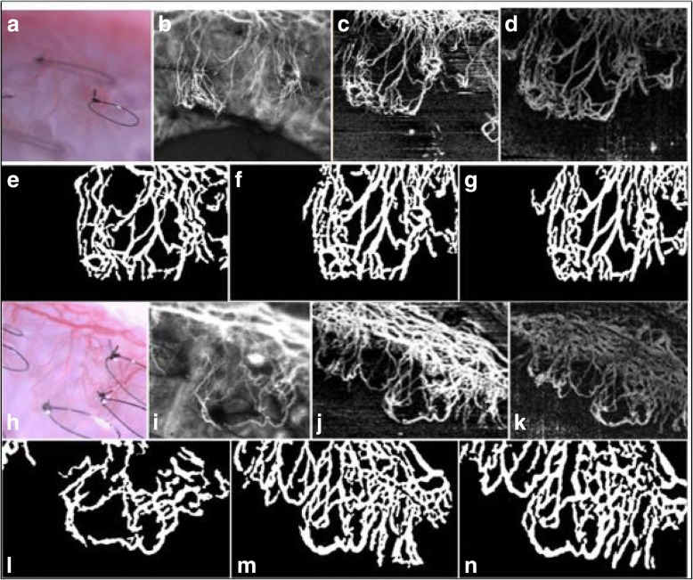
Optical coherence tomography angiography (OCTA) is a novel non-invasive angiography technology that has recently been extensively studied for its utility in anterior segment imaging. In this study, we compared a split-spectrum amplitude decorrelation angiography (SSADA) OCTA and an Complex OCT signal difference angiography [corrected] (CODAA SD) [corrected] OCTA system to current angiographic technique, indocyanine green angiography (ICGA), to assess corneal vascularisation in an animal model.

METHODS

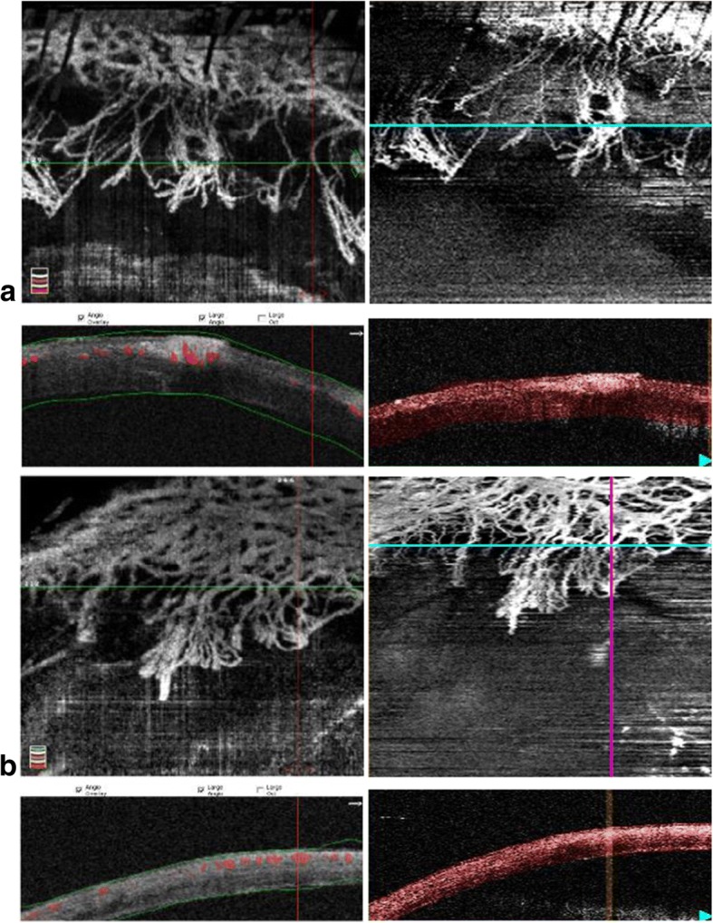
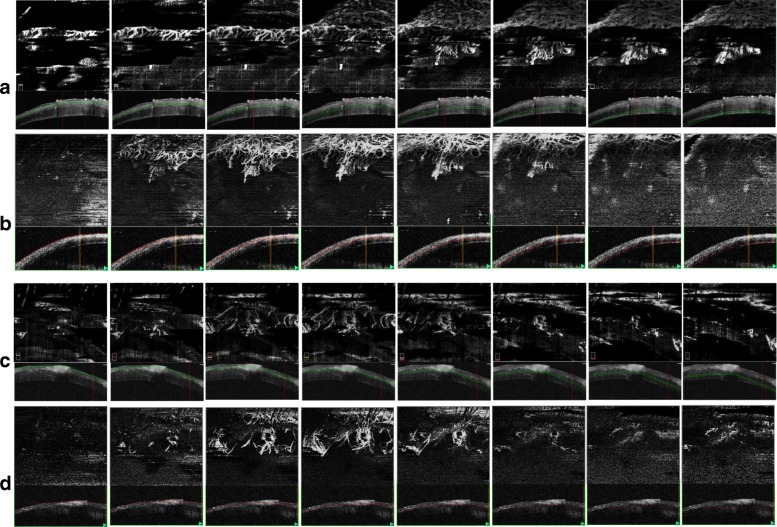
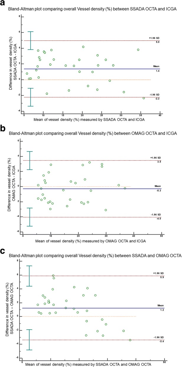
We imaged 16 rabbits, (one eye per animal) with corneal vascularisation using SSADA OCTA (AngioVue; Optovue Inc., USA), CODAA OCTA [corrected] (Angioscan; RS-3000 Nidek Co. Ltd., Japan) and ICGA in the same region of interest of the cornea at successive time-points. We then analysed all scanned images for vessel density measurements and used paired t-tests and Bland-Altman plots to examine for significant differences. The en-face segmentation images from each of the OCTA scans were also extracted and were matched at every 50 μm segmentation to be compared for vessel density at the respective depths.

RESULTS

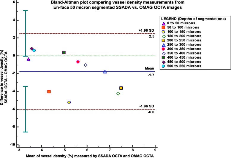
Bland-Altman plots revealed a good agreement between all three imaging techniques ( > 0.05) for all vessel density measurements computed, and the ranges of 95% limit of agreement were acceptable from a clinical perspective. No significant difference was reported, with ICGA (μ = 16.52 ± 8.94%) being more comparable to the CODAA [corrected] OCTA (μ = 16.23 ± 9.51%;  = 0.50) than the SSADA OCTA (μ = 17.09 ± 7.34%;  = 0.33) system. Also, a good correlation value ( > 0.9) was obtained when comparing the vessel density measurements of the en-face segmentations between the OCTA systems.

CONCLUSIONS

Comparable vessel density quantification between the two OCTA systems, and with ICGA was obtained. Segmentation analysis of the vasculature at different depths showed varied performance in the two OCTA systems relative to each other. The implications of the study may help to aid in the development of better OCTA algorithms for the anterior segment and its use in clinical translational research.

摘要

背景

光学相干断层扫描血管造影(OCTA)是一种新型无创血管造影技术,最近已对其在前节成像中的应用进行了广泛研究。在本研究中,我们将分裂谱幅度去相关血管造影(SSADA)OCTA和复OCT信号差异血管造影(CODAA SD)OCTA系统与当前血管造影技术吲哚菁绿血管造影(ICGA)进行比较,以评估动物模型中的角膜血管化情况。

方法

我们使用SSADA OCTA(AngioVue;美国Optovue公司)、CODAA OCTA(Angioscan;日本尼德克公司RS - 3000)和ICGA在16只患有角膜血管化的兔子(每只动物一只眼)的角膜同一感兴趣区域连续时间点进行成像。然后我们分析所有扫描图像以测量血管密度,并使用配对t检验和布兰德 - 奥特曼图来检查是否存在显著差异。还提取了每个OCTA扫描的正面分割图像,并在每50μm分割处进行匹配,以比较各自深度处的血管密度。

结果

布兰德 - 奥特曼图显示,对于所有计算的血管密度测量值,三种成像技术之间均具有良好的一致性(>0.05),并且从临床角度来看,95%一致性界限的范围是可接受的。未报告显著差异,ICGA(μ = 16.52±8.94%)与CODAA OCTA(μ = 16.23±9.51%;= 0.50)比与SSADA OCTA(μ = 17.09±7.34%;= 0.33)系统更具可比性。此外,在比较OCTA系统之间正面分割的血管密度测量值时,获得了良好的相关性值(>0.9)。

结论

获得了两种OCTA系统之间以及与ICGA相当的血管密度定量结果。在不同深度对脉管系统进行分割分析显示,两种OCTA系统相对于彼此的性能有所不同。该研究的意义可能有助于开发更好的用于前节的OCTA算法及其在临床转化研究中的应用。

https://cdn.ncbi.nlm.nih.gov/pmc/blobs/1a4a/6330743/dc959f74cc5f/40662_2018_128_Fig5_HTML.jpg
https://cdn.ncbi.nlm.nih.gov/pmc/blobs/1a4a/6330743/415c357a8638/40662_2018_128_Fig1_HTML.jpg
https://cdn.ncbi.nlm.nih.gov/pmc/blobs/1a4a/6330743/b9d99eb79515/40662_2018_128_Fig2_HTML.jpg
https://cdn.ncbi.nlm.nih.gov/pmc/blobs/1a4a/6330743/109b08514377/40662_2018_128_Fig3_HTML.jpg
https://cdn.ncbi.nlm.nih.gov/pmc/blobs/1a4a/6330743/ce8480969b42/40662_2018_128_Fig4_HTML.jpg
https://cdn.ncbi.nlm.nih.gov/pmc/blobs/1a4a/6330743/dc959f74cc5f/40662_2018_128_Fig5_HTML.jpg

相似文献

1
Vessel density and En-face segmentation of optical coherence tomography angiography to analyse corneal vascularisation in an animal model.光学相干断层扫描血管造影术的血管密度和正面分割以分析动物模型中的角膜血管生成
Eye Vis (Lond). 2019 Jan 8;6:2. doi: 10.1186/s40662-018-0128-8. eCollection 2019.
2
Comparison of anterior segment optical coherence tomography angiography systems for corneal vascularisation.眼前节光学相干断层扫描血管造影系统在角膜血管化中的比较。
Br J Ophthalmol. 2018 Jul;102(7):873-877. doi: 10.1136/bjophthalmol-2017-311072. Epub 2017 Sep 22.
3
Optical coherence tomography angiography and indocyanine green angiography for corneal vascularisation.光学相干断层扫描血管造影术和吲哚菁绿血管造影术用于角膜血管化
Br J Ophthalmol. 2016 Nov;100(11):1557-1563. doi: 10.1136/bjophthalmol-2015-307706. Epub 2016 Jan 28.
4
Imaging of Corneal Neovascularization: Optical Coherence Tomography Angiography and Fluorescence Angiography.角膜新生血管的影像学检查:光学相干断层扫描血管造影和荧光血管造影。
Invest Ophthalmol Vis Sci. 2018 Mar 1;59(3):1263-1269. doi: 10.1167/iovs.17-22035.
5
A pilot study investigating anterior segment optical coherence tomography angiography as a non-invasive tool in evaluating corneal vascularisation.一项研究角膜血管化的初步研究,探讨前节光学相干断层扫描血管造影作为一种非侵入性工具的应用。
Sci Rep. 2021 Jan 13;11(1):1212. doi: 10.1038/s41598-020-80099-2.
6
Optical Coherence Tomography Angiography Imaging to monitor Anti-VEGF treatment of Corneal Vascularization in a Rabbit Model.光学相干断层扫描血管造影成像监测兔模型角膜血管化的抗血管内皮生长因子治疗。
Sci Rep. 2019 Nov 26;9(1):17576. doi: 10.1038/s41598-019-54171-5.
7
Comparison of Optical Coherence Tomography Angiography to Indocyanine Green Angiography and Slit Lamp Photography for Corneal Vascularization in an Animal Model.比较光相干断层扫描血管造影术与吲哚菁绿血管造影术和裂隙灯照相术在动物模型中的角膜血管化评估。
Sci Rep. 2018 Jul 31;8(1):11493. doi: 10.1038/s41598-018-29752-5.
8
Serial optical coherence tomography angiography for corneal vascularization.用于角膜血管化的连续光学相干断层扫描血管造影术
Graefes Arch Clin Exp Ophthalmol. 2017 Jan;255(1):135-139. doi: 10.1007/s00417-016-3505-9. Epub 2016 Oct 8.
9
En face optical coherence tomography angiography for corneal neovascularisation.用于角膜新生血管的正面光学相干断层扫描血管造影术。
Br J Ophthalmol. 2016 May;100(5):616-21. doi: 10.1136/bjophthalmol-2015-307338. Epub 2015 Aug 26.
10
Compensation for Reflectance Variation in Vessel Density Quantification by Optical Coherence Tomography Angiography.光学相干断层扫描血管造影术在血管密度定量分析中对反射率变化的补偿
Invest Ophthalmol Vis Sci. 2016 Aug 1;57(10):4485-92. doi: 10.1167/iovs.16-20080.

引用本文的文献

1
Non-invasive quantification of corneal vascularization using anterior segment optical coherence tomography angiography.应用眼前节光学相干断层扫描血管造影术对角膜血管进行无创定量分析。
Sci Rep. 2024 Jan 24;14(1):2124. doi: 10.1038/s41598-024-52598-z.
2
Current clinical applications of anterior segment optical coherence tomography angiography: a review.眼前节光学相干断层扫描血管造影术的当前临床应用:综述
Graefes Arch Clin Exp Ophthalmol. 2023 Oct;261(10):2729-2741. doi: 10.1007/s00417-023-05997-3. Epub 2023 Mar 2.
3
Automated Image Threshold Method Comparison for Conjunctival Vessel Quantification on Optical Coherence Tomography Angiography.

本文引用的文献

1
The role of OCT-A in retinal disease management.光学相干断层扫描血管造影(OCT-A)在视网膜疾病管理中的作用。
Graefes Arch Clin Exp Ophthalmol. 2018 Nov;256(11):2019-2026. doi: 10.1007/s00417-018-4109-3. Epub 2018 Sep 3.
2
Comparison of Optical Coherence Tomography Angiography to Indocyanine Green Angiography and Slit Lamp Photography for Corneal Vascularization in an Animal Model.比较光相干断层扫描血管造影术与吲哚菁绿血管造影术和裂隙灯照相术在动物模型中的角膜血管化评估。
Sci Rep. 2018 Jul 31;8(1):11493. doi: 10.1038/s41598-018-29752-5.
3
Imaging of Corneal Neovascularization: Optical Coherence Tomography Angiography and Fluorescence Angiography.
基于光学相干断层扫描血管造影的结膜血管量化的自动图像阈值方法比较。
Transl Vis Sci Technol. 2022 Jul 8;11(7):15. doi: 10.1167/tvst.11.7.15.
4
Role of Anterior Segment-Optical Coherence Tomography Angiography in Acute Ocular Burns.眼前节光学相干断层扫描血管造影在急性眼烧伤中的作用
Diagnostics (Basel). 2022 Feb 28;12(3):607. doi: 10.3390/diagnostics12030607.
5
An end-to-end network for segmenting the vasculature of three retinal capillary plexuses from OCT angiographic volumes.一种用于从光学相干断层扫描血管造影体积中分割三个视网膜毛细血管丛血管系统的端到端网络。
Biomed Opt Express. 2021 Jul 16;12(8):4889-4900. doi: 10.1364/BOE.431888. eCollection 2021 Aug 1.
6
Role of anterior segment optical coherence tomography angiography in the assessment of acute chemical ocular injury: a pilot animal model study.眼前节光学相干断层扫描血管造影在急性化学性眼部损伤评估中的作用:一项初步的动物模型研究。
Sci Rep. 2021 Aug 17;11(1):16625. doi: 10.1038/s41598-021-96086-0.
7
The role of magnification correction in macular vessel density assessment: a contralateral eye study in anisometropia patients.放大率校正在黄斑血管密度评估中的作用:一项针对屈光参差患者对侧眼的研究。
Ann Transl Med. 2021 Mar;9(5):380. doi: 10.21037/atm-20-5698.
8
Automatic 3D adaptive vessel segmentation based on linear relationship between intensity and complex-decorrelation in optical coherence tomography angiography.基于光学相干断层扫描血管造影中强度与复相关之间线性关系的自动三维自适应血管分割
Quant Imaging Med Surg. 2021 Mar;11(3):895-906. doi: 10.21037/qims-20-868.
9
A pilot study investigating anterior segment optical coherence tomography angiography as a non-invasive tool in evaluating corneal vascularisation.一项研究角膜血管化的初步研究,探讨前节光学相干断层扫描血管造影作为一种非侵入性工具的应用。
Sci Rep. 2021 Jan 13;11(1):1212. doi: 10.1038/s41598-020-80099-2.
10
Optical Coherence Tomography Angiography Imaging to monitor Anti-VEGF treatment of Corneal Vascularization in a Rabbit Model.光学相干断层扫描血管造影成像监测兔模型角膜血管化的抗血管内皮生长因子治疗。
Sci Rep. 2019 Nov 26;9(1):17576. doi: 10.1038/s41598-019-54171-5.
角膜新生血管的影像学检查:光学相干断层扫描血管造影和荧光血管造影。
Invest Ophthalmol Vis Sci. 2018 Mar 1;59(3):1263-1269. doi: 10.1167/iovs.17-22035.
4
Optical coherence tomography angiography: a review of current and future clinical applications.光学相干断层扫描血管造影术:当前及未来临床应用综述
Graefes Arch Clin Exp Ophthalmol. 2018 Feb;256(2):237-245. doi: 10.1007/s00417-017-3896-2. Epub 2018 Jan 9.
5
Complex-based OCT angiography algorithm recovers microvascular information better than amplitude- or phase-based algorithms in phase-stable systems.基于复数的 OCT 血管造影算法在相位稳定系统中比基于幅度或相位的算法能更好地恢复微血管信息。
Phys Med Biol. 2017 Dec 19;63(1):015023. doi: 10.1088/1361-6560/aa94bc.
6
Comparison of anterior segment optical coherence tomography angiography systems for corneal vascularisation.眼前节光学相干断层扫描血管造影系统在角膜血管化中的比较。
Br J Ophthalmol. 2018 Jul;102(7):873-877. doi: 10.1136/bjophthalmol-2017-311072. Epub 2017 Sep 22.
7
OCT-angiography: A qualitative and quantitative comparison of 4 OCT-A devices.光学相干断层扫描血管造影:4种光学相干断层扫描血管造影设备的定性和定量比较。
PLoS One. 2017 May 10;12(5):e0177059. doi: 10.1371/journal.pone.0177059. eCollection 2017.
8
Swept Source Optical Coherence Tomography Angiography for Contact Lens-Related Corneal Vascularization.扫频源光学相干断层扫描血管造影术用于隐形眼镜相关的角膜血管化
J Ophthalmol. 2016;2016:9685297. doi: 10.1155/2016/9685297. Epub 2016 Sep 26.
9
Serial optical coherence tomography angiography for corneal vascularization.用于角膜血管化的连续光学相干断层扫描血管造影术
Graefes Arch Clin Exp Ophthalmol. 2017 Jan;255(1):135-139. doi: 10.1007/s00417-016-3505-9. Epub 2016 Oct 8.
10
Optical Coherence Tomography Angiography.光学相干断层扫描血管造影术
Invest Ophthalmol Vis Sci. 2016 Jul 1;57(9):OCT27-36. doi: 10.1167/iovs.15-19043.