Singapore Eye Research Institute, Singapore, Singapore.
Department of Ophthalmology and Visual Science, Duke-National University of Singapore (NUS), Graduate Medical School, Singapore, Singapore.
Sci Rep. 2021 Jan 13;11(1):1212. doi: 10.1038/s41598-020-80099-2.
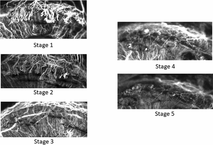
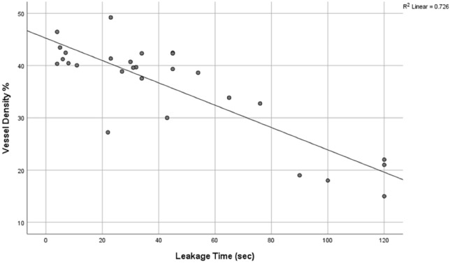
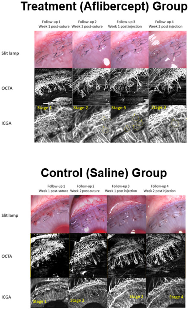
The current assessment of corneal vascularisation (CV) relies on slit-lamp examination, which may be subjective. Dye-based angiographies, like indocyanine green angiography (ICGA), allows for good visualisation of anterior segment blood vessels. However, ICGA is invasive and can be associated with systemic adverse effects. Anterior segment optical coherence tomography angiography (AS-OCTA) is a non-invasive tool that has been shown to successfully delineate CV. However, there are no previous studies that have reported if AS-OCTA can determine CV stage and activity. We used an established CV model in rabbits to examine serial AS-OCTA scans of CV development and regression following treatment with anti-vascular endothelial growth factor. We compared AS-OCTA derived vascular measurements to that of ICGA determined vessel leakage and CV staging. Our results showed that AS-OCTA vessel densities and vessel branch area significantly correlated with the severity of CV based on ICGA (all p ≤ 0.05). We also found that AS-OCTA vessel densities correlated with ICGA vessel leakage time, following an inverse linear relationship (r = - 0.726, p < 0.01). Changes in aqueous levels of CXCL-12 and PIGF cytokines significantly correlated with AS-OCTA vessel densities (r = 0.736 and r = 0.731 respectively, all p < 0.05). In summary, we found that AS-OCTA derived vessel parameters may be useful for assessing CV severity, while vessel density correlates with CV activity and leakage. Thus, our pilot animal model study suggests that AS-OCTA may be a useful non-invasive imaging tool to provide objective assessment of CV to examine progression or response in treatment, which requires confirmation in clinical studies.
目前,角膜血管新生(CV)的评估依赖于裂隙灯检查,这种方法可能具有主观性。染料基血管造影,如吲哚菁绿血管造影(ICGA),可以很好地显示前节血管。然而,ICGA 具有侵入性,并可能与全身不良反应相关。前节光学相干断层扫描血管造影(AS-OCTA)是一种非侵入性工具,已被证明可成功描绘 CV。然而,目前尚无研究报道 AS-OCTA 是否可以确定 CV 分期和活动度。我们使用已建立的兔 CV 模型,检查了抗血管内皮生长因子治疗后 CV 发展和消退的一系列 AS-OCTA 扫描。我们将 AS-OCTA 得出的血管测量值与 ICGA 确定的血管渗漏和 CV 分期进行了比较。我们的研究结果表明,AS-OCTA 血管密度和血管分支面积与基于 ICGA 的 CV 严重程度显著相关(均 p≤0.05)。我们还发现,AS-OCTA 血管密度与 ICGA 血管渗漏时间呈负线性相关(r=-0.726,p<0.01)。CXCL-12 和 PIGF 细胞因子的房水水平变化与 AS-OCTA 血管密度显著相关(r=0.736 和 r=0.731,均 p<0.05)。总之,我们发现 AS-OCTA 衍生的血管参数可能有助于评估 CV 的严重程度,而血管密度与 CV 的活动度和渗漏相关。因此,我们的动物模型研究表明,AS-OCTA 可能是一种有用的非侵入性成像工具,可用于客观评估 CV,以检查治疗的进展或反应,这需要在临床研究中得到证实。