• 文献检索
  • 文档翻译
  • 深度研究
  • 学术资讯
  • Suppr Zotero 插件Zotero 插件
  • 邀请有礼
  • 套餐&价格
  • 历史记录
应用&插件
Suppr Zotero 插件Zotero 插件浏览器插件Mac 客户端Windows 客户端微信小程序
定价
高级版会员购买积分包购买API积分包
服务
文献检索文档翻译深度研究API 文档MCP 服务
关于我们
关于 Suppr公司介绍联系我们用户协议隐私条款
关注我们

Suppr 超能文献

核心技术专利:CN118964589B侵权必究
粤ICP备2023148730 号-1Suppr @ 2026

文献检索

告别复杂PubMed语法,用中文像聊天一样搜索,搜遍4000万医学文献。AI智能推荐,让科研检索更轻松。

立即免费搜索

文件翻译

保留排版,准确专业,支持PDF/Word/PPT等文件格式,支持 12+语言互译。

免费翻译文档

深度研究

AI帮你快速写综述,25分钟生成高质量综述,智能提取关键信息,辅助科研写作。

立即免费体验

光学相干断层扫描血管造影成像监测兔模型角膜血管化的抗血管内皮生长因子治疗。

Optical Coherence Tomography Angiography Imaging to monitor Anti-VEGF treatment of Corneal Vascularization in a Rabbit Model.

机构信息

Singapore Eye Research Institute, Singapore National Eye Center, Singapore, Singapore.

Eye-ACP, Duke-NUS Graduate Medical School, Singapore, Singapore.

出版信息

Sci Rep. 2019 Nov 26;9(1):17576. doi: 10.1038/s41598-019-54171-5.

DOI:10.1038/s41598-019-54171-5
PMID:31772259
原文链接:https://pmc.ncbi.nlm.nih.gov/articles/PMC6879475/
Abstract

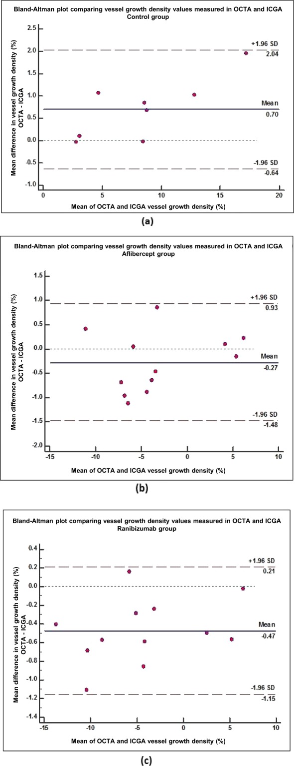
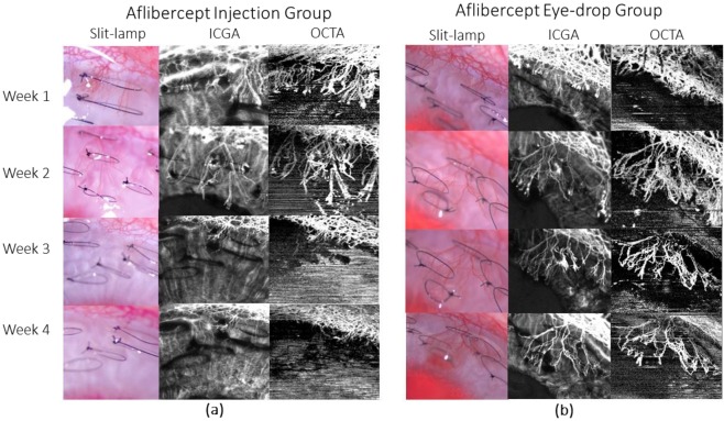
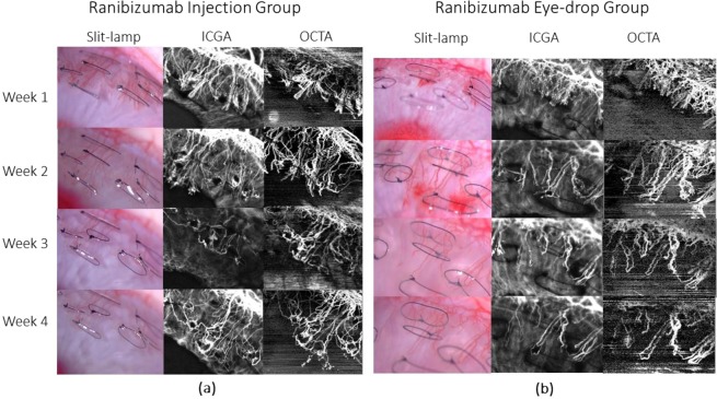
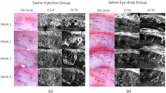
Optical coherence tomography angiography (OCTA) is a well-established non-invasive retinal vascular imaging technique. It has been recently adapted to image the anterior segment and has shown good potential to image corneal vascularisation. The purpose of the study is to evaluate the usefulness of OCTA to monitor regression of corneal vessels following anti-VEGF (vascular endothelial growth factor) treatment using a previously established corneal vascularisation rabbit model. The regression of vessels following the treatment with aflibercept and ranibizumab anti-VEGFs using both topical instillation and sub-conjunctival injection was quantified using OCTA and compared with ICGA (indocyanine green angiography). Overall vessel density measurements using OCTA showed good correlation (r = 0.988, p < 0.001) with ICGA, with no significant difference between the two treatment groups (p = 0.795). It was also shown that OCTA provided good repeatability outcomes of the quantitative measurements. Using Bland-Altman plots, vessel growth density values between anti-VEGF treatments were compared to control saline group. It was observed that aflibercept provided longer lasting effect than ranibizumab. We also observed that in both drugs, the topical route of administration topical provided longer regression outcomes compared to one-time sub-conjunctival injection. Thereby, with this pilot study, it was demonstrated that OCTA is a reliable imaging technique to follow-up and monitor corneal vascularisation and its treatment quantitatively.

摘要

光学相干断层扫描血管造影术(OCTA)是一种成熟的非侵入性视网膜血管成像技术。它最近已被应用于眼前段成像,并显示出很好的成像角膜血管化的潜力。本研究的目的是评估 OCTA 在使用先前建立的角膜血管化兔模型监测抗血管内皮生长因子(VEGF)治疗后角膜血管消退中的作用。使用 OCTA 定量评估和脉络膜荧光血管造影(ICGA)比较了玻璃体内注射和局部滴眼两种方式给予阿柏西普和雷珠单抗抗 VEGF 治疗后血管的消退情况。OCTA 的整体血管密度测量与 ICGA 具有良好的相关性(r=0.988,p<0.001),两组治疗之间无显著差异(p=0.795)。结果还表明,OCTA 可提供定量测量的良好可重复性。使用 Bland-Altman 图,将抗 VEGF 治疗之间的血管生长密度值与对照组生理盐水组进行比较。结果观察到阿柏西普的作用持续时间长于雷珠单抗。我们还观察到,在两种药物中,局部给药途径比一次性结膜下注射提供更长的消退效果。因此,通过这项初步研究,证明了 OCTA 是一种可靠的成像技术,可用于定量随访和监测角膜血管化及其治疗。

https://cdn.ncbi.nlm.nih.gov/pmc/blobs/3d28/6879475/041f083e5b8a/41598_2019_54171_Fig7_HTML.jpg
https://cdn.ncbi.nlm.nih.gov/pmc/blobs/3d28/6879475/5fb3174ea9a0/41598_2019_54171_Fig1_HTML.jpg
https://cdn.ncbi.nlm.nih.gov/pmc/blobs/3d28/6879475/0df70369bdfd/41598_2019_54171_Fig2_HTML.jpg
https://cdn.ncbi.nlm.nih.gov/pmc/blobs/3d28/6879475/95381de4221e/41598_2019_54171_Fig3_HTML.jpg
https://cdn.ncbi.nlm.nih.gov/pmc/blobs/3d28/6879475/9cdea584d849/41598_2019_54171_Fig4_HTML.jpg
https://cdn.ncbi.nlm.nih.gov/pmc/blobs/3d28/6879475/18daf7d2425d/41598_2019_54171_Fig5_HTML.jpg
https://cdn.ncbi.nlm.nih.gov/pmc/blobs/3d28/6879475/3db72f2d3089/41598_2019_54171_Fig6_HTML.jpg
https://cdn.ncbi.nlm.nih.gov/pmc/blobs/3d28/6879475/041f083e5b8a/41598_2019_54171_Fig7_HTML.jpg

相似文献

1
Optical Coherence Tomography Angiography Imaging to monitor Anti-VEGF treatment of Corneal Vascularization in a Rabbit Model.光学相干断层扫描血管造影成像监测兔模型角膜血管化的抗血管内皮生长因子治疗。
Sci Rep. 2019 Nov 26;9(1):17576. doi: 10.1038/s41598-019-54171-5.
2
Optical coherence tomography angiography and indocyanine green angiography for corneal vascularisation.光学相干断层扫描血管造影术和吲哚菁绿血管造影术用于角膜血管化
Br J Ophthalmol. 2016 Nov;100(11):1557-1563. doi: 10.1136/bjophthalmol-2015-307706. Epub 2016 Jan 28.
3
A pilot study investigating anterior segment optical coherence tomography angiography as a non-invasive tool in evaluating corneal vascularisation.一项研究角膜血管化的初步研究,探讨前节光学相干断层扫描血管造影作为一种非侵入性工具的应用。
Sci Rep. 2021 Jan 13;11(1):1212. doi: 10.1038/s41598-020-80099-2.
4
Comparison of Optical Coherence Tomography Angiography to Indocyanine Green Angiography and Slit Lamp Photography for Corneal Vascularization in an Animal Model.比较光相干断层扫描血管造影术与吲哚菁绿血管造影术和裂隙灯照相术在动物模型中的角膜血管化评估。
Sci Rep. 2018 Jul 31;8(1):11493. doi: 10.1038/s41598-018-29752-5.
5
Observation of treated iris neovascularization by swept-source-based en-face anterior-segment optical coherence tomography angiography.应用扫频源光相干断层扫描血管成像技术观察治疗性虹膜新生血管。
Sci Rep. 2019 Jul 16;9(1):10262. doi: 10.1038/s41598-019-46514-z.
6
OPTICAL COHERENCE TOMOGRAPHY ANGIOGRAPHY CHANGES IN EARLY TYPE 3 NEOVASCULARIZATION AFTER ANTI-VASCULAR ENDOTHELIAL GROWTH FACTOR TREATMENT.抗血管内皮生长因子治疗后早期3型新生血管形成的光学相干断层扫描血管造影变化
Retina. 2017 Oct;37(10):1873-1879. doi: 10.1097/IAE.0000000000001447.
7
Vessel density and En-face segmentation of optical coherence tomography angiography to analyse corneal vascularisation in an animal model.光学相干断层扫描血管造影术的血管密度和正面分割以分析动物模型中的角膜血管生成
Eye Vis (Lond). 2019 Jan 8;6:2. doi: 10.1186/s40662-018-0128-8. eCollection 2019.
8
Optical coherence tomography angiography of type 3 neovascularisation in age-related macular degeneration after antiangiogenic therapy.抗血管生成治疗后年龄相关性黄斑变性3型新生血管形成的光学相干断层扫描血管造影
Br J Ophthalmol. 2017 May;101(5):597-602. doi: 10.1136/bjophthalmol-2016-308815. Epub 2016 Aug 8.
9
Variable response of subretinal hyperreflective material to anti-vascular endothelial growth factor classified with optical coherence tomography angiography.视网膜下高反射物质对抗血管内皮生长因子的可变反应,通过光学相干断层扫描血管造影进行分类。
Graefes Arch Clin Exp Ophthalmol. 2018 Nov;256(11):2089-2096. doi: 10.1007/s00417-018-4121-7. Epub 2018 Sep 1.
10
Responses of Types 1 and 2 Neovascularization in Age-Related Macular Degeneration to Anti-Vascular Endothelial Growth Factor Treatment: Optical Coherence Tomography Angiography Analysis.年龄相关性黄斑变性中1型和2型新生血管对抗血管内皮生长因子治疗的反应:光学相干断层扫描血管造影分析
Semin Ophthalmol. 2019;34(3):168-176. doi: 10.1080/08820538.2019.1620791. Epub 2019 May 27.

引用本文的文献

1
Optical coherence tomography for noninvasive monitoring of drug delivery.用于药物递送无创监测的光学相干断层扫描技术
Adv Drug Deliv Rev. 2025 May;220:115571. doi: 10.1016/j.addr.2025.115571. Epub 2025 Mar 24.
2
Anterior Segment Optical Coherence Tomography Angiography: A Review of Applications for the Cornea and Ocular Surface.眼前节光学相干断层扫描血管造影:角膜和眼表面应用的综述。
Medicina (Kaunas). 2024 Sep 28;60(10):1597. doi: 10.3390/medicina60101597.
3
Sustained and Efficient Delivery of Antivascular Endothelial Growth Factor by the Adeno-associated Virus for the Treatment of Corneal Neovascularization: An Outlook for Its Clinical Translation.

本文引用的文献

1
Vessel density and En-face segmentation of optical coherence tomography angiography to analyse corneal vascularisation in an animal model.光学相干断层扫描血管造影术的血管密度和正面分割以分析动物模型中的角膜血管生成
Eye Vis (Lond). 2019 Jan 8;6:2. doi: 10.1186/s40662-018-0128-8. eCollection 2019.
2
Comparison of Optical Coherence Tomography Angiography to Indocyanine Green Angiography and Slit Lamp Photography for Corneal Vascularization in an Animal Model.比较光相干断层扫描血管造影术与吲哚菁绿血管造影术和裂隙灯照相术在动物模型中的角膜血管化评估。
Sci Rep. 2018 Jul 31;8(1):11493. doi: 10.1038/s41598-018-29752-5.
3
Anterior segment optical coherence tomography.
腺相关病毒持续高效递送抗血管内皮生长因子治疗角膜新生血管:临床转化前景
J Ophthalmol. 2024 Sep 9;2024:5487973. doi: 10.1155/2024/5487973. eCollection 2024.
4
Molecular and cellular imaging of the eye.眼睛的分子与细胞成像
Biomed Opt Express. 2023 Dec 21;15(1):360-386. doi: 10.1364/BOE.502350. eCollection 2024 Jan 1.
5
Current clinical applications of anterior segment optical coherence tomography angiography: a review.眼前节光学相干断层扫描血管造影术的当前临床应用:综述
Graefes Arch Clin Exp Ophthalmol. 2023 Oct;261(10):2729-2741. doi: 10.1007/s00417-023-05997-3. Epub 2023 Mar 2.
6
Recent advances in optical coherence tomography for anterior segment imaging in small animals and their clinical implications.小动物眼前节成像的光学相干断层扫描的最新进展及其临床意义。
Ocul Surf. 2022 Oct;26:222-233. doi: 10.1016/j.jtos.2022.08.011. Epub 2022 Oct 3.
7
Imaging of iris vasculature: current limitations and future perspective.虹膜血管成像:当前的局限性和未来展望。
Eye (Lond). 2022 May;36(5):930-940. doi: 10.1038/s41433-021-01809-2. Epub 2021 Oct 14.
8
Efficacious, safe, and stable inhibition of corneal neovascularization by AAV-vectored anti-VEGF therapeutics.通过腺相关病毒载体抗血管内皮生长因子疗法有效、安全且稳定地抑制角膜新生血管形成。
Mol Ther Methods Clin Dev. 2021 Jun 24;22:107-121. doi: 10.1016/j.omtm.2021.06.007. eCollection 2021 Sep 10.
9
Role of anterior segment optical coherence tomography angiography in the assessment of acute chemical ocular injury: a pilot animal model study.眼前节光学相干断层扫描血管造影在急性化学性眼部损伤评估中的作用:一项初步的动物模型研究。
Sci Rep. 2021 Aug 17;11(1):16625. doi: 10.1038/s41598-021-96086-0.
10
A pilot study investigating anterior segment optical coherence tomography angiography as a non-invasive tool in evaluating corneal vascularisation.一项研究角膜血管化的初步研究,探讨前节光学相干断层扫描血管造影作为一种非侵入性工具的应用。
Sci Rep. 2021 Jan 13;11(1):1212. doi: 10.1038/s41598-020-80099-2.
眼前节光学相干断层扫描。
Prog Retin Eye Res. 2018 Sep;66:132-156. doi: 10.1016/j.preteyeres.2018.04.002. Epub 2018 Apr 7.
4
Therapeutic approaches for corneal neovascularization.角膜新生血管的治疗方法。
Eye Vis (Lond). 2017 Dec 10;4:28. doi: 10.1186/s40662-017-0094-6. eCollection 2017.
5
Comparison of anterior segment optical coherence tomography angiography systems for corneal vascularisation.眼前节光学相干断层扫描血管造影系统在角膜血管化中的比较。
Br J Ophthalmol. 2018 Jul;102(7):873-877. doi: 10.1136/bjophthalmol-2017-311072. Epub 2017 Sep 22.
6
Topical versus subconjunctival anti-vascular endothelial growth factor therapy (Bevacizumab, Ranibizumab and Aflibercept) for treatment of corneal neovascularization.局部应用与结膜下注射抗血管内皮生长因子疗法(贝伐单抗、雷珠单抗和阿柏西普)治疗角膜新生血管化
Saudi J Ophthalmol. 2017 Apr-Jun;31(2):99-105. doi: 10.1016/j.sjopt.2017.02.008. Epub 2017 Mar 8.
7
Topical Delivery of Anti-VEGF Drugs to the Ocular Posterior Segment Using Cell-Penetrating Peptides.使用细胞穿透肽将抗血管内皮生长因子药物局部递送至眼后段
Invest Ophthalmol Vis Sci. 2017 May 1;58(5):2578-2590. doi: 10.1167/iovs.16-20072.
8
En face optical coherence tomography angiography for corneal neovascularisation.用于角膜新生血管的正面光学相干断层扫描血管造影术。
Br J Ophthalmol. 2016 May;100(5):616-21. doi: 10.1136/bjophthalmol-2015-307338. Epub 2015 Aug 26.
9
Complex differential variance algorithm for optical coherence tomography angiography.光学相干断层扫描血管造影的复杂差分方差算法
Biomed Opt Express. 2014 Oct 7;5(11):3822-32. doi: 10.1364/BOE.5.003822. eCollection 2014 Nov 1.
10
Intravitreal aflibercept for diabetic macular edema.玻璃体内注射阿柏西普治疗糖尿病性黄斑水肿。
Ophthalmology. 2014 Nov;121(11):2247-54. doi: 10.1016/j.ophtha.2014.05.006. Epub 2014 Jul 8.