Vascular and Genomic Center, Changhua Christian Hospital, Changhua, 50094, Taiwan.
Division of General Surgery, Department of Surgery, Changhua Christian Hospital, Changhua, 50094, Taiwan.
J Exp Clin Cancer Res. 2019 Jan 23;38(1):30. doi: 10.1186/s13046-019-1028-z.
The transfer of whole mitochondria that occurs during cell contact has been found to support cancer progression. However, the regulatory role of mitochondria alone is difficult to elucidate due to the complex microenvironment. Currently, mitochondrial transplantation is an available approach for restoring mitochondrial function in mitochondrial diseases but remains unclear in breast cancer. Herein, effects of mitochondrial transplantation via different approaches in breast cancer were investigated.
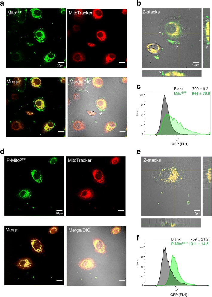
Whole mitochondria (approximately 10.5 μg/ml) were transported into MCF-7 breast cancer cells via passive uptake or Pep-1-mediated delivery. Fresh mitochondria isolated from homeoplasmic 143B osteosarcoma cybrids containing mitochondrial DNA (mtDNA) derived from health individuals (Mito) or mtDNA with the A8344G mutation (Mito) were conjugated with cell-penetrating peptide Pep-1 (P-Mito) or not conjugated prior to cell co-culture. Before isolation, mitochondria were stained with MitoTracker dye as the tracking label. After 3 days of treatment, cell viability, proliferation, oxidative stress, drug sensitivity to Doxorubicin/Paclitaxel and mitochondrial function were assessed.
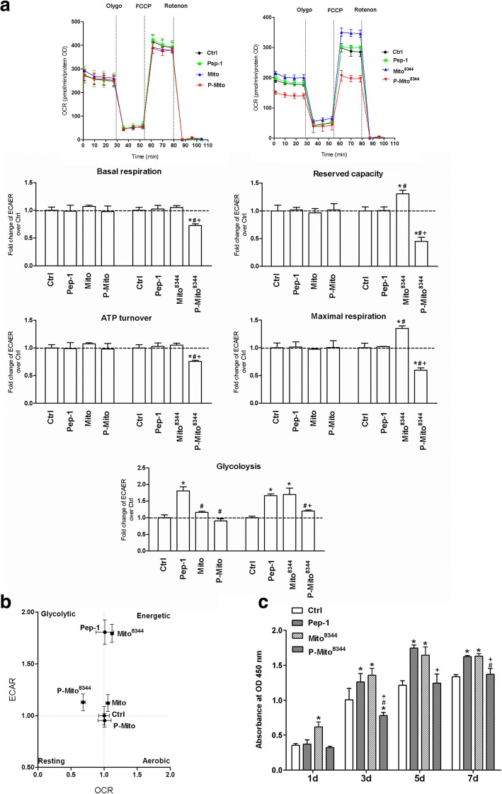
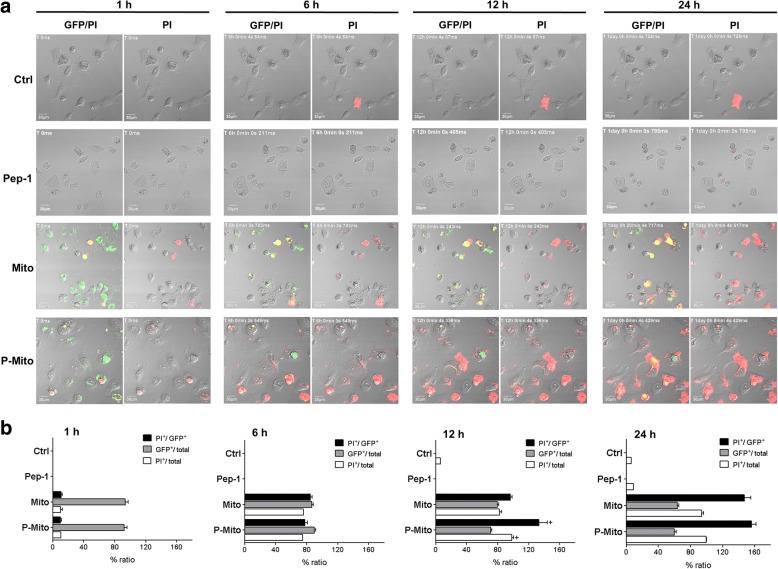
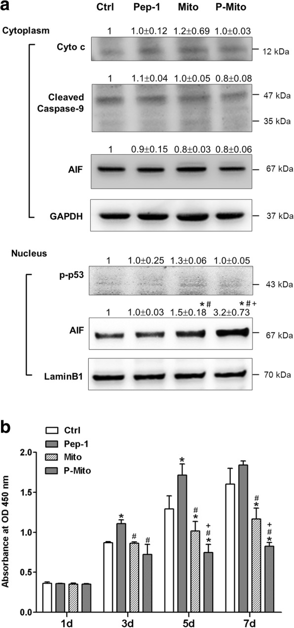
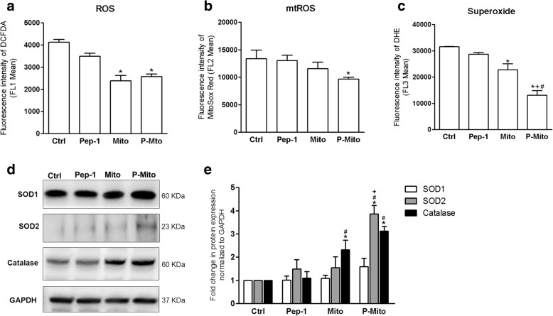
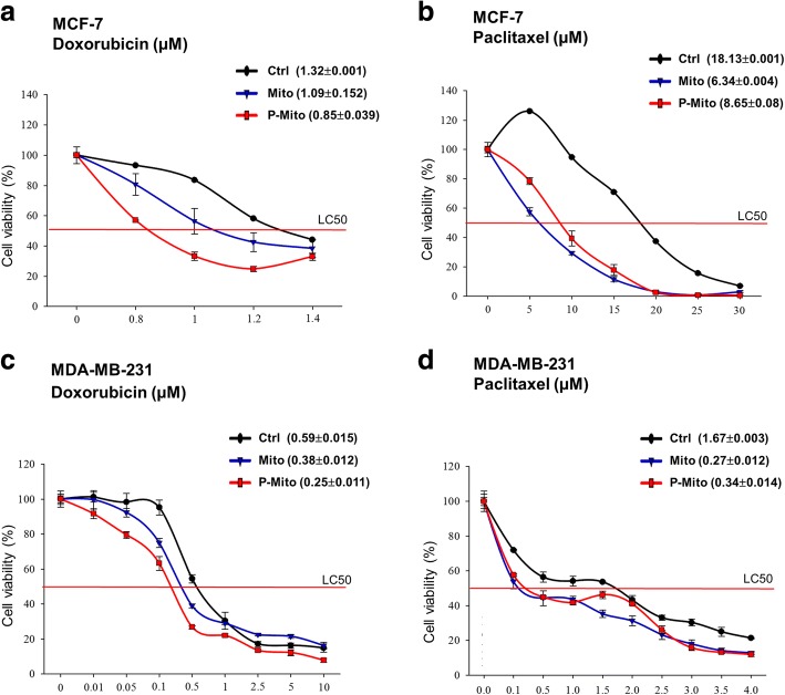
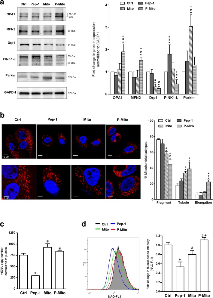
Compared with P-Mito, a small portion of Mito adhered to the cell membrane, and this was accompanied by a slightly lower fluorescent signal by foreign mitochondria in MCF-7 cells. Both transplantations induced cell apoptosis by increasing the nuclear translocation of apoptosis-inducing factor; inhibited cell growth and decreased oxidative stress in MCF-7 cells; and increased the cellular susceptibility of both the MCF-7 and MDA-MB-231 cell lines to Doxorubicin and Paclitaxel. Mitochondrial transplantation also consistently decreased Drp-1, which resulted in an enhancement of the tubular mitochondrial network, but a distinct machinery through the increase of parkin and mitochondrial fusion proteins was observed in the Mito and P-Mito groups, respectively. Furthermore, although there were no differences in energy metabolism after transplantation of normal mitochondria, metabolism was switched to the energetic and glycolytic phenotypes when the mitochondria were replaced with dysfunctional mitochondria, namely, Mito and P-Mito, due to dramatically induced glycolysis and reduced mitochondrial respiration, respectively. Consequently, transplant-induced growth inhibition was abolished, and cell growth in the Mito group was even higher than that in the control group.
This study reveals the antitumour potential of mitochondrial transplantation in breast cancer via distinct regulation of mitochondrial function.
细胞接触过程中发生的整个线粒体转移被发现可支持癌症进展。然而,由于复杂的微环境,仅调节线粒体本身的作用是难以阐明的。目前,线粒体移植是一种可用于恢复线粒体疾病中线粒体功能的方法,但在乳腺癌中尚不清楚。在此,研究了通过不同方法进行线粒体移植对乳腺癌的影响。
通过被动摄取或 Pep-1 介导的递送来将大约 10.5μg/ml 的完整线粒体输送到 MCF-7 乳腺癌细胞中。从含有来自健康个体的线粒体 DNA(mtDNA)的同源 143B 骨肉瘤杂种细胞中分离出的新鲜线粒体(Mito)或具有 A8344G 突变的 mtDNA(Mito)与细胞穿透肽 Pep-1(P-Mito)共轭或不共轭,然后进行细胞共培养。在分离之前,用 MitoTracker 染料对线粒体进行染色作为示踪标记。治疗 3 天后,评估细胞活力、增殖、氧化应激、对多柔比星/紫杉醇的药物敏感性以及线粒体功能。
与 P-Mito 相比,一小部分 Mito 附着在细胞膜上,并且这伴随着 MCF-7 细胞中外来线粒体的荧光信号略有降低。两种转染均通过增加凋亡诱导因子的核易位诱导细胞凋亡;抑制 MCF-7 细胞的生长并降低氧化应激;并增加 MCF-7 和 MDA-MB-231 细胞系对多柔比星和紫杉醇的细胞敏感性。线粒体移植还一致降低了 Drp-1,导致管状线粒体网络增强,但在 Mito 和 P-Mito 组中观察到不同的机制,分别是 Parkin 和线粒体融合蛋白的增加。此外,尽管正常线粒体移植后代谢没有差异,但当用功能失调的线粒体(即 Mito 和 P-Mito)替换线粒体时,代谢会切换到能量和糖酵解表型,因为分别急剧诱导糖酵解和减少线粒体呼吸。因此,移植诱导的生长抑制被消除,并且 Mito 组的细胞生长甚至高于对照组。
本研究通过对线粒体功能的不同调节揭示了乳腺癌中线粒体移植的抗肿瘤潜力。