Dept. of Psychology, University of Toronto Mississauga, Mississauga, ON, M9A1C5, Canada.
Cell and Systems Biology, University of Toronto Mississauga, Mississauga, ON, M9A1C5, Canada.
Sci Rep. 2019 Jan 23;9(1):359. doi: 10.1038/s41598-018-36897-w.
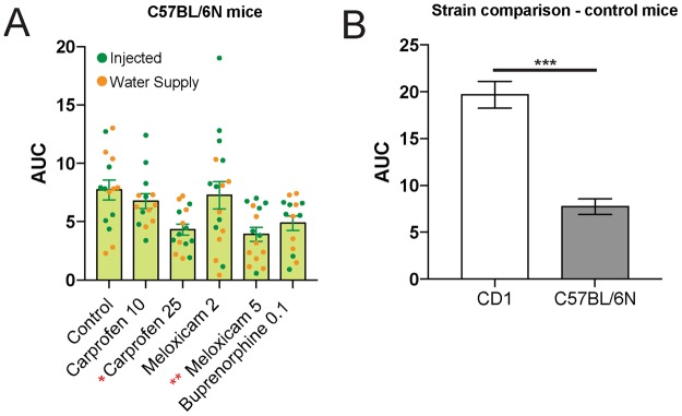
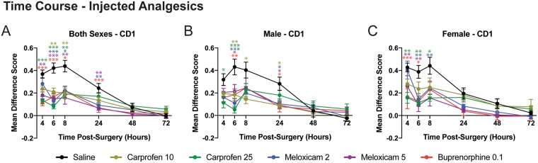
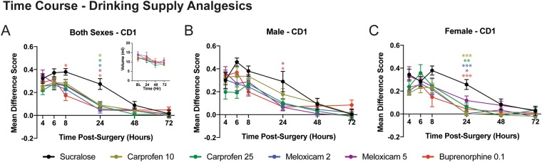
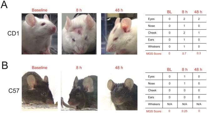
Most research laboratories abide by guidelines and mandates set by their research institution regarding the administration of analgesics to control pain during the postoperative period. Unfortunately, measuring pain originating from the head is difficult, making adequate decisions regarding pain control following stereotaxic surgery problematic. In addition, most postsurgical analgesia protocols require multiple injections over several days, which may cause stress and distress during a critical recovery period. Here we sought to (1) assess the degree of postoperative pain following craniotomy in mice, (2) compare the efficacy of three common rodent analgesics (carprofen, meloxicam and buprenorphine) for reducing this pain and (3) determine whether the route of administration (injected or self-administered through the drinking supply) influenced pain relief post-craniotomy. Using the mouse grimace scale (MGS), we found that injectable analgesics were significantly more effective at relieving post-craniotomy pain, however, both routes of administration decreased pain scores in the first 24 h postsurgery. Specifically, buprenorphine administered independently of administration route was the most effective at reducing MGS scores, however, female mice showed greater sensitivity to carprofen when administered through the water supply. Although it is necessary to provide laboratory animals with analgesics after an invasive procedure, there remains a gap in the literature regarding the degree of craniotomy-related pain in rodents and the efficacy of alternative routes of administration. Our study highlights the limitations of administering drugs through the drinking supply, even at doses that are considered to be higher than those currently recommended by most research institutions for treating pain of mild to moderate severity.
大多数研究实验室都遵守其研究机构制定的关于在术后期间管理镇痛药以控制疼痛的准则和规定。不幸的是,测量源自头部的疼痛是困难的,这使得在立体定向手术后针对疼痛控制做出充分的决策成为问题。此外,大多数术后镇痛方案需要在数天内多次注射,这可能会在关键的恢复期引起应激和痛苦。在这里,我们试图:(1)评估小鼠开颅手术后的术后疼痛程度;(2)比较三种常见的啮齿动物镇痛药(卡洛芬、美洛昔康和丁丙诺啡)减轻这种疼痛的效果;(3)确定给药途径(注射或通过饮用水自行给药)是否会影响开颅术后的疼痛缓解。使用小鼠痛苦表情量表(MGS),我们发现注射用镇痛药在缓解开颅术后疼痛方面更有效,然而,两种给药途径都能在术后 24 小时内降低疼痛评分。具体而言,丁丙诺啡通过独立于给药途径给药时最有效地降低 MGS 评分,然而,雌性小鼠在通过饮用水供应给药时对卡洛芬表现出更高的敏感性。尽管在侵入性手术后为实验动物提供镇痛药是必要的,但在啮齿动物开颅相关疼痛的程度和替代给药途径的有效性方面,文献中仍存在差距。我们的研究强调了通过饮用水供应给药的局限性,即使在剂量被认为高于大多数研究机构目前推荐的用于治疗轻度至中度疼痛的剂量时也是如此。