• 文献检索
  • 文档翻译
  • 深度研究
  • 学术资讯
  • Suppr Zotero 插件Zotero 插件
  • 邀请有礼
  • 套餐&价格
  • 历史记录
应用&插件
Suppr Zotero 插件Zotero 插件浏览器插件Mac 客户端Windows 客户端微信小程序
定价
高级版会员购买积分包购买API积分包
服务
文献检索文档翻译深度研究API 文档MCP 服务
关于我们
关于 Suppr公司介绍联系我们用户协议隐私条款
关注我们

Suppr 超能文献

核心技术专利:CN118964589B侵权必究
粤ICP备2023148730 号-1Suppr @ 2026

文献检索

告别复杂PubMed语法,用中文像聊天一样搜索,搜遍4000万医学文献。AI智能推荐,让科研检索更轻松。

立即免费搜索

文件翻译

保留排版,准确专业,支持PDF/Word/PPT等文件格式,支持 12+语言互译。

免费翻译文档

深度研究

AI帮你快速写综述,25分钟生成高质量综述,智能提取关键信息,辅助科研写作。

立即免费体验

真正的心脏解剖结构是怎样的?

What is the real cardiac anatomy?

作者信息

Mori Shumpei, Tretter Justin T, Spicer Diane E, Bolender David L, Anderson Robert H

机构信息

Division of Cardiovascular Medicine, Department of Internal Medicine, Kobe University Graduate School of Medicine, Kobe, Japan.

Heart Institute, Cincinnati Children's Hospital Medical Center, Cincinnati, Ohio.

出版信息

Clin Anat. 2019 Apr;32(3):288-309. doi: 10.1002/ca.23340. Epub 2019 Feb 13.

DOI:10.1002/ca.23340
PMID:30675928
原文链接:https://pmc.ncbi.nlm.nih.gov/articles/PMC6849845/
Abstract

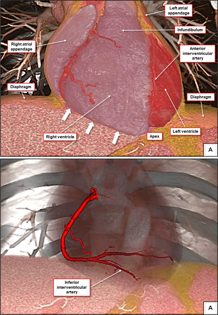
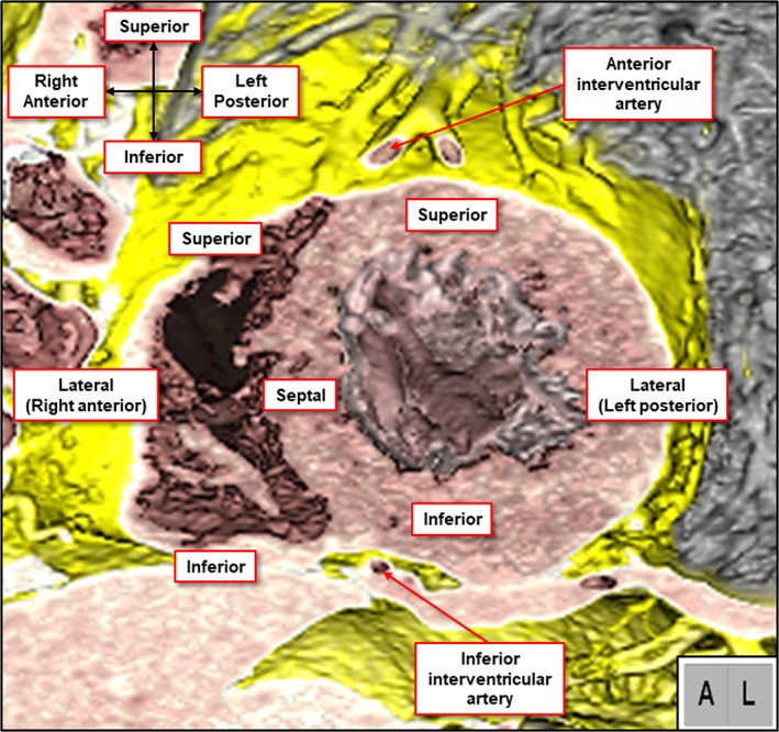
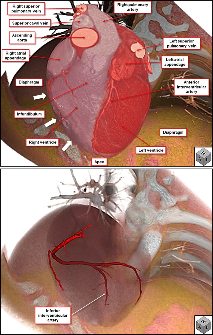
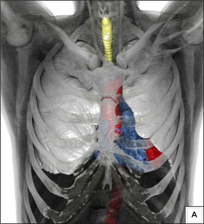
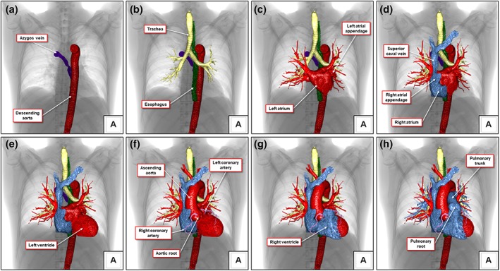
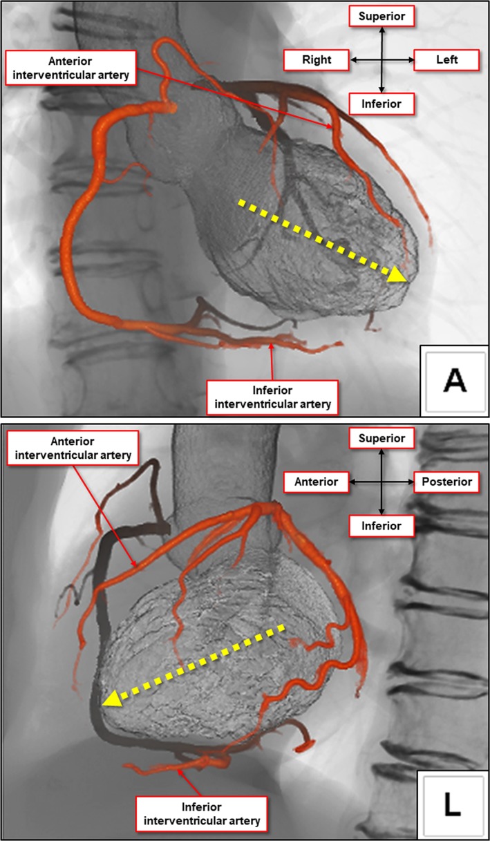
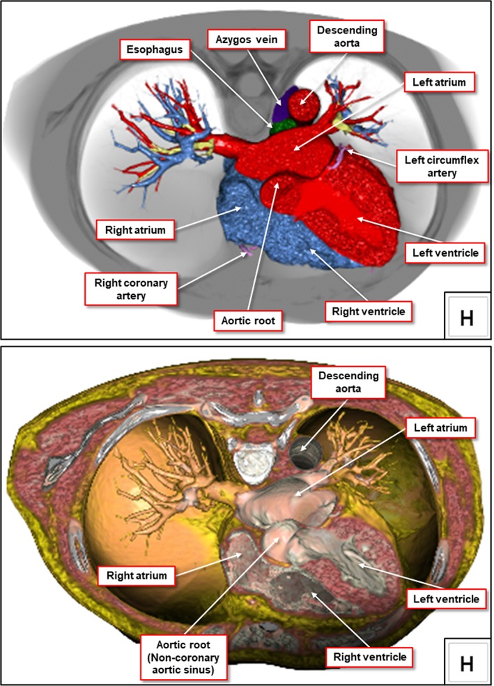
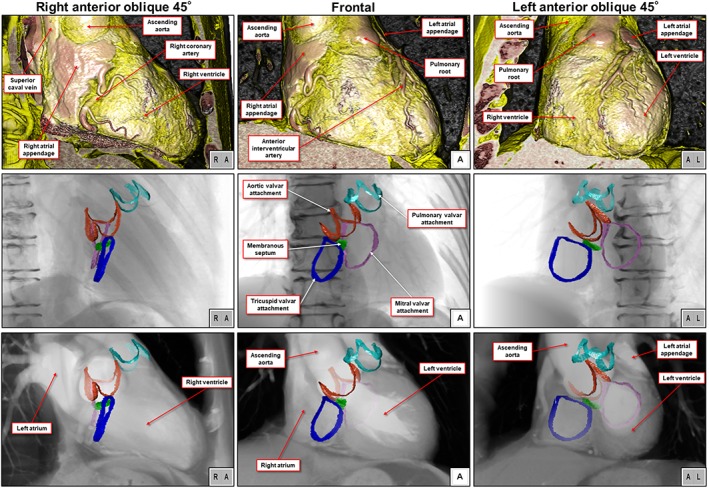
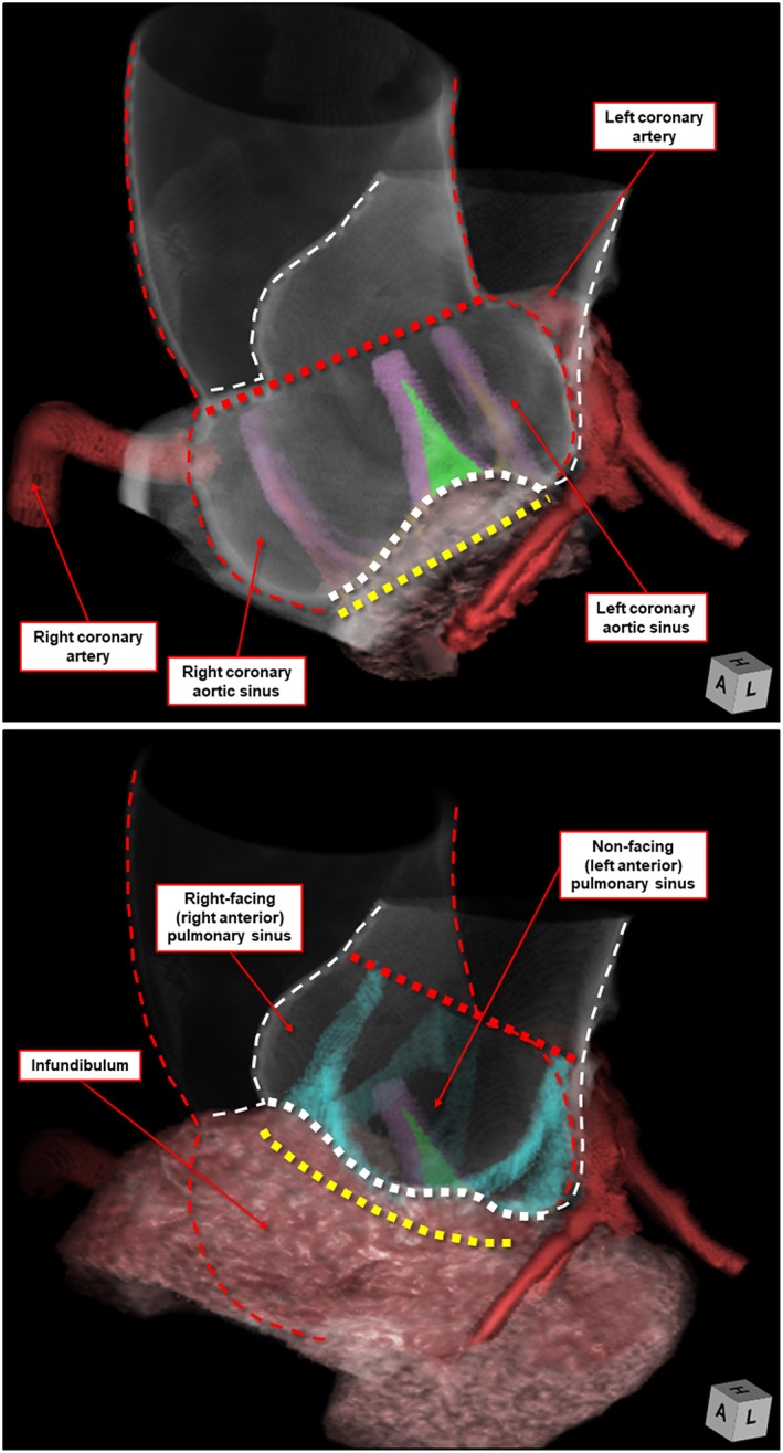
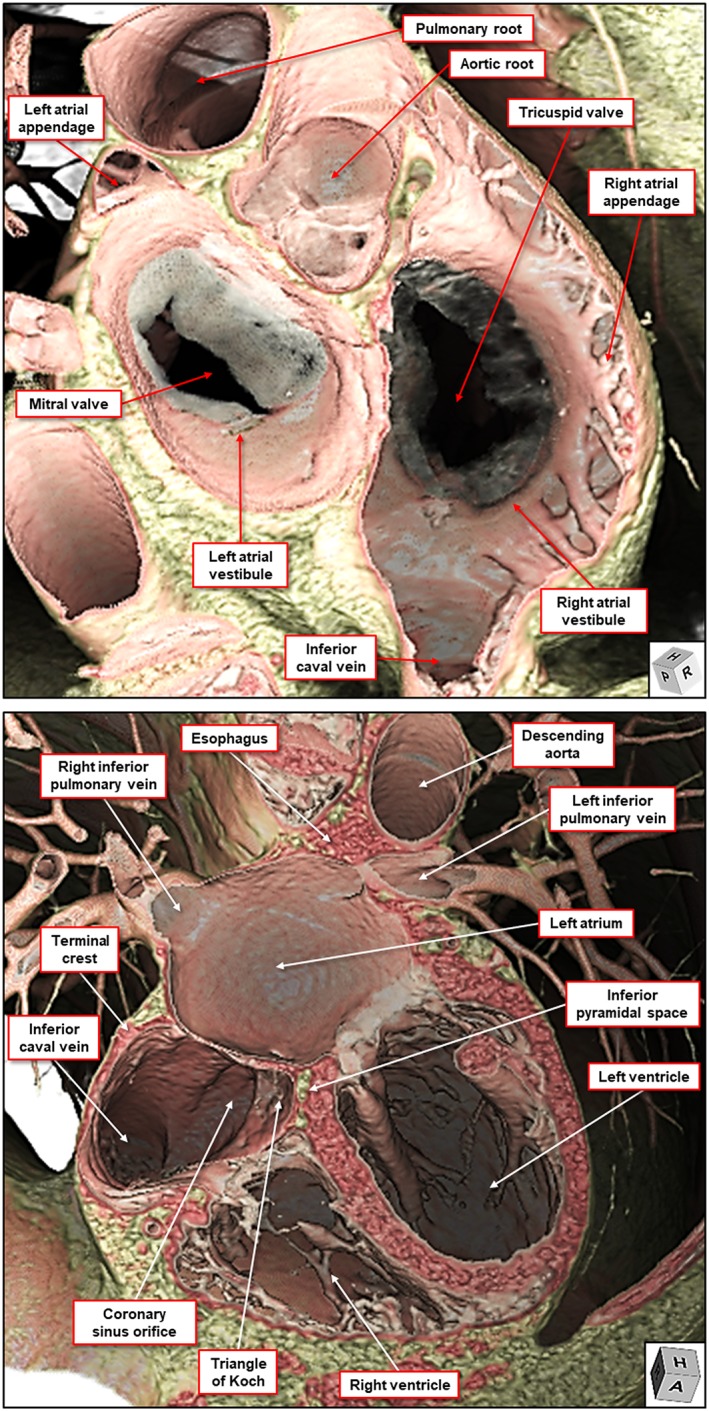
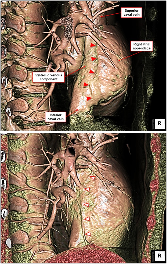
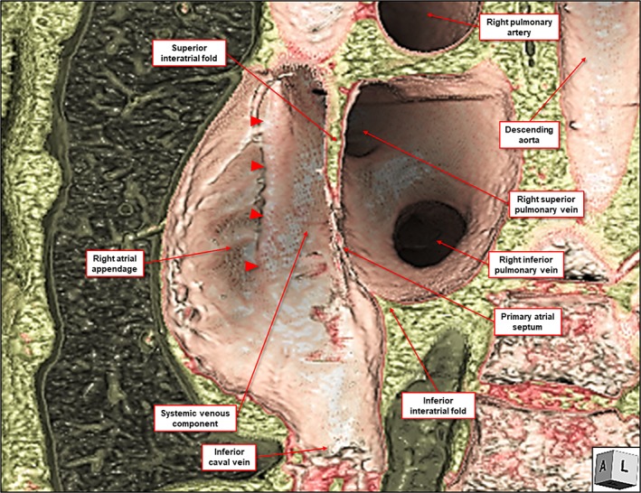
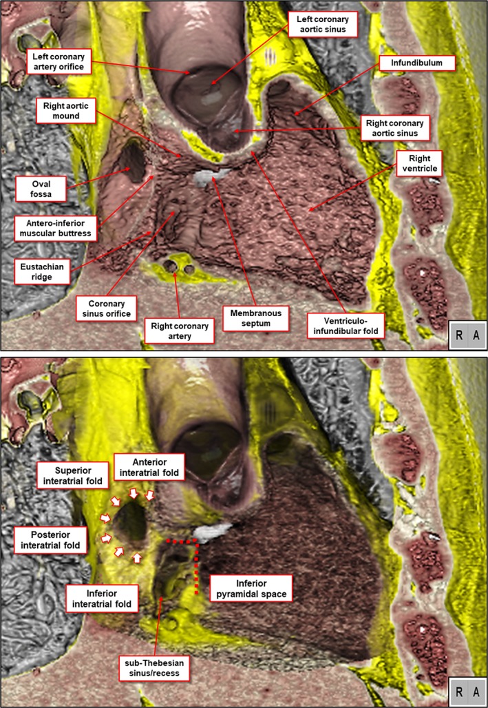
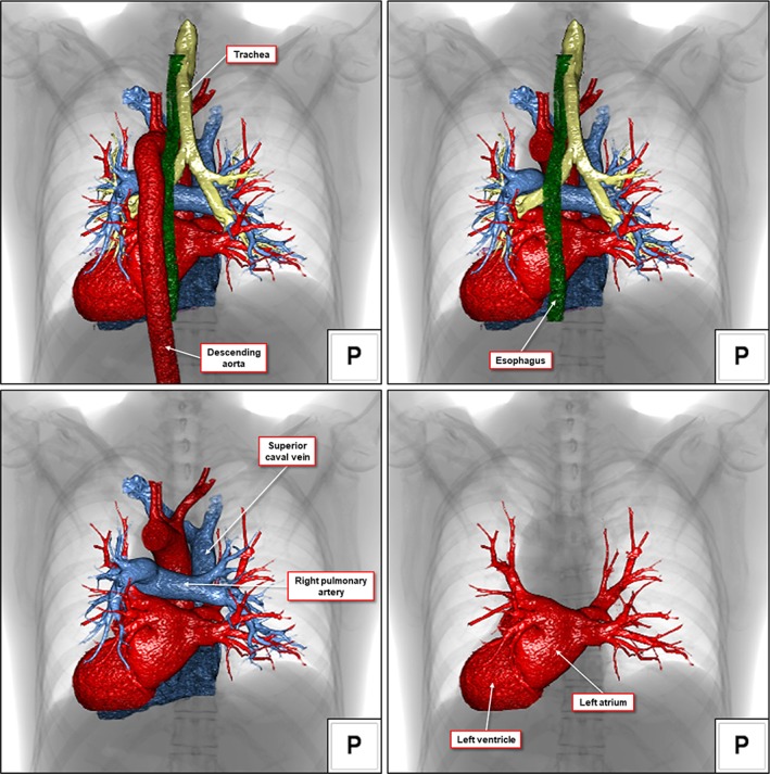
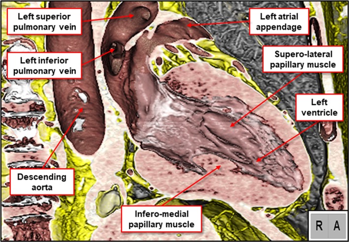
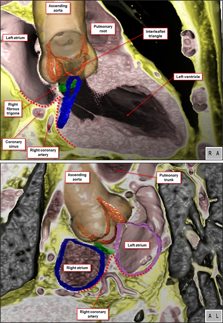
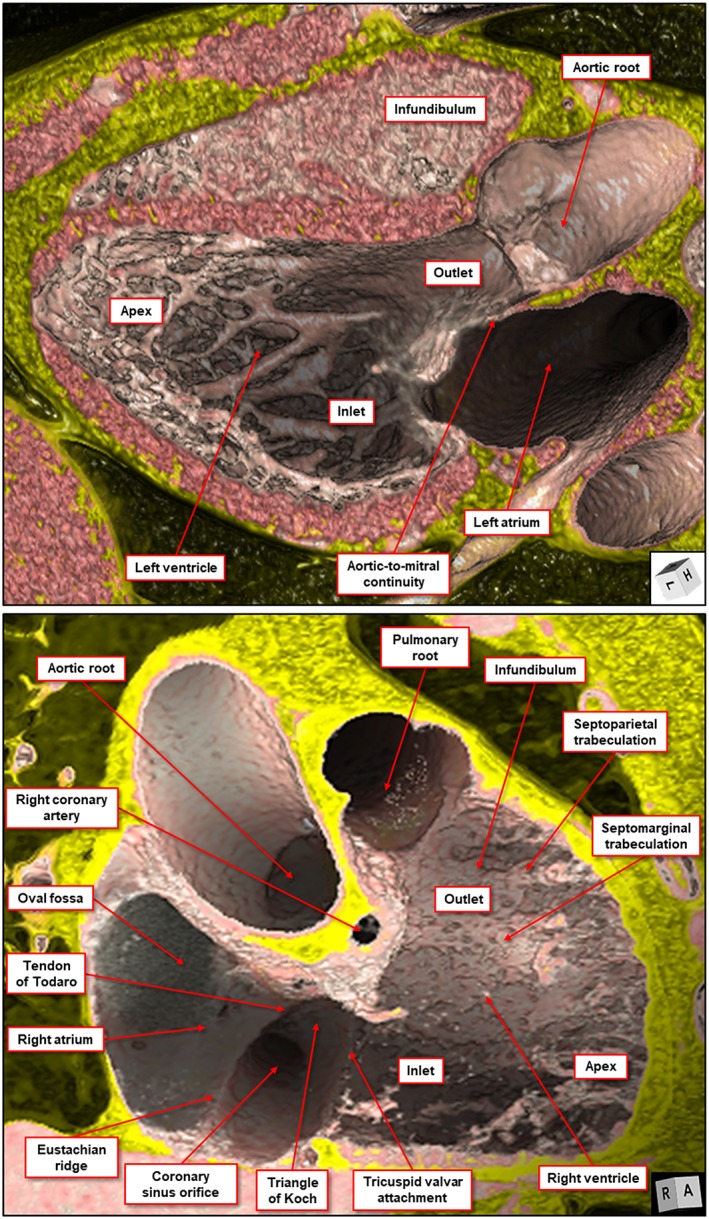
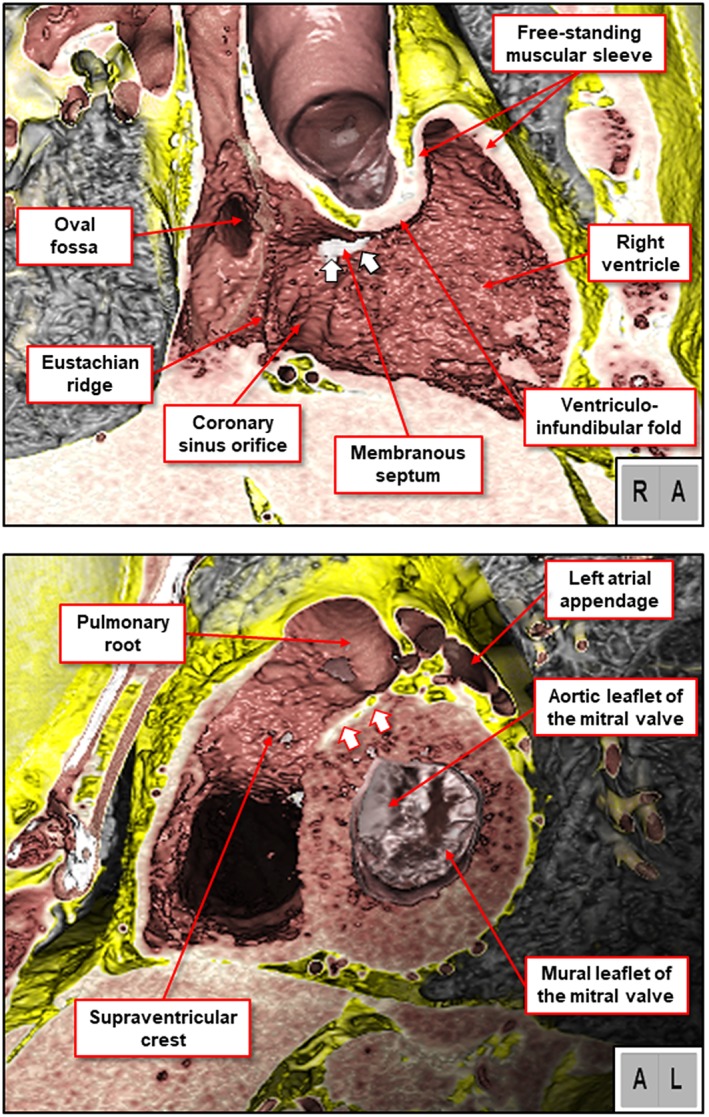
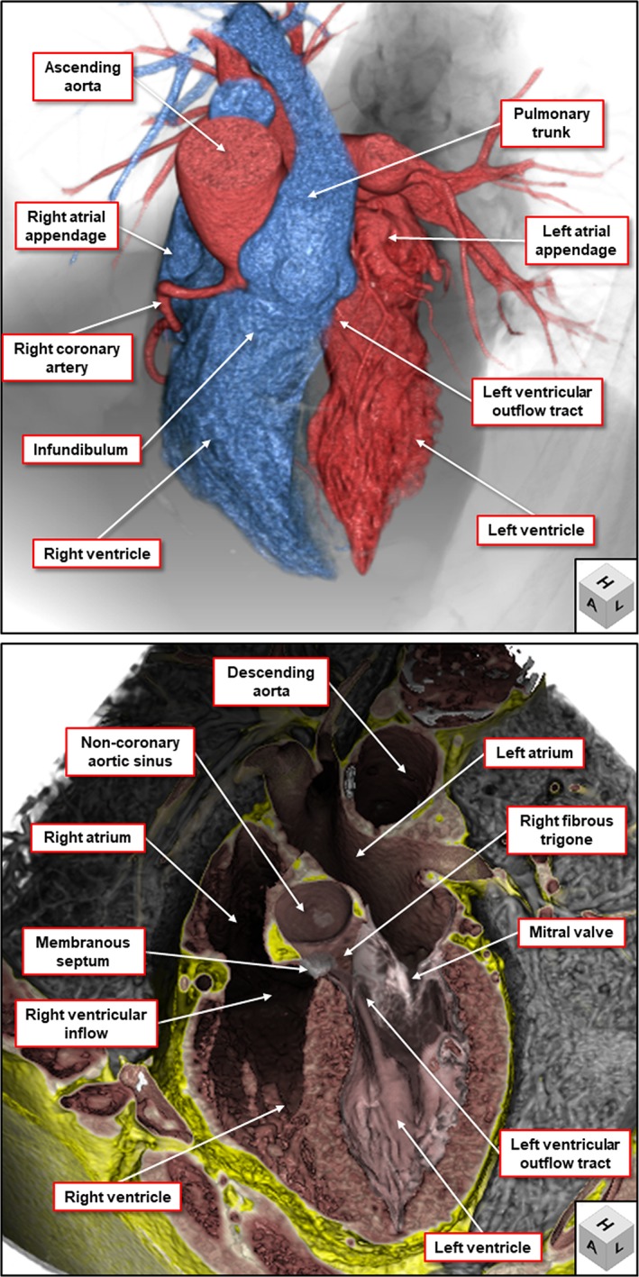
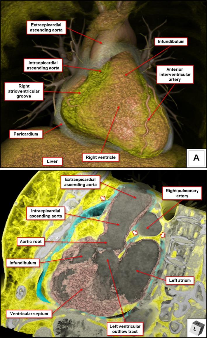
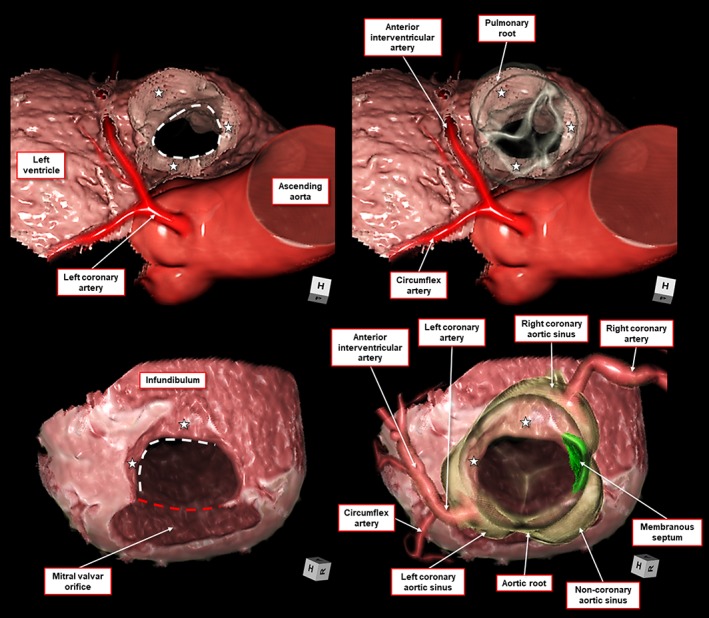
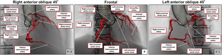
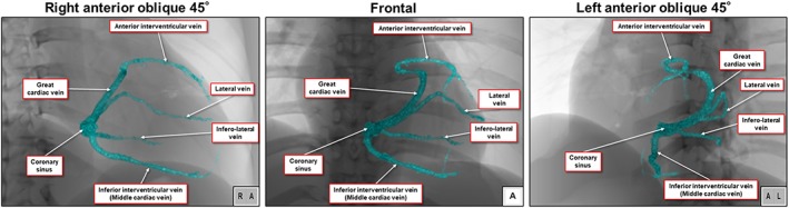
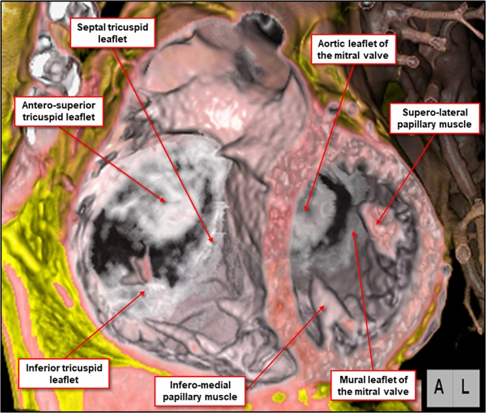
The heart is a remarkably complex organ. Teaching its details to medical students and clinical trainees can be very difficult. Despite the complexity, accurate recognition of these details is a pre-requisite for the subsequent understanding of clinical cardiologists and cardiac surgeons. A recent publication promoted the benefits of virtual reconstructions in facilitating the initial understanding achieved by medical students. If such teaching is to achieve its greatest value, the datasets used to provide the virtual images should themselves be anatomically accurate. They should also take note of a basic rule of human anatomy, namely that components of all organs should be described as they are normally situated within the body. It is almost universal at present for textbooks of anatomy to illustrate the heart as if removed from the body and positioned on its apex, the so-called Valentine situation. In the years prior to the emergence of interventional techniques to treat cardiac diseases, this approach was of limited significance. Nowadays, therapeutic interventions are commonplace worldwide. Advances in three-dimensional imaging technology, furthermore, now mean that the separate components of the heart can readily be segmented, and then shown in attitudinally appropriate fashion. In this review, we demonstrate how such virtual dissection of computed tomographic datasets in attitudinally appropriate fashion reveals the true details of cardiac anatomy. The virtual approach to teaching the arrangement of the cardiac components has much to commend it. If it is to be used, nonetheless, the anatomical details on which the reconstructions are based must be accurate. Clin. Anat. 32:288-309, 2019. © 2019 The Authors. Clinical Anatomy published by Wiley Periodicals, Inc. on behalf of American Association of Clinical Anatomists.

摘要

心脏是一个极其复杂的器官。向医学生和临床实习生讲授其细节可能非常困难。尽管复杂,但准确识别这些细节是临床心脏病学家和心脏外科医生后续理解的先决条件。最近的一篇出版物宣扬了虚拟重建在促进医学生初步理解方面的益处。如果这种教学要实现其最大价值,用于提供虚拟图像的数据集本身在解剖学上应该是准确的。它们还应注意人体解剖学的一条基本规则,即所有器官的组成部分都应按照其在体内的正常位置来描述。目前,解剖学教科书几乎普遍将心脏描绘成好像从身体中取出并置于心尖上,即所谓的“情人节姿势”。在治疗心脏病的介入技术出现之前的那些年里,这种方法的意义有限。如今,治疗性干预在全球都很常见。此外,三维成像技术的进步现在意味着心脏的各个组成部分可以很容易地被分割,然后以姿态合适的方式显示出来。在这篇综述中,我们展示了以姿态合适的方式对计算机断层扫描数据集进行这种虚拟解剖如何揭示心脏解剖学的真实细节。以虚拟方式教授心脏各组成部分的排列有很多值得称赞之处。然而,如果要使用它,重建所基于的解剖学细节必须准确。《临床解剖学》2019年第32卷:288 - 309页。© 2019作者。《临床解剖学》由威利期刊公司代表美国临床解剖学家协会出版。

https://cdn.ncbi.nlm.nih.gov/pmc/blobs/6e44/6849845/0bd23f79e3d9/CA-32-288-g025.jpg
https://cdn.ncbi.nlm.nih.gov/pmc/blobs/6e44/6849845/d0766884d2ef/CA-32-288-g001.jpg
https://cdn.ncbi.nlm.nih.gov/pmc/blobs/6e44/6849845/a79d77c2a990/CA-32-288-g002.jpg
https://cdn.ncbi.nlm.nih.gov/pmc/blobs/6e44/6849845/9595b3c603c2/CA-32-288-g003.jpg
https://cdn.ncbi.nlm.nih.gov/pmc/blobs/6e44/6849845/0648a9d682ab/CA-32-288-g004.jpg
https://cdn.ncbi.nlm.nih.gov/pmc/blobs/6e44/6849845/c1428b554ce8/CA-32-288-g005.jpg
https://cdn.ncbi.nlm.nih.gov/pmc/blobs/6e44/6849845/c60777f86bb9/CA-32-288-g006.jpg
https://cdn.ncbi.nlm.nih.gov/pmc/blobs/6e44/6849845/9f1f2e950818/CA-32-288-g007.jpg
https://cdn.ncbi.nlm.nih.gov/pmc/blobs/6e44/6849845/7a99f32ae0e4/CA-32-288-g008.jpg
https://cdn.ncbi.nlm.nih.gov/pmc/blobs/6e44/6849845/c66a5ea1659c/CA-32-288-g009.jpg
https://cdn.ncbi.nlm.nih.gov/pmc/blobs/6e44/6849845/2b170578b844/CA-32-288-g010.jpg
https://cdn.ncbi.nlm.nih.gov/pmc/blobs/6e44/6849845/4c46b79b21cc/CA-32-288-g011.jpg
https://cdn.ncbi.nlm.nih.gov/pmc/blobs/6e44/6849845/6a1c18b4e866/CA-32-288-g012.jpg
https://cdn.ncbi.nlm.nih.gov/pmc/blobs/6e44/6849845/512bea9b30d6/CA-32-288-g013.jpg
https://cdn.ncbi.nlm.nih.gov/pmc/blobs/6e44/6849845/177e2ad8d3a0/CA-32-288-g014.jpg
https://cdn.ncbi.nlm.nih.gov/pmc/blobs/6e44/6849845/a9ce81bf5198/CA-32-288-g015.jpg
https://cdn.ncbi.nlm.nih.gov/pmc/blobs/6e44/6849845/c7b44294a0dd/CA-32-288-g016.jpg
https://cdn.ncbi.nlm.nih.gov/pmc/blobs/6e44/6849845/71311786e42a/CA-32-288-g017.jpg
https://cdn.ncbi.nlm.nih.gov/pmc/blobs/6e44/6849845/14528915a95c/CA-32-288-g018.jpg
https://cdn.ncbi.nlm.nih.gov/pmc/blobs/6e44/6849845/3a41f7896d80/CA-32-288-g019.jpg
https://cdn.ncbi.nlm.nih.gov/pmc/blobs/6e44/6849845/48ae8e6f9e56/CA-32-288-g020.jpg
https://cdn.ncbi.nlm.nih.gov/pmc/blobs/6e44/6849845/ce74b0ffb479/CA-32-288-g021.jpg
https://cdn.ncbi.nlm.nih.gov/pmc/blobs/6e44/6849845/553e6f3023a7/CA-32-288-g022.jpg
https://cdn.ncbi.nlm.nih.gov/pmc/blobs/6e44/6849845/a5ef4274d0db/CA-32-288-g023.jpg
https://cdn.ncbi.nlm.nih.gov/pmc/blobs/6e44/6849845/353fc2ec6d1d/CA-32-288-g024.jpg
https://cdn.ncbi.nlm.nih.gov/pmc/blobs/6e44/6849845/0bd23f79e3d9/CA-32-288-g025.jpg

相似文献

1
What is the real cardiac anatomy?真正的心脏解剖结构是怎样的?
Clin Anat. 2019 Apr;32(3):288-309. doi: 10.1002/ca.23340. Epub 2019 Feb 13.
2
Why do we break one of the first rules of anatomy when describing the components of the heart?为什么在描述心脏的组成部分时,我们违背了解剖学的首要规则之一?
Clin Anat. 2019 May;32(4):585-596. doi: 10.1002/ca.23356. Epub 2019 Mar 9.
3
The importance of attitudinally appropriate description of cardiac anatomy.对心脏解剖结构进行态度恰当描述的重要性。
Clin Anat. 2009 Jan;22(1):47-51. doi: 10.1002/ca.20741.
4
Revisiting the Anatomy of the Living Heart.重新审视活体心脏的解剖结构。
Circ J. 2016;80(1):24-33. doi: 10.1253/circj.CJ-15-1147. Epub 2015 Dec 17.
5
Describing the cardiac components--attitudinally appropriate nomenclature.描述心脏结构部分——采用恰当态度的命名法。
J Cardiovasc Transl Res. 2013 Apr;6(2):118-23. doi: 10.1007/s12265-012-9434-z. Epub 2012 Dec 8.
6
Take away body parts! An investigation into the use of 3D-printed anatomical models in undergraduate anatomy education.拿走身体部位!3D 打印解剖模型在本科解剖学教育中的应用研究。
Anat Sci Educ. 2018 Jan;11(1):44-53. doi: 10.1002/ase.1718. Epub 2017 Jul 28.
7
Demonstration of living anatomy clarifies the morphology of interatrial communications.活体解剖演示阐明了房间隔交通的形态。
Heart. 2018 Dec;104(24):2003-2009. doi: 10.1136/heartjnl-2018-313758. Epub 2018 Sep 4.
8
Virtual reality and cardiac anatomy: Exploring immersive three-dimensional cardiac imaging, a pilot study in undergraduate medical anatomy education.虚拟现实与心脏解剖:探索沉浸式三维心脏成像,一项本科医学解剖学教育的试点研究。
Clin Anat. 2019 Mar;32(2):238-243. doi: 10.1002/ca.23292. Epub 2018 Oct 31.
9
Cardiac anatomy revisited.心脏解剖学再探讨。
J Anat. 2004 Sep;205(3):159-77. doi: 10.1111/j.0021-8782.2004.00330.x.
10
Using 3D Modeling Techniques to Enhance Teaching of Difficult Anatomical Concepts.运用3D建模技术加强难解剖概念的教学。
Acad Radiol. 2016 Apr;23(4):507-16. doi: 10.1016/j.acra.2015.12.012. Epub 2016 Feb 17.

引用本文的文献

1
Age-associated changes in the heart: implications for COVID-19 therapies.心脏的年龄相关变化:对COVID-19治疗的影响。
Aging (Albany NY). 2025 May 13;17(5):1340-1367. doi: 10.18632/aging.206251.
2
High Mobility Group Box 1 and Cardiovascular Diseases: Study of Act and Connect.高迁移率族蛋白 1 与心血管疾病:Act 和 Connect 的研究。
Cardiovasc Toxicol. 2024 Nov;24(11):1268-1286. doi: 10.1007/s12012-024-09919-5. Epub 2024 Sep 6.
3
Left ventricular trabeculation in Hominidae: divergence of the human cardiac phenotype.人科左心室小梁化:人类心脏表型的分歧。

本文引用的文献

1
Virtual Reality Perhaps, but Is this Real Cardiac Anatomy?虚拟现实?也许吧,但这是真实的心脏解剖结构吗?
Clin Anat. 2019 May;32(4):468. doi: 10.1002/ca.23306. Epub 2018 Dec 27.
2
Virtual reality and cardiac anatomy: Exploring immersive three-dimensional cardiac imaging, a pilot study in undergraduate medical anatomy education.虚拟现实与心脏解剖:探索沉浸式三维心脏成像,一项本科医学解剖学教育的试点研究。
Clin Anat. 2019 Mar;32(2):238-243. doi: 10.1002/ca.23292. Epub 2018 Oct 31.
3
Alarming decline in recognition of anatomical structures amongst medical students and physicians.
Commun Biol. 2024 Jun 14;7(1):682. doi: 10.1038/s42003-024-06280-9.
4
Revisiting the anatomy of the left ventricle in the light of knowledge of its development.重新审视左心室的解剖结构,了解其发育知识。
J Anat. 2024 Aug;245(2):201-216. doi: 10.1111/joa.14048. Epub 2024 Apr 17.
5
Development of the arterial roots and ventricular outflow tracts.动脉干和心室流出道的发育。
J Anat. 2024 Mar;244(3):497-513. doi: 10.1111/joa.13973. Epub 2023 Nov 13.
6
Revisiting the anatomy of the right ventricle in the light of knowledge of its development.重新审视右心室的解剖结构,结合其发育知识。
J Anat. 2024 Feb;244(2):297-311. doi: 10.1111/joa.13960. Epub 2023 Oct 9.
7
An Overview of Traditional and Advanced Visualization Techniques Applied to Anatomical Instruction Involving Cadaveric Dissection.传统和先进的可视化技术在涉及尸体解剖的解剖教学中的应用概述。
Adv Exp Med Biol. 2023;1431:35-63. doi: 10.1007/978-3-031-36727-4_3.
8
Use of a two-handed model to improve comprehension of ventricular outflow tract anatomy.使用双手模型提高对心室流出道解剖结构的理解。
BMC Med Educ. 2023 Feb 8;23(1):101. doi: 10.1186/s12909-023-04083-w.
9
Letter: The time has come to use attitudinally appropriate terminology when describing cardiac anatomy.信:现在是时候在描述心脏解剖结构时使用态度恰当的术语了。
EuroIntervention. 2022 Apr 22;17(18):1538. doi: 10.4244/EIJ-D-21-01077L.
10
Pathophysiological, Cellular, and Molecular Events of the Vascular System in Anaphylaxis.过敏反应中血管系统的病理生理学、细胞和分子事件。
Front Immunol. 2022 Mar 8;13:836222. doi: 10.3389/fimmu.2022.836222. eCollection 2022.
医学生和医生对解剖结构的认知令人担忧地下降。
Ann Anat. 2019 Jan;221:48-56. doi: 10.1016/j.aanat.2018.09.004. Epub 2018 Sep 20.
4
Demonstration of living anatomy clarifies the morphology of interatrial communications.活体解剖演示阐明了房间隔交通的形态。
Heart. 2018 Dec;104(24):2003-2009. doi: 10.1136/heartjnl-2018-313758. Epub 2018 Sep 4.
5
The anatomic substrates for outflow tract arrhythmias.流出道心律失常的解剖学基础。
Heart Rhythm. 2019 Feb;16(2):290-297. doi: 10.1016/j.hrthm.2018.08.014. Epub 2018 Aug 18.
6
Living anatomy of the pulmonary root.肺根的活体解剖学
J Cardiovasc Electrophysiol. 2018 Sep;29(9):1238-1240. doi: 10.1111/jce.13691. Epub 2018 Jul 20.
7
Relationship between the membranous septum and the virtual basal ring of the aortic root in candidates for transcatheter implantation of the aortic valve.经导管主动脉瓣植入术候选者中膜性间隔与主动脉根部虚拟基环的关系
Clin Anat. 2018 May;31(4):525-534. doi: 10.1002/ca.23071. Epub 2018 Mar 25.
8
Variations in rotation of the aortic root and membranous septum with implications for transcatheter valve implantation.主动脉根部和膜性间隔旋转的变化及其对经导管瓣膜植入术的影响。
Heart. 2018 Jun;104(12):999-1005. doi: 10.1136/heartjnl-2017-312390. Epub 2017 Nov 16.
9
Isomerism in the setting of the so-called "heterotaxy": The usefulness of computed tomographic analysis.所谓“内脏异位”情况下的异构现象:计算机断层扫描分析的作用
Ann Pediatr Cardiol. 2017 May-Aug;10(2):175-186. doi: 10.4103/apc.APC_171_16.
10
The association between wedging of the aorta and cardiac structural anatomy as revealed using multidetector-row computed tomography.使用多排螺旋计算机断层扫描揭示的主动脉楔入与心脏结构解剖之间的关联。
J Anat. 2017 Jul;231(1):110-120. doi: 10.1111/joa.12611. Epub 2017 Apr 11.