• 文献检索
  • 文档翻译
  • 深度研究
  • 学术资讯
  • Suppr Zotero 插件Zotero 插件
  • 邀请有礼
  • 套餐&价格
  • 历史记录
应用&插件
Suppr Zotero 插件Zotero 插件浏览器插件Mac 客户端Windows 客户端微信小程序
定价
高级版会员购买积分包购买API积分包
服务
文献检索文档翻译深度研究API 文档MCP 服务
关于我们
关于 Suppr公司介绍联系我们用户协议隐私条款
关注我们

Suppr 超能文献

核心技术专利:CN118964589B侵权必究
粤ICP备2023148730 号-1Suppr @ 2026

文献检索

告别复杂PubMed语法,用中文像聊天一样搜索,搜遍4000万医学文献。AI智能推荐,让科研检索更轻松。

立即免费搜索

文件翻译

保留排版,准确专业,支持PDF/Word/PPT等文件格式,支持 12+语言互译。

免费翻译文档

深度研究

AI帮你快速写综述,25分钟生成高质量综述,智能提取关键信息,辅助科研写作。

立即免费体验

定量扩散加权成像预测神经母细胞瘤患儿的无事件生存期:一项回顾性队列研究的初步结果

Quantitative DWI predicts event-free survival in children with neuroblastic tumours: preliminary findings from a retrospective cohort study.

作者信息

Peschmann Anna-Lydia, Beer Meinrad, Ammann Bettina, Dreyhaupt Jens, Kneer Katharina, Beer Ambros J, Beltinger Christian, Steinbach Daniel, Cario Holger, Neubauer Henning

机构信息

Department of Diagnostic and Interventional Radiology, University Hospital Ulm, Albert-Schweitzer-Allee 23, 89081, Ulm, Germany.

Department of Biometrics, University Hospital Ulm, 89081, Ulm, Germany.

出版信息

Eur Radiol Exp. 2019 Jan 30;3(1):6. doi: 10.1186/s41747-019-0087-4.

DOI:10.1186/s41747-019-0087-4
PMID:30701332
原文链接:https://pmc.ncbi.nlm.nih.gov/articles/PMC6353978/
Abstract

BACKGROUND

Quantitative diffusion-weighted imaging (DWI) probes into tissue microstructure in solid tumours. In this retrospective ethically approved study, we investigated DWI as a potential non-invasive predictor of tumour dignity and prognosis in paediatric patients with neuroblastic tumours.

METHODS

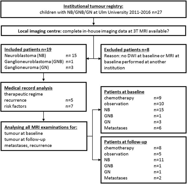
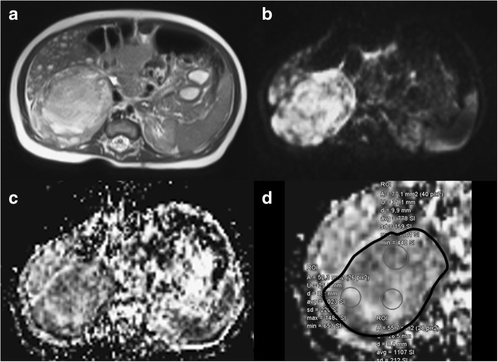
Nineteen consecutive patients with neuroblastoma (NB, n = 15), ganglioneuroblastoma (GNB, n = 1) and ganglioneuroma (GN, n = 3) underwent 3-T magnetic resonance imaging at first diagnosis and after 3-month follow-up, following a protocol including DWI (b = 50 and 800 s/mm) in addition to standard sequences. All DWI scans were analysed for tumour volume assessment and apparent diffusion coefficient (ADC) calculation. Correlation with tumour pathology and risk factors (bone-marrow metastases, MYCN-amplification and 1p-deletion), therapeutic regime (observation versus chemotherapy) and clinical follow-up was evaluated.

RESULTS

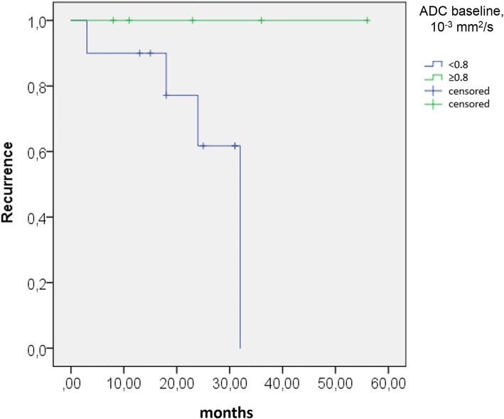
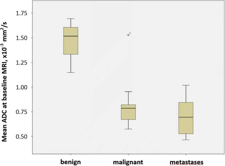
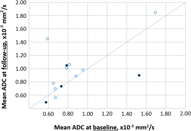
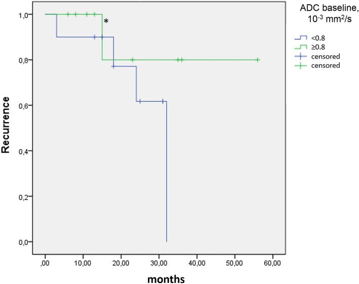
At baseline, mean ADC in NB was lower than in GNB/GN (0.76 vs. 1.47 × 10 mm/s, p = 0.003). An ADC cutoff ≤ 1.05 identified malignant disease with 100.0% sensitivity (95% confidence interval [CI] 29.2-100.0%) and 93.8% specificity (95% CI 69.8-99.8%). Initial ADC was < 0.80 in all NB patients with eventual tumour relapse. During follow-up, tumour ADC values increased in the observation group (NB/GN) without relapse (p = 0.043). In eventually relapsing tumours, ADC values at follow-up tended to decrease further despite reduction in tumour volume.

CONCLUSIONS

ADC values at first presentation differed significantly between malignant and benign neuroblastic tumours. Low baseline ADC was predictive of tumour progression and relapse in NB patients. With therapy, increasing ADC values appeared to predict relapse-free survival, while a decreasing ADC during therapy was an indicator of poor prognosis.

摘要

背景

定量扩散加权成像(DWI)可探究实体瘤的组织微观结构。在这项经伦理批准的回顾性研究中,我们调查了DWI作为小儿神经母细胞瘤患者肿瘤特性和预后的潜在非侵入性预测指标。

方法

19例连续的神经母细胞瘤(NB,n = 15)、神经节神经母细胞瘤(GNB,n = 1)和神经节瘤(GN,n = 3)患者在初次诊断时及3个月随访后接受了3-T磁共振成像检查,检查方案除标准序列外还包括DWI(b = 50和800 s/mm²)。对所有DWI扫描进行分析以评估肿瘤体积并计算表观扩散系数(ADC)。评估其与肿瘤病理及危险因素(骨髓转移、MYCN扩增和1p缺失)、治疗方案(观察与化疗)及临床随访的相关性。

结果

基线时,NB的平均ADC低于GNB/GN(0.76对1.47×10⁻³mm²/s,p = 0.003)。ADC截止值≤1.05可识别恶性疾病,敏感性为100.0%(95%置信区间[CI] 29.2 - 100.0%),特异性为93.8%(95% CI 69.8 - 99.8%)。所有最终肿瘤复发的NB患者初始ADC均<0.80。随访期间,未复发的观察组(NB/GN)肿瘤ADC值升高(p = 0.043)。在最终复发的肿瘤中,尽管肿瘤体积减小,但随访时的ADC值仍倾向于进一步降低。

结论

初次就诊时恶性和良性神经母细胞瘤的ADC值存在显著差异。低基线ADC可预测NB患者的肿瘤进展和复发。随着治疗,ADC值升高似乎可预测无复发生存,而治疗期间ADC值降低则提示预后不良。

https://cdn.ncbi.nlm.nih.gov/pmc/blobs/f1ac/6353978/55150048133d/41747_2019_87_Fig7_HTML.jpg
https://cdn.ncbi.nlm.nih.gov/pmc/blobs/f1ac/6353978/aa2c4d374df7/41747_2019_87_Fig1_HTML.jpg
https://cdn.ncbi.nlm.nih.gov/pmc/blobs/f1ac/6353978/ccc01a4649e1/41747_2019_87_Fig2_HTML.jpg
https://cdn.ncbi.nlm.nih.gov/pmc/blobs/f1ac/6353978/ccc01a4649e1/41747_2019_87_Fig3_HTML.jpg
https://cdn.ncbi.nlm.nih.gov/pmc/blobs/f1ac/6353978/a9159f99ad02/41747_2019_87_Fig4_HTML.jpg
https://cdn.ncbi.nlm.nih.gov/pmc/blobs/f1ac/6353978/abc3271f199f/41747_2019_87_Fig5_HTML.jpg
https://cdn.ncbi.nlm.nih.gov/pmc/blobs/f1ac/6353978/a1c9336b1cb3/41747_2019_87_Fig6_HTML.jpg
https://cdn.ncbi.nlm.nih.gov/pmc/blobs/f1ac/6353978/55150048133d/41747_2019_87_Fig7_HTML.jpg

相似文献

1
Quantitative DWI predicts event-free survival in children with neuroblastic tumours: preliminary findings from a retrospective cohort study.定量扩散加权成像预测神经母细胞瘤患儿的无事件生存期:一项回顾性队列研究的初步结果
Eur Radiol Exp. 2019 Jan 30;3(1):6. doi: 10.1186/s41747-019-0087-4.
2
Role of diffusion-weighted imaging in distinguishing thoracoabdominal neuroblastic tumours of various histological types and differentiation grades.扩散加权成像在鉴别不同组织学类型和分化程度的胸腹部神经母细胞瘤中的作用
J Med Imaging Radiat Oncol. 2017 Dec;61(6):718-724. doi: 10.1111/1754-9485.12615. Epub 2017 May 16.
3
Diagnostic Value of Diffusion-Weighted MRI for Tumor Characterization, Differentiation and Monitoring in Pediatric Patients with Neuroblastic Tumors.扩散加权磁共振成像对小儿神经母细胞瘤肿瘤特征、鉴别及监测的诊断价值
Rofo. 2017 Jul;189(7):640-650. doi: 10.1055/s-0043-108993. Epub 2017 May 16.
4
Feasibility and possible value of quantitative semi-automated diffusion weighted imaging volumetry of neuroblastic tumors.神经母细胞瘤定量半自动化弥散加权成像容积测量的可行性和可能价值。
Cancer Imaging. 2020 Dec 17;20(1):89. doi: 10.1186/s40644-020-00366-3.
5
Diffusion weighted imaging in differentiating malignant and benign neuroblastic tumors.扩散加权成像在鉴别恶性和良性神经母细胞瘤中的应用
Jpn J Radiol. 2016 Sep;34(9):620-4. doi: 10.1007/s11604-016-0565-z. Epub 2016 Jul 14.
6
Diffusion-weighted MRI for differentiation of neuroblastoma and ganglioneuroblastoma/ganglioneuroma.弥散加权 MRI 鉴别神经母细胞瘤与神经节母细胞瘤/神经节细胞瘤。
Eur J Radiol. 2011 Sep;79(3):443-6. doi: 10.1016/j.ejrad.2010.04.005. Epub 2010 May 13.
7
Role of Functional Magnetic Resonance Imaging Derived Parameters as Imaging Biomarkers and Correlation with Clinicopathological Features in Carcinoma of Uterine Cervix.功能磁共振成像衍生参数作为宫颈癌成像生物标志物的作用及其与临床病理特征的相关性
J Clin Diagn Res. 2017 Aug;11(8):XC06-XC11. doi: 10.7860/JCDR/2017/29165.10426. Epub 2017 Aug 1.
8
Diffusion MRI of uterine and ovarian masses: identifying the benign lesions.磁共振弥散加权成像在子宫及卵巢肿块中的应用:良性病变的鉴别。
Abdom Radiol (NY). 2016 Dec;41(12):2466-2475. doi: 10.1007/s00261-016-0909-2.
9
Differentiation of Benign and Malignant Head and Neck Lesions With Diffusion Tensor Imaging and DWI.利用扩散张量成像和扩散加权成像鉴别头颈部良恶性病变
AJR Am J Roentgenol. 2017 May;208(5):1110-1115. doi: 10.2214/AJR.16.16486. Epub 2017 Feb 28.
10
Why prostate tumour delineation based on apparent diffusion coefficient is challenging: an exploration of the tissue microanatomy.为什么基于表观扩散系数的前列腺肿瘤勾画具有挑战性:对组织微观解剖结构的探索。
Acta Oncol. 2013 Nov;52(8):1629-36. doi: 10.3109/0284186X.2013.787164. Epub 2013 Apr 28.

引用本文的文献

1
Quantitative MRI in Childhood Neuroblastoma: Beyond the Assessment of Image-defined Risk Factors.儿童神经母细胞瘤的定量 MRI:超越图像定义危险因素的评估。
Radiol Imaging Cancer. 2024 Nov;6(6):e240089. doi: 10.1148/rycan.240089.
2
Image-defined risk factors associated with MYCN oncogene amplification in neuroblastoma and their association with overall survival.与神经母细胞瘤中MYCN癌基因扩增相关的图像定义风险因素及其与总生存期的关联。
Abdom Radiol (NY). 2024 Jun;49(6):1949-1960. doi: 10.1007/s00261-024-04196-w. Epub 2024 Mar 4.
3
Local MRI before and after Tumor Resection in Neuroblastoma: Impact of Residual Disease on Event Free Survival.

本文引用的文献

1
Incidence, Survival, and Treatment of Localized and Metastatic Neuroblastoma in Germany 1979-2015.1979 - 2015年德国局限性和转移性神经母细胞瘤的发病率、生存率及治疗情况
Paediatr Drugs. 2017 Dec;19(6):577-593. doi: 10.1007/s40272-017-0251-3.
2
2017 GPOH Guidelines for Diagnosis and Treatment of Patients with Neuroblastic Tumors.2017年德国儿童肿瘤学会神经母细胞瘤患者诊断和治疗指南
Klin Padiatr. 2017 May;229(3):147-167. doi: 10.1055/s-0043-103086. Epub 2017 May 30.
3
Diagnostic Value of Diffusion-Weighted MRI for Tumor Characterization, Differentiation and Monitoring in Pediatric Patients with Neuroblastic Tumors.
神经母细胞瘤肿瘤切除前后的局部磁共振成像:残留疾病对无事件生存期的影响
J Clin Med. 2023 Nov 24;12(23):7297. doi: 10.3390/jcm12237297.
4
Combined Metabolic and Functional Tumor Volumes on [F]FDG-PET/MRI in Neuroblastoma Using Voxel-Wise Analysis.使用体素分析在神经母细胞瘤中通过[F]FDG-PET/MRI测量联合代谢和功能肿瘤体积
J Clin Med. 2023 Sep 15;12(18):5976. doi: 10.3390/jcm12185976.
5
Imaging biomarkers and radiomics in pediatric oncology: a view from the PRIMAGE (PRedictive In silico Multiscale Analytics to support cancer personalized diaGnosis and prognosis, Empowered by imaging biomarkers) project.儿科肿瘤学中的影像生物标志物和放射组学:来自 PRIMAGE(基于预测性计算多尺度分析以支持癌症个体化诊断和预后,由影像生物标志物赋能)项目的观点。
Pediatr Radiol. 2024 Apr;54(4):562-570. doi: 10.1007/s00247-023-05770-y. Epub 2023 Sep 25.
6
[Radiological imaging of neuroblastoma].[神经母细胞瘤的放射影像学]
Radiologe. 2021 Jul;61(7):639-648. doi: 10.1007/s00117-021-00875-7. Epub 2021 Jun 22.
7
Feasibility and possible value of quantitative semi-automated diffusion weighted imaging volumetry of neuroblastic tumors.神经母细胞瘤定量半自动化弥散加权成像容积测量的可行性和可能价值。
Cancer Imaging. 2020 Dec 17;20(1):89. doi: 10.1186/s40644-020-00366-3.
8
Comparison Between Diffusion-Weighted MRI and I-mIBG Uptake in Primary High-Risk Neuroblastoma.弥散加权 MRI 与 I-mIBG 摄取在原发性高危神经母细胞瘤中的比较。
J Magn Reson Imaging. 2021 May;53(5):1486-1497. doi: 10.1002/jmri.27458. Epub 2020 Dec 6.
9
Whole-body magnetic resonance imaging in pediatric oncology - recommendations by the Oncology Task Force of the ESPR.全身磁共振成像在儿科肿瘤学中的应用-ESP 肿瘤学工作组的建议。
Pediatr Radiol. 2020 Jul;50(8):1162-1174. doi: 10.1007/s00247-020-04683-4. Epub 2020 May 28.
扩散加权磁共振成像对小儿神经母细胞瘤肿瘤特征、鉴别及监测的诊断价值
Rofo. 2017 Jul;189(7):640-650. doi: 10.1055/s-0043-108993. Epub 2017 May 16.
4
Role of diffusion-weighted imaging in distinguishing thoracoabdominal neuroblastic tumours of various histological types and differentiation grades.扩散加权成像在鉴别不同组织学类型和分化程度的胸腹部神经母细胞瘤中的作用
J Med Imaging Radiat Oncol. 2017 Dec;61(6):718-724. doi: 10.1111/1754-9485.12615. Epub 2017 May 16.
5
Diffusion weighted imaging in differentiating malignant and benign neuroblastic tumors.扩散加权成像在鉴别恶性和良性神经母细胞瘤中的应用
Jpn J Radiol. 2016 Sep;34(9):620-4. doi: 10.1007/s11604-016-0565-z. Epub 2016 Jul 14.
6
Gadolinium deposition within the dentate nucleus and globus pallidus after repeated administrations of gadolinium-based contrast agents-current status.多次给予钆基造影剂后钆在齿状核和苍白球中的沉积——现状
Neuroradiology. 2016 May;58(5):433-41. doi: 10.1007/s00234-016-1658-1. Epub 2016 Feb 12.
7
Monitorization of chemotherapy response using diffusion-weighted imaging in neuroblastoma.利用扩散加权成像监测神经母细胞瘤化疗反应
Diagn Interv Radiol. 2016 Jan-Feb;22(1):108. doi: 10.5152/dir.2015.15178.
8
Variations in apparent diffusion coefficient values following chemotherapy in pediatric neuroblastoma.小儿神经母细胞瘤化疗后表观扩散系数值的变化
Diagn Interv Radiol. 2015 Mar-Apr;21(2):184-8. doi: 10.5152/dir.2014.14187.
9
Diffusion-weighted magnetic resonance imaging for early response assessment of chemoradiotherapy in patients with nasopharyngeal carcinoma.扩散加权磁共振成像用于鼻咽癌患者放化疗早期疗效评估
Magn Reson Imaging. 2014 Jul;32(6):630-7. doi: 10.1016/j.mri.2014.02.009. Epub 2014 Feb 11.
10
The degree of tumor volume reduction during the early phase of induction chemotherapy is an independent prognostic factor in patients with high-risk neuroblastoma.诱导化疗早期肿瘤体积减少的程度是高危神经母细胞瘤患者的独立预后因素。
Cancer. 2013 Feb 1;119(3):656-64. doi: 10.1002/cncr.27775. Epub 2012 Sep 5.