• 文献检索
  • 文档翻译
  • 深度研究
  • 学术资讯
  • Suppr Zotero 插件Zotero 插件
  • 邀请有礼
  • 套餐&价格
  • 历史记录
应用&插件
Suppr Zotero 插件Zotero 插件浏览器插件Mac 客户端Windows 客户端微信小程序
定价
高级版会员购买积分包购买API积分包
服务
文献检索文档翻译深度研究API 文档MCP 服务
关于我们
关于 Suppr公司介绍联系我们用户协议隐私条款
关注我们

Suppr 超能文献

核心技术专利:CN118964589B侵权必究
粤ICP备2023148730 号-1Suppr @ 2026

文献检索

告别复杂PubMed语法,用中文像聊天一样搜索,搜遍4000万医学文献。AI智能推荐,让科研检索更轻松。

立即免费搜索

文件翻译

保留排版,准确专业,支持PDF/Word/PPT等文件格式,支持 12+语言互译。

免费翻译文档

深度研究

AI帮你快速写综述,25分钟生成高质量综述,智能提取关键信息,辅助科研写作。

立即免费体验

神经母细胞瘤定量半自动化弥散加权成像容积测量的可行性和可能价值。

Feasibility and possible value of quantitative semi-automated diffusion weighted imaging volumetry of neuroblastic tumors.

机构信息

Department of Diagnostic and Interventional Radiology, University Hospital Tuebingen, Hoppe-Seyler-Straße 3, 72076, Tuebingen, Germany.

Department of Pediatric Surgery and Pediatric Urology, University Children's Hospital Tuebingen, Tuebingen, Germany.

出版信息

Cancer Imaging. 2020 Dec 17;20(1):89. doi: 10.1186/s40644-020-00366-3.

DOI:10.1186/s40644-020-00366-3
PMID:33334369
原文链接:https://pmc.ncbi.nlm.nih.gov/articles/PMC7745476/
Abstract

BACKGROUND

To assess the feasibility and possible value of semi-automated diffusion weighted imaging (DWI) volumetry of whole neuroblastic tumors with apparent diffusion coefficient (ADC) map evaluation after neoadjuvant chemotherapy.

METHODS

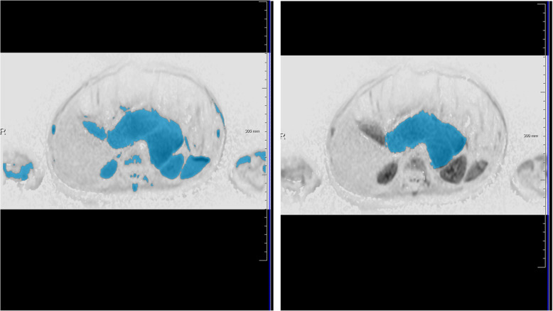
Pediatric patients who underwent surgical resection of neuroblastic tumors at our institution from 2013 to 2019 and who received a preoperative MRI scan with DWI after chemotherapy were included. Tumor volume was assessed with a semi-automated approach in DWI using a dedicated software prototype. Quantitative ADC values were calculated automatically of the total tumor volume after manual exclusion of necrosis. Manual segmentation in T1 weighted and T2 weighted sequences was used as reference standard for tumor volume comparison. The Student's t test was used for parametric data while the Wilcoxon rank sum test and the Kruskal-Wallis test were applied for non-parametric data.

RESULTS

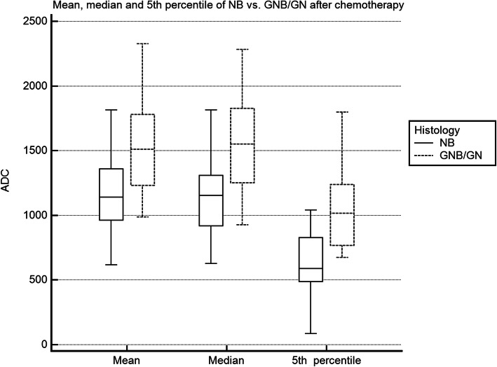
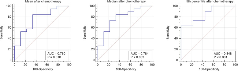
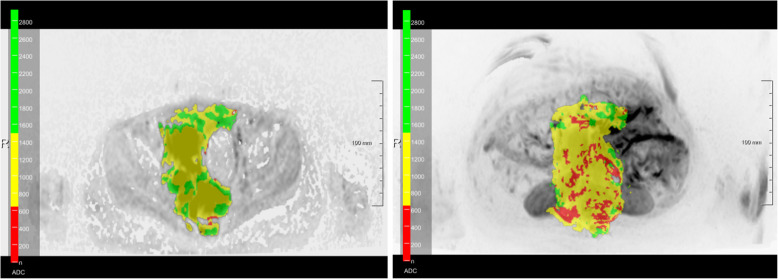
Twenty seven patients with 28 lesions (neuroblastoma (NB): n = 19, ganglioneuroblastoma (GNB): n = 7, ganglioneuroma (GN): n = 2) could be evaluated. Mean patient age was 4.5 ± 3.2 years. Median volume of standard volumetry (T1w or T2w) was 50.2 ml (interquartile range (IQR): 91.9 ml) vs. 45.1 ml (IQR: 98.4 ml) of DWI (p = 0.145). Mean ADC values (× 10 mm/s) of the total tumor volume (without necrosis) were 1187 ± 301 in NB vs. 1552 ± 114 in GNB/GN (p = 0.037). The 5th percentile of ADC values of NB (614 ± 275) and GNB/GN (1053 ± 362) provided the most significant difference (p = 0.007) with an area under the curve of 0.848 (p < 0.001).

CONCLUSIONS

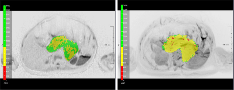
Quantitative semi-automated DWI volumetry is feasible in neuroblastic tumors with integrated analysis of tissue characteristics by providing automatically calculated ADC values of the whole tumor as well as an ADC heatmap. The 5th percentile of the ADC values of the whole tumor volume proved to be the most significant parameter for differentiation of the histopathological subtypes in our patient cohort and further investigation seems to be worthwhile.

摘要

背景

评估新辅助化疗后表观扩散系数(ADC)图评估的半自动化扩散加权成像(DWI)全神经母细胞瘤容积测量的可行性和可能价值。

方法

本研究纳入了 2013 年至 2019 年在我院行手术切除神经母细胞瘤的儿科患者,并在化疗前行 MRI 扫描,包括 DWI。使用专用软件原型在 DWI 中采用半自动方法评估肿瘤体积。在手动排除坏死后,自动计算总肿瘤体积的定量 ADC 值。T1 加权和 T2 加权序列的手动分割被用作肿瘤体积比较的参考标准。参数数据采用学生 t 检验,非参数数据采用 Wilcoxon 秩和检验和 Kruskal-Wallis 检验。

结果

共 27 例 28 处病变(神经母细胞瘤(NB):n=19,神经节母细胞瘤(GNB):n=7,神经节细胞瘤(GN):n=2)可进行评估。患者平均年龄为 4.5±3.2 岁。标准容积测量(T1w 或 T2w)的中位数体积为 50.2ml(四分位距(IQR):91.9ml),而 DWI 为 45.1ml(IQR:98.4ml)(p=0.145)。总肿瘤体积(无坏死)的平均 ADC 值(×10mm/s)在 NB 中为 1187±301,在 GNB/GN 中为 1552±114(p=0.037)。NB(614±275)和 GNB/GN(1053±362)的 ADC 值的第 5 百分位数差异最大(p=0.007),曲线下面积为 0.848(p<0.001)。

结论

半自动化 DWI 容积测量在神经母细胞瘤中是可行的,通过对整个肿瘤进行组织特征的综合分析,可以提供自动计算的 ADC 值以及 ADC 热图。在我们的患者队列中,整个肿瘤体积 ADC 值的第 5 百分位数被证明是区分组织学亚型的最显著参数,进一步的研究似乎是值得的。

https://cdn.ncbi.nlm.nih.gov/pmc/blobs/37b2/7745476/9d6616f87ceb/40644_2020_366_Fig6_HTML.jpg
https://cdn.ncbi.nlm.nih.gov/pmc/blobs/37b2/7745476/c6343a604006/40644_2020_366_Fig1_HTML.jpg
https://cdn.ncbi.nlm.nih.gov/pmc/blobs/37b2/7745476/735b8cb2ced7/40644_2020_366_Fig2_HTML.jpg
https://cdn.ncbi.nlm.nih.gov/pmc/blobs/37b2/7745476/ae706a5aaeca/40644_2020_366_Fig3_HTML.jpg
https://cdn.ncbi.nlm.nih.gov/pmc/blobs/37b2/7745476/4c140eaf3e2e/40644_2020_366_Fig4_HTML.jpg
https://cdn.ncbi.nlm.nih.gov/pmc/blobs/37b2/7745476/34dbb19767fc/40644_2020_366_Fig5_HTML.jpg
https://cdn.ncbi.nlm.nih.gov/pmc/blobs/37b2/7745476/9d6616f87ceb/40644_2020_366_Fig6_HTML.jpg

相似文献

1
Feasibility and possible value of quantitative semi-automated diffusion weighted imaging volumetry of neuroblastic tumors.神经母细胞瘤定量半自动化弥散加权成像容积测量的可行性和可能价值。
Cancer Imaging. 2020 Dec 17;20(1):89. doi: 10.1186/s40644-020-00366-3.
2
Diagnostic Value of Diffusion-Weighted MRI for Tumor Characterization, Differentiation and Monitoring in Pediatric Patients with Neuroblastic Tumors.扩散加权磁共振成像对小儿神经母细胞瘤肿瘤特征、鉴别及监测的诊断价值
Rofo. 2017 Jul;189(7):640-650. doi: 10.1055/s-0043-108993. Epub 2017 May 16.
3
Role of diffusion-weighted imaging in distinguishing thoracoabdominal neuroblastic tumours of various histological types and differentiation grades.扩散加权成像在鉴别不同组织学类型和分化程度的胸腹部神经母细胞瘤中的作用
J Med Imaging Radiat Oncol. 2017 Dec;61(6):718-724. doi: 10.1111/1754-9485.12615. Epub 2017 May 16.
4
Diffusion weighted imaging in differentiating malignant and benign neuroblastic tumors.扩散加权成像在鉴别恶性和良性神经母细胞瘤中的应用
Jpn J Radiol. 2016 Sep;34(9):620-4. doi: 10.1007/s11604-016-0565-z. Epub 2016 Jul 14.
5
Diffusion-weighted MRI for differentiation of neuroblastoma and ganglioneuroblastoma/ganglioneuroma.弥散加权 MRI 鉴别神经母细胞瘤与神经节母细胞瘤/神经节细胞瘤。
Eur J Radiol. 2011 Sep;79(3):443-6. doi: 10.1016/j.ejrad.2010.04.005. Epub 2010 May 13.
6
Retrospective accuracy analysis of MRI based lesion size measurement in neuroblastic tumors: which sequence should we choose?基于 MRI 的神经母细胞瘤病灶大小测量的回顾性准确性分析:我们应该选择哪种序列?
BMC Med Imaging. 2020 Sep 10;20(1):105. doi: 10.1186/s12880-020-00503-1.
7
Quantitative DWI predicts event-free survival in children with neuroblastic tumours: preliminary findings from a retrospective cohort study.定量扩散加权成像预测神经母细胞瘤患儿的无事件生存期:一项回顾性队列研究的初步结果
Eur Radiol Exp. 2019 Jan 30;3(1):6. doi: 10.1186/s41747-019-0087-4.
8
Diffusion-weighted imaging (DWI) with apparent diffusion coefficient (ADC) mapping as a quantitative imaging biomarker for prediction of immunohistochemical receptor status, proliferation rate, and molecular subtypes of breast cancer.弥散加权成像(DWI)及其表观扩散系数(ADC)图作为一种定量影像学生物标志物,可预测乳腺癌的免疫组织化学受体状态、增殖率和分子亚型。
J Magn Reson Imaging. 2019 Sep;50(3):836-846. doi: 10.1002/jmri.26697. Epub 2019 Feb 27.
9
Feasibility study of reduced field of view diffusion-weighted magnetic resonance imaging in head and neck tumors.头颈肿瘤中缩小视野扩散加权磁共振成像的可行性研究
Acta Radiol. 2017 Mar;58(3):292-300. doi: 10.1177/0284185116652014. Epub 2016 Jul 20.
10
Whole-tumor radiomics analysis of T2-weighted imaging in differentiating neuroblastoma from ganglioneuroblastoma/ganglioneuroma in children: an exploratory study.儿童T2加权成像全肿瘤放射组学分析在鉴别神经母细胞瘤与神经节神经母细胞瘤/神经节神经瘤中的应用:一项探索性研究
Abdom Radiol (NY). 2023 Apr;48(4):1372-1382. doi: 10.1007/s00261-023-03862-9. Epub 2023 Mar 9.

引用本文的文献

1
White paper on the current state of imaging biomarkers in paediatric oncology.儿科肿瘤学中影像生物标志物现状白皮书。
Pediatr Radiol. 2025 Sep 2. doi: 10.1007/s00247-025-06372-6.
2
Local MRI before and after Tumor Resection in Neuroblastoma: Impact of Residual Disease on Event Free Survival.神经母细胞瘤肿瘤切除前后的局部磁共振成像:残留疾病对无事件生存期的影响
J Clin Med. 2023 Nov 24;12(23):7297. doi: 10.3390/jcm12237297.
3
Combined Metabolic and Functional Tumor Volumes on [F]FDG-PET/MRI in Neuroblastoma Using Voxel-Wise Analysis.

本文引用的文献

1
Quantitative DWI predicts event-free survival in children with neuroblastic tumours: preliminary findings from a retrospective cohort study.定量扩散加权成像预测神经母细胞瘤患儿的无事件生存期:一项回顾性队列研究的初步结果
Eur Radiol Exp. 2019 Jan 30;3(1):6. doi: 10.1186/s41747-019-0087-4.
2
A review of neuroblastoma image-defined risk factors on magnetic resonance imaging.磁共振成像上神经母细胞瘤图像定义风险因素的综述。
Pediatr Radiol. 2018 Aug;48(9):1337-1347. doi: 10.1007/s00247-018-4117-9. Epub 2018 Aug 4.
3
Staging and following common pediatric malignancies: MRI versus CT versus functional imaging.
使用体素分析在神经母细胞瘤中通过[F]FDG-PET/MRI测量联合代谢和功能肿瘤体积
J Clin Med. 2023 Sep 15;12(18):5976. doi: 10.3390/jcm12185976.
4
Imaging biomarkers and radiomics in pediatric oncology: a view from the PRIMAGE (PRedictive In silico Multiscale Analytics to support cancer personalized diaGnosis and prognosis, Empowered by imaging biomarkers) project.儿科肿瘤学中的影像生物标志物和放射组学:来自 PRIMAGE(基于预测性计算多尺度分析以支持癌症个体化诊断和预后,由影像生物标志物赋能)项目的观点。
Pediatr Radiol. 2024 Apr;54(4):562-570. doi: 10.1007/s00247-023-05770-y. Epub 2023 Sep 25.
5
Independent Validation of a Deep Learning nnU-Net Tool for Neuroblastoma Detection and Segmentation in MR Images.用于磁共振图像中神经母细胞瘤检测与分割的深度学习nnU-Net工具的独立验证
Cancers (Basel). 2023 Mar 6;15(5):1622. doi: 10.3390/cancers15051622.
6
Whole-tumor radiomics analysis of T2-weighted imaging in differentiating neuroblastoma from ganglioneuroblastoma/ganglioneuroma in children: an exploratory study.儿童T2加权成像全肿瘤放射组学分析在鉴别神经母细胞瘤与神经节神经母细胞瘤/神经节神经瘤中的应用:一项探索性研究
Abdom Radiol (NY). 2023 Apr;48(4):1372-1382. doi: 10.1007/s00261-023-03862-9. Epub 2023 Mar 9.
7
Value of quantitative apparent diffusion coefficients in differentiating low-grade gliomas from mixed neuronal-glial tumors.定量表观扩散系数在鉴别低级别胶质瘤与混合性神经元-胶质肿瘤中的价值。
World Neurosurg X. 2023 Jan 28;18:100159. doi: 10.1016/j.wnsx.2023.100159. eCollection 2023 Apr.
8
Comparative Multicentric Evaluation of Inter-Observer Variability in Manual and Automatic Segmentation of Neuroblastic Tumors in Magnetic Resonance Images.磁共振图像中神经母细胞瘤手动与自动分割的观察者间变异性的多中心比较评估
Cancers (Basel). 2022 Jul 27;14(15):3648. doi: 10.3390/cancers14153648.
9
Two-cavities approach for resection of pediatric abdominal neuroblastic tumors: experience of a national reference pediatric onco-surgical center.双腔入路切除小儿腹部神经母细胞瘤:国家小儿肿瘤外科参考中心的经验。
J Cancer Res Clin Oncol. 2023 Apr;149(4):1485-1493. doi: 10.1007/s00432-022-04027-9. Epub 2022 May 6.
10
[Radiological imaging of neuroblastoma].[神经母细胞瘤的放射影像学]
Radiologe. 2021 Jul;61(7):639-648. doi: 10.1007/s00117-021-00875-7. Epub 2021 Jun 22.
儿童常见恶性肿瘤的分期与随访:磁共振成像(MRI)、计算机断层扫描(CT)与功能成像的比较
Pediatr Radiol. 2018 Aug;48(9):1324-1336. doi: 10.1007/s00247-018-4162-4. Epub 2018 Aug 4.
4
Guidelines on nuclear medicine imaging in neuroblastoma.神经母细胞瘤核医学影像学指南。
Eur J Nucl Med Mol Imaging. 2018 Oct;45(11):2009-2024. doi: 10.1007/s00259-018-4070-8.
5
Updates in Diagnosis, Management, and Treatment of Neuroblastoma.神经母细胞瘤的诊断、治疗和管理的新进展。
Radiographics. 2018 Mar-Apr;38(2):566-580. doi: 10.1148/rg.2018170132.
6
Diffusion-weighted MRI for differentiating Wilms tumor from neuroblastoma.用于鉴别肾母细胞瘤与神经母细胞瘤的扩散加权磁共振成像
Diagn Interv Radiol. 2017 Sep-Oct;23(5):403-406. doi: 10.5152/dir.2017.16541.
7
Diagnostic Value of Diffusion-Weighted MRI for Tumor Characterization, Differentiation and Monitoring in Pediatric Patients with Neuroblastic Tumors.扩散加权磁共振成像对小儿神经母细胞瘤肿瘤特征、鉴别及监测的诊断价值
Rofo. 2017 Jul;189(7):640-650. doi: 10.1055/s-0043-108993. Epub 2017 May 16.
8
Image-defined risk factors in unresectable neuroblastoma: SIOPEN study on incidence, chemotherapy-induced variation, and impact on surgical outcomes.不可切除神经母细胞瘤的影像定义风险因素:国际小儿肿瘤学会(SIOPEN)关于发病率、化疗引起的变化及对手术结果影响的研究
Pediatr Blood Cancer. 2017 Nov;64(11). doi: 10.1002/pbc.26605. Epub 2017 Apr 25.
9
Risk Stratification of Pediatric Patients With Neuroblastoma Using Volumetric Parameters of 18F-FDG and 18F-DOPA PET/CT.使用 18F-FDG 和 18F-DOPA PET/CT 的容积参数对神经母细胞瘤患儿进行风险分层。
Clin Nucl Med. 2017 Mar;42(3):e142-e148. doi: 10.1097/RLU.0000000000001529.
10
Diffusion-weighted Imaging Using Readout-segmented EPI Reveals Bony Metastases from Neuroblastoma.使用读出分段回波平面成像的扩散加权成像揭示神经母细胞瘤的骨转移。
J Pediatr Hematol Oncol. 2016 Oct;38(7):e263-6. doi: 10.1097/MPH.0000000000000643.