Jensen Carina, Carl Jesper, Boesen Lars, Langkilde Niels Christian, Østergaard Lasse Riis
Department of Medical Physics, Oncology, Aalborg University Hospital, Aalborg, Denmark.
Department of Oncology, Naestved Sygehus, Zealand University Hospital, Roskilde, Denmark.
J Appl Clin Med Phys. 2019 Feb;20(2):146-153. doi: 10.1002/acm2.12542. Epub 2019 Feb 3.
To automatically assess the aggressiveness of prostate cancer (PCa) lesions using zonal-specific image features extracted from diffusion weighted imaging (DWI) and T2W MRI.
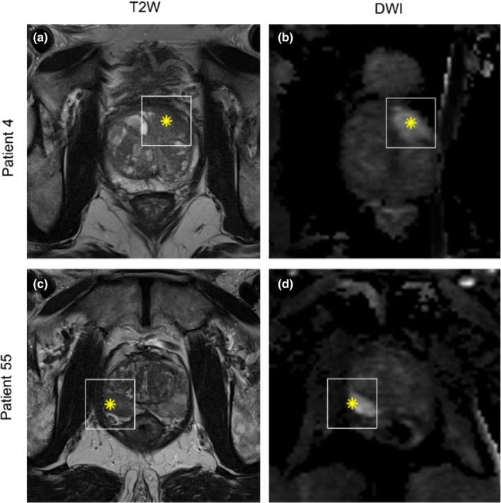
Region of interest was extracted from DWI (peripheral zone) and T2W MRI (transitional zone and anterior fibromuscular stroma) around the center of 112 PCa lesions from 99 patients. Image histogram and texture features, 38 in total, were used together with a k-nearest neighbor classifier to classify lesions into their respective prognostic Grade Group (GG) (proposed by the International Society of Urological Pathology 2014 consensus conference). A semi-exhaustive feature search was performed (1-6 features in each feature set) and validated using threefold stratified cross validation in a one-versus-rest classification setup.
Classifying PCa lesions into GGs resulted in AUC of 0.87, 0.88, 0.96, 0.98, and 0.91 for GG1, GG2, GG1 + 2, GG3, and GG4 + 5 for the peripheral zone, respectively. The results for transitional zone and anterior fibromuscular stroma were AUC of 0.85, 0.89, 0.83, 0.94, and 0.86 for GG1, GG2, GG1 + 2, GG3, and GG4 + 5, respectively.
This study showed promising results with reasonable AUC values for classification of all GG indicating that zonal-specific imaging features from DWI and T2W MRI can be used to differentiate between PCa lesions of various aggressiveness.
利用从扩散加权成像(DWI)和T2加权磁共振成像(T2W MRI)中提取的区域特异性图像特征,自动评估前列腺癌(PCa)病变的侵袭性。
从99例患者的112个PCa病变中心周围的DWI(外周带)和T2W MRI(移行带和前部纤维肌基质)中提取感兴趣区域。总共38个图像直方图和纹理特征与k近邻分类器一起用于将病变分类到各自的预后分级组(GG)(由国际泌尿病理学会2014年共识会议提出)。进行了半穷举特征搜索(每个特征集1 - 6个特征),并在一对其余分类设置中使用三倍分层交叉验证进行验证。
将PCa病变分类到GG中,外周带GG1、GG2、GG1 + 2、GG3和GG4 + 5的曲线下面积(AUC)分别为0.87、0.88、0.96、0.98和0.91。移行带和前部纤维肌基质的结果是,GG1、GG2、GG1 + 2、GG3和GG4 + 5的AUC分别为0.85、0.89、0.83、0.94和0.86。
本研究显示出有前景的结果,所有GG分类的AUC值合理,表明来自DWI和T2W MRI的区域特异性成像特征可用于区分不同侵袭性的PCa病变。