• 文献检索
  • 文档翻译
  • 深度研究
  • 学术资讯
  • Suppr Zotero 插件Zotero 插件
  • 邀请有礼
  • 套餐&价格
  • 历史记录
应用&插件
Suppr Zotero 插件Zotero 插件浏览器插件Mac 客户端Windows 客户端微信小程序
定价
高级版会员购买积分包购买API积分包
服务
文献检索文档翻译深度研究API 文档MCP 服务
关于我们
关于 Suppr公司介绍联系我们用户协议隐私条款
关注我们

Suppr 超能文献

核心技术专利:CN118964589B侵权必究
粤ICP备2023148730 号-1Suppr @ 2026

文献检索

告别复杂PubMed语法,用中文像聊天一样搜索,搜遍4000万医学文献。AI智能推荐,让科研检索更轻松。

立即免费搜索

文件翻译

保留排版,准确专业,支持PDF/Word/PPT等文件格式,支持 12+语言互译。

免费翻译文档

深度研究

AI帮你快速写综述,25分钟生成高质量综述,智能提取关键信息,辅助科研写作。

立即免费体验

T 加权 MRI 纹理分析在评估外周区前列腺癌侵袭性中的应用:一项单臂、多中心研究。

Utility of T-weighted MRI texture analysis in assessment of peripheral zone prostate cancer aggressiveness: a single-arm, multicenter study.

机构信息

Department of Circulation and Medical Imaging, NTNU-Norwegian University of Science and Technology, Trondheim, Norway.

Department of Radiology and Nuclear Medicine, St. Olavs Hospital, Trondheim University Hospital, Trondheim, Norway.

出版信息

Sci Rep. 2021 Jan 22;11(1):2085. doi: 10.1038/s41598-021-81272-x.

DOI:10.1038/s41598-021-81272-x
PMID:33483545
原文链接:https://pmc.ncbi.nlm.nih.gov/articles/PMC7822867/
Abstract

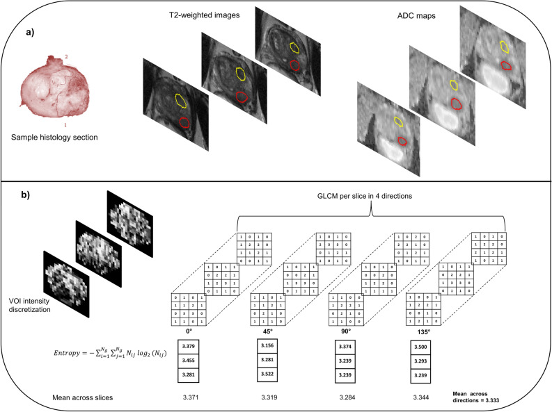
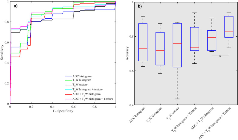
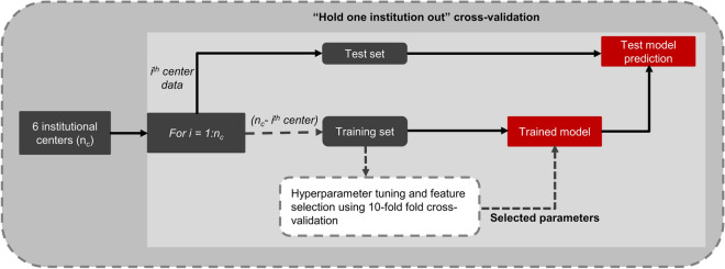
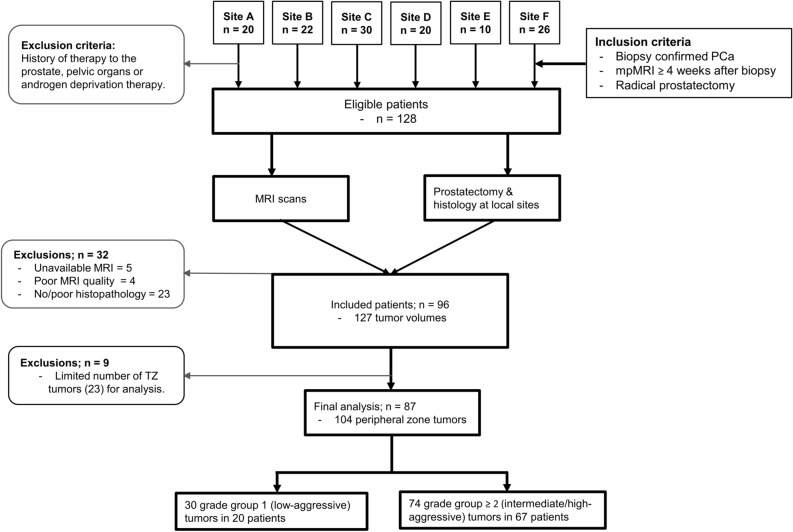
T-weighted (TW) MRI provides high spatial resolution and tissue-specific contrast, but it is predominantly used for qualitative evaluation of prostate anatomy and anomalies. This retrospective multicenter study evaluated the potential of TW image-derived textural features for quantitative assessment of peripheral zone prostate cancer (PCa) aggressiveness. A standardized preoperative multiparametric MRI was performed on 87 PCa patients across 6 institutions. TW intensity and apparent diffusion coefficient (ADC) histogram, and TW textural features were computed from tumor volumes annotated based on whole-mount histology. Spearman correlations were used to evaluate association between textural features and PCa grade groups (i.e. 1-5). Feature utility in differentiating and classifying low-(grade group 1) vs. intermediate/high-(grade group ≥ 2) aggressive cancers was evaluated using Mann-Whitney U-tests, and a support vector machine classifier employing "hold-one-institution-out" cross-validation scheme, respectively. Textural features indicating image homogeneity and disorder/complexity correlated significantly (p < 0.05) with PCa grade groups. In the intermediate/high-aggressive cancers, textural homogeneity and disorder/complexity were significantly lower and higher, respectively, compared to the low-aggressive cancers. The mean classification accuracy across the centers was highest for the combined ADC and TW intensity-textural features (84%) compared to ADC histogram (75%), TW histogram (72%), TW textural (72%) features alone or TW histogram and texture (77%), TW and ADC histogram (79%) combined. Texture analysis of TW images provides quantitative information or features that are associated with peripheral zone PCa aggressiveness and can augment their classification.

摘要

T 加权(TW)MRI 提供了高空间分辨率和组织特异性对比,但主要用于前列腺解剖结构和异常的定性评估。本回顾性多中心研究评估了 TW 图像衍生纹理特征在定量评估外周带前列腺癌(PCa)侵袭性方面的潜力。在 6 家机构对 87 例 PCa 患者进行了标准化的术前多参数 MRI 检查。从基于全组织学的肿瘤体积标注中计算了 TW 强度和表观扩散系数(ADC)直方图以及 TW 纹理特征。使用 Spearman 相关分析评估纹理特征与 PCa 分级组(即 1-5 级)之间的相关性。使用 Mann-Whitney U 检验评估了纹理特征在区分和分类低(分级组 1)与中/高(分级组≥2)侵袭性癌症方面的作用,并使用支持向量机分类器,采用“一机构外留一机构”交叉验证方案。指示图像均匀性和无序/复杂性的纹理特征与 PCa 分级组显著相关(p<0.05)。在中/高侵袭性癌症中,与低侵袭性癌症相比,纹理均匀性和无序/复杂性分别显著降低和升高。跨中心的平均分类准确率以 ADC 和 TW 强度纹理特征的组合最高(84%),其次是 ADC 直方图(75%)、TW 直方图(72%)、TW 纹理(72%)特征单独或 TW 直方图和纹理(77%)、TW 和 ADC 直方图(79%)组合。TW 图像的纹理分析提供了与外周带 PCa 侵袭性相关的定量信息或特征,可增强其分类。

https://cdn.ncbi.nlm.nih.gov/pmc/blobs/c8f4/7822867/79c63c1d9d28/41598_2021_81272_Fig5_HTML.jpg
https://cdn.ncbi.nlm.nih.gov/pmc/blobs/c8f4/7822867/a810c6ae1a0f/41598_2021_81272_Fig1_HTML.jpg
https://cdn.ncbi.nlm.nih.gov/pmc/blobs/c8f4/7822867/7aef4f349bfe/41598_2021_81272_Fig2_HTML.jpg
https://cdn.ncbi.nlm.nih.gov/pmc/blobs/c8f4/7822867/0afa5d2fdb7d/41598_2021_81272_Fig3_HTML.jpg
https://cdn.ncbi.nlm.nih.gov/pmc/blobs/c8f4/7822867/fa7931124a13/41598_2021_81272_Fig4_HTML.jpg
https://cdn.ncbi.nlm.nih.gov/pmc/blobs/c8f4/7822867/79c63c1d9d28/41598_2021_81272_Fig5_HTML.jpg

相似文献

1
Utility of T-weighted MRI texture analysis in assessment of peripheral zone prostate cancer aggressiveness: a single-arm, multicenter study.T 加权 MRI 纹理分析在评估外周区前列腺癌侵袭性中的应用:一项单臂、多中心研究。
Sci Rep. 2021 Jan 22;11(1):2085. doi: 10.1038/s41598-021-81272-x.
2
T2-weighted MRI-derived textural features reflect prostate cancer aggressiveness: preliminary results.基于T2加权磁共振成像的纹理特征反映前列腺癌侵袭性:初步结果。
Eur Radiol. 2017 Jul;27(7):3050-3059. doi: 10.1007/s00330-016-4663-1. Epub 2016 Dec 14.
3
Transition zone prostate cancer: Logistic regression and machine-learning models of quantitative ADC, shape and texture features are highly accurate for diagnosis.移行区前列腺癌:定量 ADC、形态和纹理特征的逻辑回归和机器学习模型对诊断具有高度准确性。
J Magn Reson Imaging. 2019 Sep;50(3):940-950. doi: 10.1002/jmri.26674. Epub 2019 Jan 30.
4
Fully automated detection of prostate transition zone tumors on T2-weighted and apparent diffusion coefficient (ADC) map MR images using U-Net ensemble.基于 U-Net 集成的 T2 加权和表观扩散系数 (ADC) 图磁共振成像上前列腺移行区肿瘤的全自动检测。
Med Phys. 2021 Nov;48(11):6889-6900. doi: 10.1002/mp.15181. Epub 2021 Aug 30.
5
Comparison of Histogram-based Textural Features between Cancerous and Normal Prostatic Tissue in Multiparametric Magnetic Resonance Images.多参数磁共振图像中癌性与正常前列腺组织基于直方图的纹理特征比较
Annu Int Conf IEEE Eng Med Biol Soc. 2020 Jul;2020:1671-1674. doi: 10.1109/EMBC44109.2020.9176307.
6
Central gland and peripheral zone prostate tumors have significantly different quantitative imaging signatures on 3 Tesla endorectal, in vivo T2-weighted MR imagery.在 3.0T 直肠内活体 T2 加权磁共振成像中,中央腺体和周围区域前列腺肿瘤具有明显不同的定量成像特征。
J Magn Reson Imaging. 2012 Jul;36(1):213-24. doi: 10.1002/jmri.23618. Epub 2012 Feb 15.
7
Utility of machine learning of apparent diffusion coefficient (ADC) and T2-weighted (T2W) radiomic features in PI-RADS version 2.1 category 3 lesions to predict prostate cancer diagnosis.基于表观扩散系数(ADC)和 T2 加权(T2W)影像组学特征的机器学习在 PI-RADS 版本 2.1 分类 3 病变中预测前列腺癌诊断的效用。
Abdom Radiol (NY). 2021 Dec;46(12):5647-5658. doi: 10.1007/s00261-021-03235-0. Epub 2021 Aug 31.
8
Differentiating prostate cancer from benign prostatic hyperplasia using whole-lesion histogram and texture analysis of diffusion- and T2-weighted imaging.利用弥散加权和 T2 加权成像的全病变直方图和纹理分析鉴别前列腺癌与良性前列腺增生。
Cancer Imaging. 2021 Sep 27;21(1):54. doi: 10.1186/s40644-021-00423-5.
9
Impact of androgen deprivation therapy on apparent diffusion coefficient and T2w MRI for histogram and texture analysis with respect to focal radiotherapy of prostate cancer.雄激素剥夺治疗对前列腺癌局部放射治疗的表观扩散系数和 T2wMRI 直方图和纹理分析的影响。
Strahlenther Onkol. 2019 May;195(5):402-411. doi: 10.1007/s00066-018-1402-3. Epub 2018 Nov 26.
10
Automatic stratification of prostate tumour aggressiveness using multiparametric MRI: a horizontal comparison of texture features.基于多参数 MRI 的前列腺肿瘤侵袭性自动分层:纹理特征的横向比较。
Acta Oncol. 2019 Aug;58(8):1118-1126. doi: 10.1080/0284186X.2019.1598576. Epub 2019 Apr 17.

引用本文的文献

1
The use of ADC histogram analysis in the diagnosis and determination of aggressiveness of peripheral zone prostate cancer.表观扩散系数(ADC)直方图分析在周围区前列腺癌诊断及侵袭性判定中的应用
Pol J Radiol. 2025 Jul 28;90:e374-e383. doi: 10.5114/pjr/205459. eCollection 2025.
2
The Role of Radiomic Analysis and Different Machine Learning Models in Prostate Cancer Diagnosis.放射组学分析及不同机器学习模型在前列腺癌诊断中的作用
J Imaging. 2025 Jul 23;11(8):250. doi: 10.3390/jimaging11080250.
3
In vivo variability of MRI radiomics features in prostate lesions assessed by a test-retest study with repositioning.

本文引用的文献

1
Automated reference tissue normalization of T2-weighted MR images of the prostate using object recognition.利用目标识别实现前列腺 T2 加权磁共振图像的自动参考组织标准化。
MAGMA. 2021 Apr;34(2):309-321. doi: 10.1007/s10334-020-00871-3. Epub 2020 Jul 31.
2
The Image Biomarker Standardization Initiative: Standardized Quantitative Radiomics for High-Throughput Image-based Phenotyping.影像生物标志物标准化倡议:高通量基于影像表型的标准化定量放射组学。
Radiology. 2020 May;295(2):328-338. doi: 10.1148/radiol.2020191145. Epub 2020 Mar 10.
3
Radiomics and machine learning of multisequence multiparametric prostate MRI: Towards improved non-invasive prostate cancer characterization.
通过重新定位的重测研究评估前列腺病变中MRI影像组学特征的体内变异性。
Sci Rep. 2025 Aug 13;15(1):29703. doi: 10.1038/s41598-025-09989-7.
4
Combining multi-parametric MRI radiomics features with tumor abnormal protein to construct a machine learning-based predictive model for prostate cancer.结合多参数MRI影像组学特征与肿瘤异常蛋白构建基于机器学习的前列腺癌预测模型。
Sci Rep. 2025 Jul 2;15(1):22816. doi: 10.1038/s41598-025-05718-2.
5
Evaluating the feasibility of AI-predicted bpMRI image features for predicting prostate cancer aggressiveness: a multi-center study.评估人工智能预测的bpMRI图像特征用于预测前列腺癌侵袭性的可行性:一项多中心研究。
Insights Imaging. 2025 Jan 15;16(1):20. doi: 10.1186/s13244-024-01865-8.
6
Computational analysis of variability and uncertainty in the clinical reference on magnetic resonance imaging radiomics: modelling and performance.磁共振成像放射组学临床参考中变异性和不确定性的计算分析:建模与性能
Vis Comput Ind Biomed Art. 2024 Nov 19;7(1):28. doi: 10.1186/s42492-024-00180-9.
7
Biparametric MRI of the prostate radiomics model for prediction of pelvic lymph node metastasis in prostate cancers : a two-centre study.前列腺多参数 MRI 放射组学模型预测前列腺癌盆腔淋巴结转移:一项双中心研究。
BMC Med Imaging. 2024 Jul 25;24(1):185. doi: 10.1186/s12880-024-01372-8.
8
Evaluation of the Reliability and the Performance of Magnetic Resonance Imaging Radiomics in the Presence of Randomly Generated Irrelevant Features for Prostate Cancer.在存在随机生成的无关特征情况下对前列腺癌磁共振成像放射组学的可靠性和性能评估
Diagnostics (Basel). 2023 Dec 1;13(23):3580. doi: 10.3390/diagnostics13233580.
9
Development trends and knowledge framework in the application of magnetic resonance imaging in prostate cancer: a bibliometric analysis from 1984 to 2022.磁共振成像在前列腺癌应用中的发展趋势与知识框架:1984年至2022年的文献计量分析
Quant Imaging Med Surg. 2023 Oct 1;13(10):6761-6777. doi: 10.21037/qims-23-446. Epub 2023 Sep 11.
10
The impact of pre-processing and disease characteristics on reproducibility of T2-weighted MRI radiomics features.预处理和疾病特征对 T2 加权 MRI 放射组学特征可重复性的影响。
MAGMA. 2023 Dec;36(6):945-956. doi: 10.1007/s10334-023-01112-z. Epub 2023 Aug 9.
多序列多参数前列腺 MRI 的放射组学和机器学习:实现对前列腺癌的无创性特征分析。
PLoS One. 2019 Jul 8;14(7):e0217702. doi: 10.1371/journal.pone.0217702. eCollection 2019.
4
Repeatability of Multiparametric Prostate MRI Radiomics Features.多参数前列腺 MRI 放射组学特征的可重复性。
Sci Rep. 2019 Jul 1;9(1):9441. doi: 10.1038/s41598-019-45766-z.
5
Multisite evaluation of radiomic feature reproducibility and discriminability for identifying peripheral zone prostate tumors on MRI.用于在MRI上识别外周带前列腺肿瘤的放射组学特征可重复性和可辨别性的多中心评估。
J Med Imaging (Bellingham). 2019 Apr;6(2):024502. doi: 10.1117/1.JMI.6.2.024502. Epub 2019 Jun 14.
6
Repeatability and reproducibility of MRI-based radiomic features in cervical cancer.基于 MRI 的宫颈癌放射组学特征的可重复性和可再现性。
Radiother Oncol. 2019 Jun;135:107-114. doi: 10.1016/j.radonc.2019.03.001. Epub 2019 Mar 19.
7
A Single-Arm, Multicenter Validation Study of Prostate Cancer Localization and Aggressiveness With a Quantitative Multiparametric Magnetic Resonance Imaging Approach.一种基于定量多参数磁共振成像的前列腺癌定位和侵袭性的单臂、多中心验证研究。
Invest Radiol. 2019 Jul;54(7):437-447. doi: 10.1097/RLI.0000000000000558.
8
Prostate Imaging Reporting and Data System Version 2.1: 2019 Update of Prostate Imaging Reporting and Data System Version 2.前列腺影像报告和数据系统第 2.1 版:前列腺影像报告和数据系统第 2 版 2019 年更新。
Eur Urol. 2019 Sep;76(3):340-351. doi: 10.1016/j.eururo.2019.02.033. Epub 2019 Mar 18.
9
Stability of radiomics features in apparent diffusion coefficient maps from a multi-centre test-retest trial.多中心复测试验中表观扩散系数图的放射组学特征的稳定性。
Sci Rep. 2019 Mar 18;9(1):4800. doi: 10.1038/s41598-019-41344-5.
10
Head-to-head Comparison of Transrectal Ultrasound-guided Prostate Biopsy Versus Multiparametric Prostate Resonance Imaging with Subsequent Magnetic Resonance-guided Biopsy in Biopsy-naïve Men with Elevated Prostate-specific Antigen: A Large Prospective Multicenter Clinical Study.经直肠超声引导前列腺活检与多参数前列腺磁共振成像引导活检在前列腺特异性抗原升高的初次活检男性中的头对头比较:一项大型前瞻性多中心临床研究。
Eur Urol. 2019 Apr;75(4):570-578. doi: 10.1016/j.eururo.2018.11.023. Epub 2018 Nov 23.