Department of Biomedical Engineering and Health Systems, KTH Royal Institute of Technology, Stockholm, Sweden.
Department of Clinical Sciences, Karolinska Institutet, Stockholm, Sweden.
Sci Rep. 2019 Feb 4;9(1):1375. doi: 10.1038/s41598-018-37714-0.
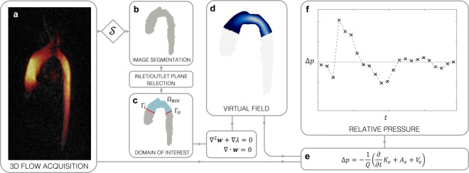
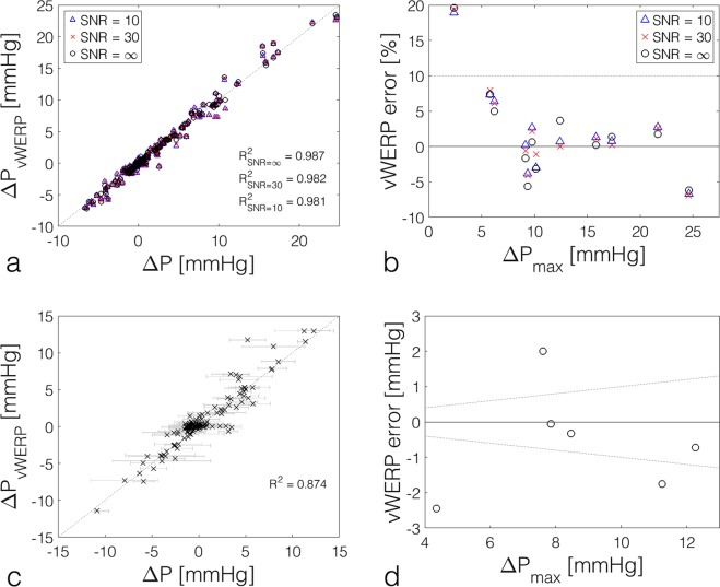
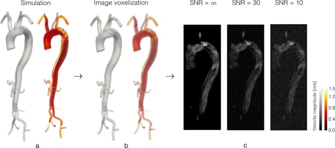
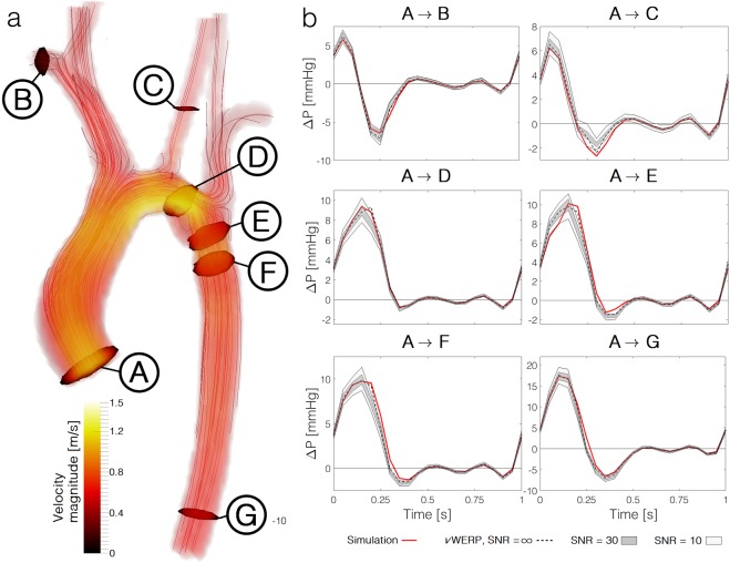
Many cardiovascular diseases lead to local increases in relative pressure, reflecting the higher costs of driving blood flow. The utility of this biomarker for stratifying the severity of disease has thus driven the development of methods to measure these relative pressures. While intravascular catheterisation remains the most direct measure, its invasiveness limits clinical application in many instances. Non-invasive Doppler ultrasound estimates have partially addressed this gap; however only provide relative pressure estimates for a range of constricted cardiovascular conditions. Here we introduce a non-invasive method that enables arbitrary interrogation of relative pressures throughout an imaged vascular structure, leveraging modern phase contrast magnetic resonance imaging, the virtual work-energy equations, and a virtual field to provide robust and accurate estimates. The versatility and accuracy of the method is verified in a set of complex patient-specific cardiovascular models, where relative pressures into previously inaccessible flow regions are assessed. The method is further validated within a cohort of congenital heart disease patients, providing a novel tool for probing relative pressures in-vivo.
许多心血管疾病会导致局部相对压力升高,这反映了驱动血流的成本更高。因此,这种生物标志物在疾病严重程度分层方面的实用性推动了测量这些相对压力的方法的发展。虽然血管内导管插入术仍然是最直接的测量方法,但由于其侵入性,在许多情况下限制了临床应用。非侵入性多普勒超声估计在一定程度上弥补了这一差距;然而,它只能为一系列心血管狭窄疾病提供相对压力估计。在这里,我们介绍了一种非侵入性方法,它可以利用现代相衬磁共振成像、虚拟功-能方程和虚拟场,在整个成像血管结构中任意询问相对压力,从而提供稳健和准确的估计。该方法在一组复杂的特定于患者的心血管模型中得到了验证,这些模型评估了先前无法进入的血流区域的相对压力。该方法还在一组先天性心脏病患者中进行了验证,为体内探测相对压力提供了一种新工具。