Kotrbová Anna, Štěpka Karel, Maška Martin, Pálenik Jakub Jozef, Ilkovics Ladislav, Klemová Dobromila, Kravec Marek, Hubatka František, Dave Zankruti, Hampl Aleš, Bryja Vítězslav, Matula Pavel, Pospíchalová Vendula
Department of Experimental Biology, Faculty of Science, Masaryk University, Brno, Czech Republic.
Centre for Biomedical Image Analysis, Faculty of Informatics, Masaryk University, Brno, Czech Republic.
J Extracell Vesicles. 2019 Jan 21;8(1):1560808. doi: 10.1080/20013078.2018.1560808. eCollection 2019.
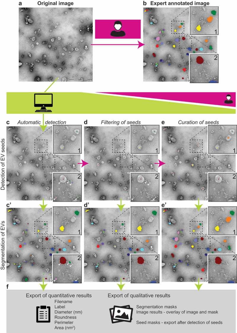
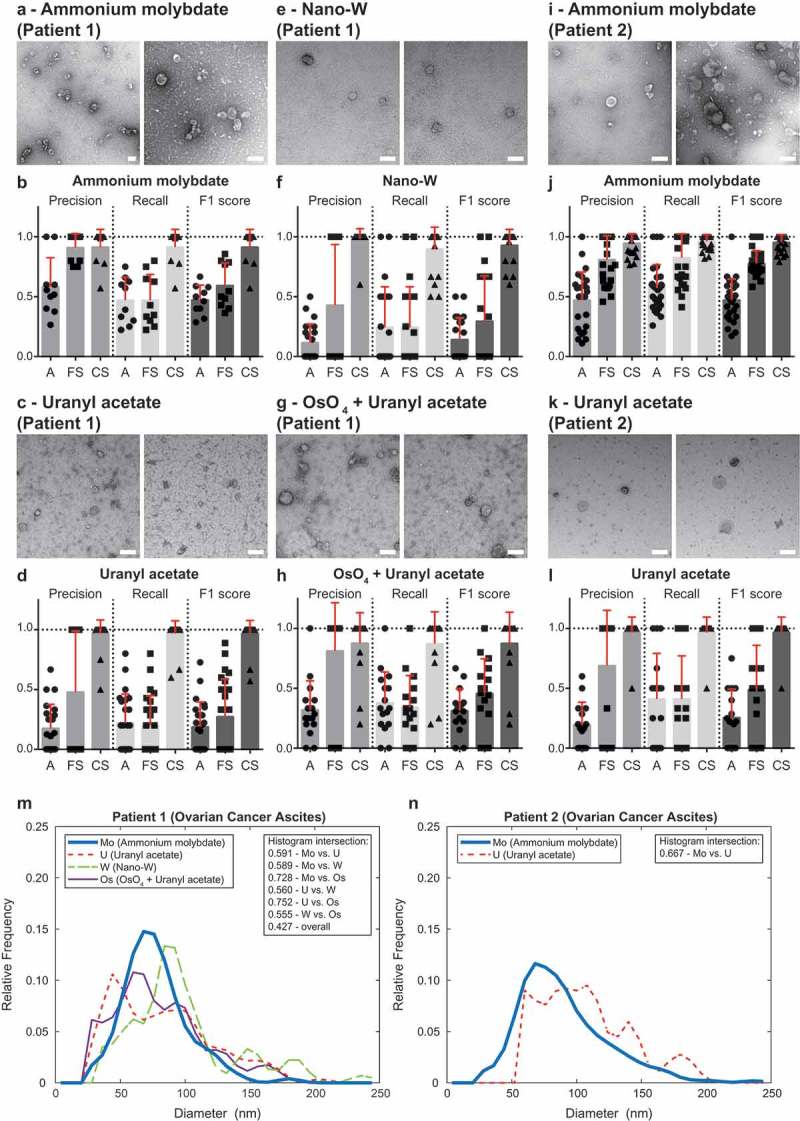
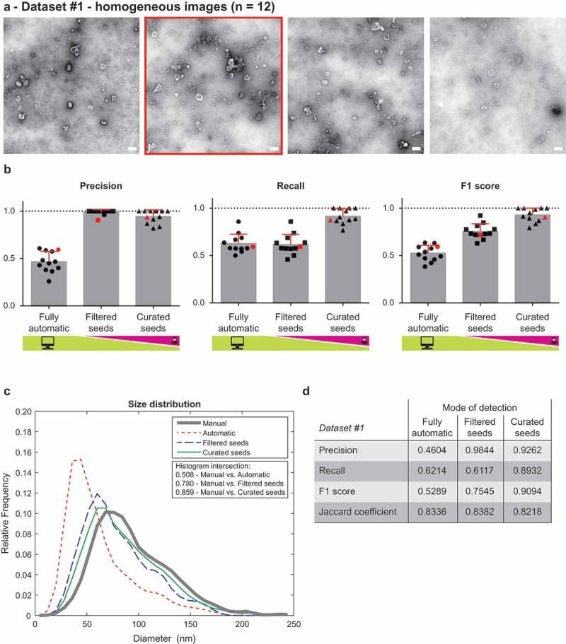
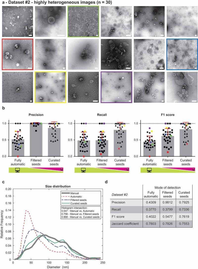
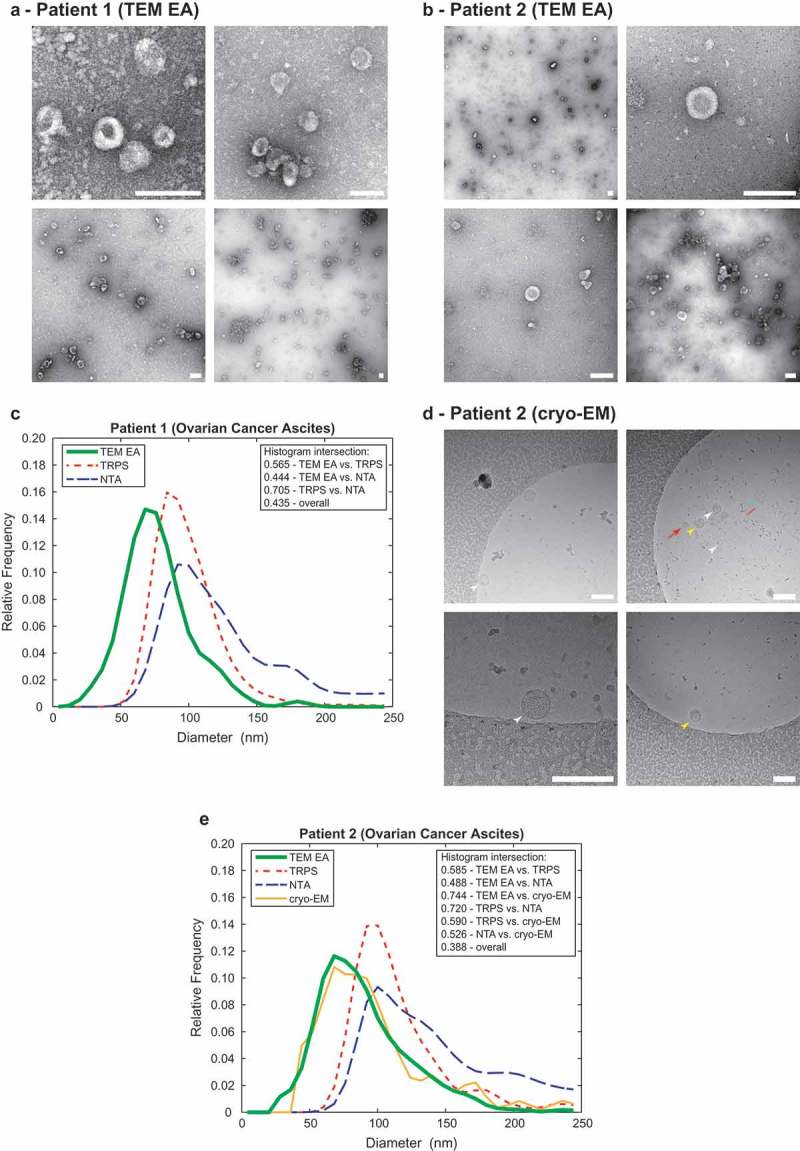
Extracellular vesicles (EVs) function as important conveyers of information between cells and thus can be exploited as drug delivery systems or disease biomarkers. Transmission electron microscopy (TEM) remains the gold standard method for visualisation of EVs, however the analysis of individual EVs in TEM images is time-consuming if performed manually. Therefore, we present here a software tool for computer-assisted evaluation of EVs in TEM images. TEM ExosomeAnalyzer detects EVs based on their shape and edge contrast criteria and subsequently analyses their size and roundness. The software tool is compatible with common negative staining protocols and isolation methods used in the field of EV research; even with challenging TEM images (EVs both lighter and darker than the background, images containing artefacts or precipitated stain, .). If the fully-automatic analysis fails to produce correct results, users can promptly adjust the detected seeds of EVs as well as their boundaries manually. The performance of our tool was evaluated for three different modes with variable levels of human interaction, using two datasets with various heterogeneity. The semi-automatic mode analyses EVs with high success rate in the homogenous dataset (F1 score 0.9094, Jaccard coefficient 0.8218) as well as in the highly heterogeneous dataset containing EVs isolated from cell culture medium and patient samples (F1 score 0.7619, Jaccard coefficient 0.7553). Moreover, the extracted size distribution profiles of EVs isolated from malignant ascites of ovarian cancer patients overlap with those derived by cryo-EM and are comparable to NTA- and TRPS-derived data. In summary, TEM ExosomeAnalyzer is an easy-to-use software tool for evaluation of many types of vesicular microparticles and is available at http://cbia.fi.muni.cz/exosome-analyzer free of charge for non-commercial and research purposes. The web page contains also detailed description how to use the software tool including a video tutorial.
细胞外囊泡(EVs)是细胞间重要的信息传递载体,因此可用作药物递送系统或疾病生物标志物。透射电子显微镜(TEM)仍是可视化EVs的金标准方法,然而,在TEM图像中手动分析单个EVs非常耗时。因此,我们在此展示一种用于计算机辅助评估TEM图像中EVs的软件工具。TEM外泌体分析仪根据EVs的形状和边缘对比度标准检测它们,随后分析其大小和圆度。该软件工具与EV研究领域常用的负染色方案和分离方法兼容;即使是具有挑战性的TEM图像(EVs比背景亮或暗、包含伪影或沉淀污渍的图像等)也适用。如果全自动分析未能产生正确结果,用户可以立即手动调整检测到的EVs种子及其边界。我们使用两个具有不同异质性的数据集,针对三种不同程度的人机交互模式评估了我们工具的性能。半自动模式在同质数据集中(F1分数为0.9094,杰卡德系数为0.8218)以及在包含从细胞培养基和患者样本中分离出的EVs的高度异质数据集中(F1分数为0.7619,杰卡德系数为0.7553)都能以高成功率分析EVs。此外,从卵巢癌患者恶性腹水中分离出的EVs的提取大小分布曲线与通过冷冻电镜获得的曲线重叠,并且与基于纳米颗粒跟踪分析(NTA)和可调电阻脉冲传感(TRPS)获得的数据相当。总之,TEM外泌体分析仪是一种易于使用的软件工具,可用于评估多种类型的囊泡微粒,可从http://cbia.fi.muni.cz/exosome-analyzer免费获取,供非商业和研究目的使用。该网页还包含如何使用该软件工具的详细说明,包括视频教程。