• 文献检索
  • 文档翻译
  • 深度研究
  • 学术资讯
  • Suppr Zotero 插件Zotero 插件
  • 邀请有礼
  • 套餐&价格
  • 历史记录
应用&插件
Suppr Zotero 插件Zotero 插件浏览器插件Mac 客户端Windows 客户端微信小程序
定价
高级版会员购买积分包购买API积分包
服务
文献检索文档翻译深度研究API 文档MCP 服务
关于我们
关于 Suppr公司介绍联系我们用户协议隐私条款
关注我们

Suppr 超能文献

核心技术专利:CN118964589B侵权必究
粤ICP备2023148730 号-1Suppr @ 2026

文献检索

告别复杂PubMed语法,用中文像聊天一样搜索,搜遍4000万医学文献。AI智能推荐,让科研检索更轻松。

立即免费搜索

文件翻译

保留排版,准确专业,支持PDF/Word/PPT等文件格式,支持 12+语言互译。

免费翻译文档

深度研究

AI帮你快速写综述,25分钟生成高质量综述,智能提取关键信息,辅助科研写作。

立即免费体验

基于相对脑血容量(rCBV)和表观扩散系数(ADC)的脑膜瘤分级及对新兴分子诊断的展望

rCBV- and ADC-based Grading of Meningiomas With Glimpse Into Emerging Molecular Diagnostics.

作者信息

Rohilla Seema, Garg Harender K, Singh Ishwar, Yadav Rohtas K, Dhaulakhandi Dhara B

机构信息

Department of Radiodiagnosis & Imaging, Post Graduate Institute of Medical Sciences, Sharma University of Health Sciences, Rohtak, Haryana, India.

Department of Neurosurgery, Post Graduate Institute of Medical Sciences, Sharma University of Health Sciences, Rohtak, Haryana, India.

出版信息

Basic Clin Neurosci. 2018 Nov-Dec;9(6):417-428. doi: 10.32598/bcn.9.6.417. Epub 2018 Nov 1.

DOI:10.32598/bcn.9.6.417
PMID:30719256
原文链接:https://pmc.ncbi.nlm.nih.gov/articles/PMC6359681/
Abstract

INTRODUCTION

This study was conducted to grade meningiomas based on relative Cerebral Blood Volume (rCBV) and Apparent Diffusion Coefficient (ADC) to help surgeons plan the approach and extent of operation as well as decide on the need of any adjuvant radio/chemo therapy. The current and evolving genomic, proteomic, and spectroscopic technologies are also discussed which can supplement the current radiologic methods and procedures in grading meningiomas.

METHODS

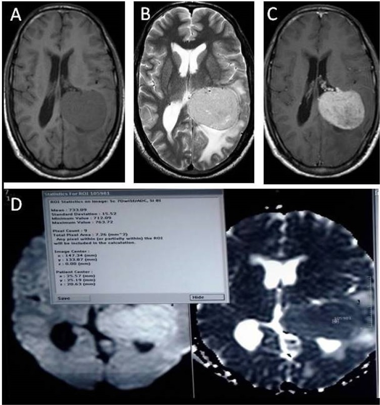
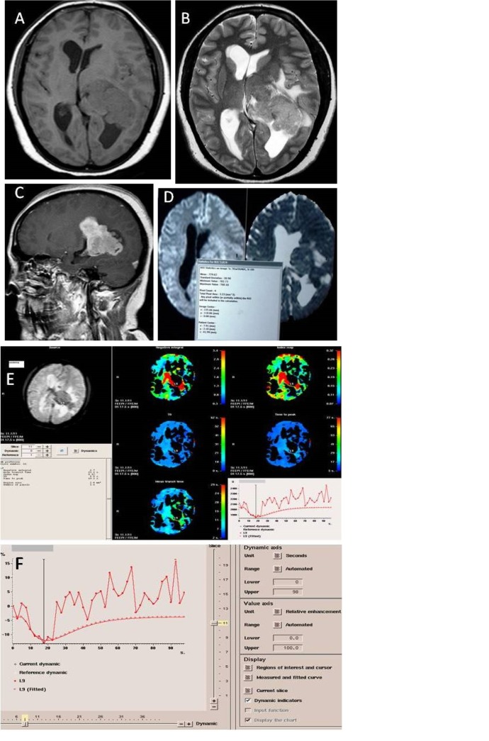
A total of 35 patients with meningioma prospectively underwent basic MR sequences (T1W, T2W, T2W/FLAIR) in axial, sagittal and coronal planes followed by Diffusion Weighted (DW) imaging having b value of 1000 (minimum ADC values used for analysis). Then, gadobenate dimeglumine/meglumine gadoterate was administered (0.1 mmol/kg at a rate of 4 mL/s) followed by saline flush (20 mL at a rate of 4 mL/s). Next, T*W/FFE dynamic images were acquired; dynamics showing maximum fall in intensity was used for creating rCBV and relative Cerebral Blood Flow (rCBF) maps and calculating rCBV.

RESULTS

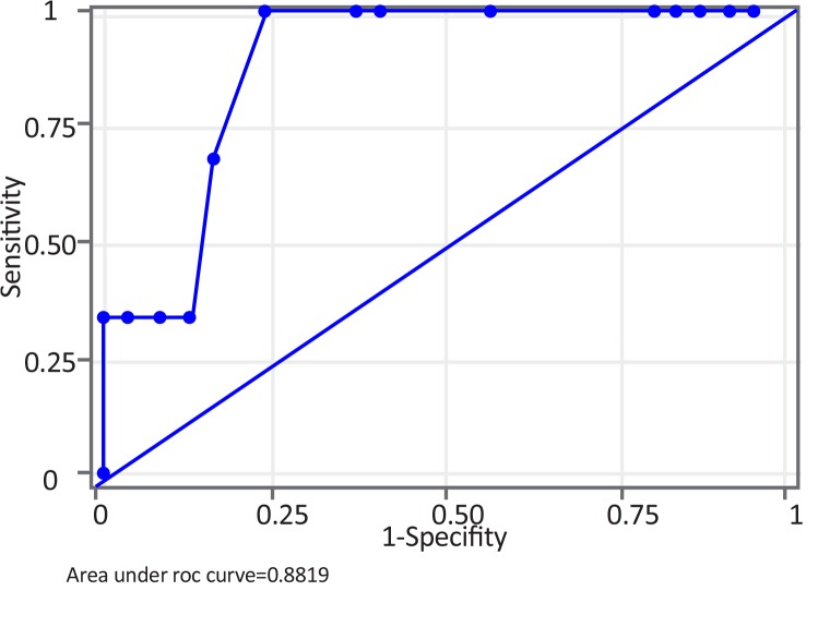
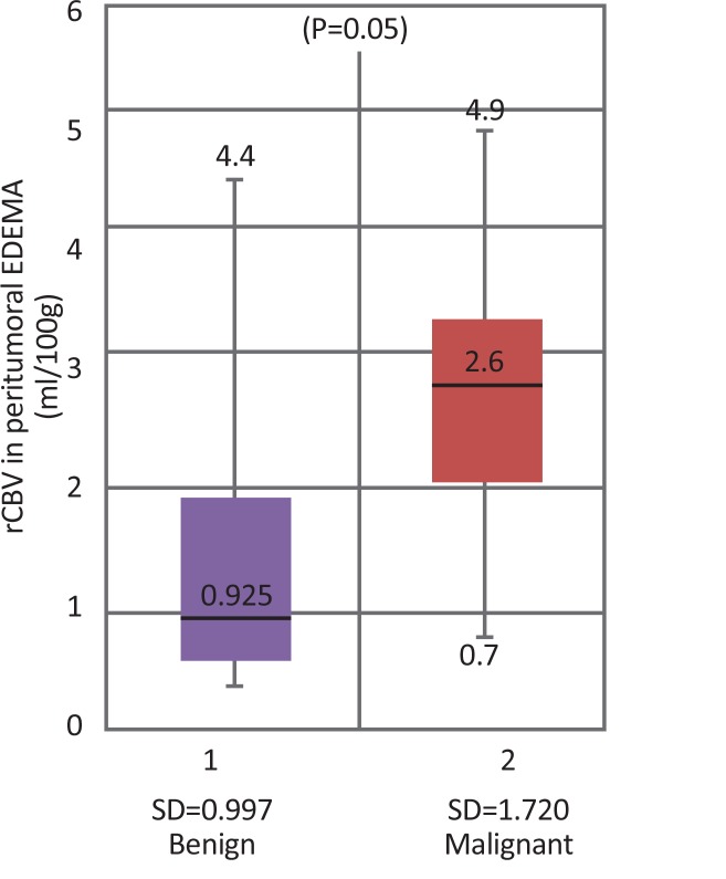
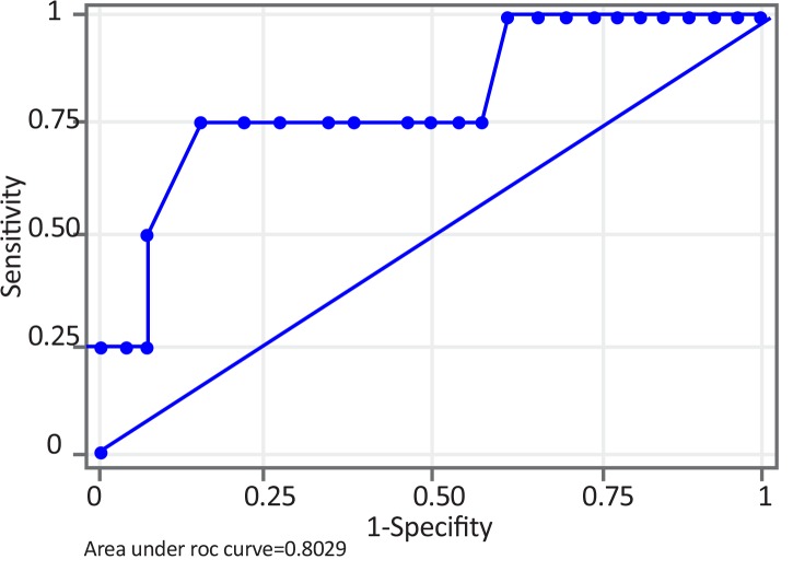
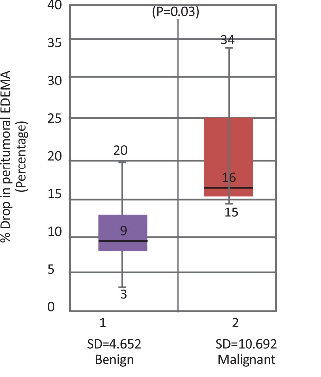
Both maximum rCBV and minimum ADC within the tumor were not significant for differentiating benign from malignant meningiomas. A cut-off maximum rCBV of 2.5 mL/100 g in peritumoral edema was 75% sensitive, 84.6% specific, and 83.3% accurate in differentiating benign from malignant meningiomas.

CONCLUSION

Benign and malignant meningiomas can be differentiated based on maximum rCBV in peritumoral edema but ADC values within the tumor are insignificant in differentiating benign and malignant tumors. rCBV values within tumor, however, may be helpful in subtyping meningiomas, especially transitional and meningothelial meningiomas.

摘要

引言

本研究旨在根据相对脑血容量(rCBV)和表观扩散系数(ADC)对脑膜瘤进行分级,以帮助外科医生规划手术入路和范围,并决定是否需要任何辅助放疗/化疗。还讨论了当前和不断发展的基因组学、蛋白质组学和光谱技术,这些技术可以补充当前在脑膜瘤分级中的放射学方法和程序。

方法

总共35例脑膜瘤患者前瞻性地接受了轴位、矢状位和冠状位的基本MR序列(T1W、T2W、T2W/FLAIR),随后进行b值为1000的扩散加权(DW)成像(分析时使用最小ADC值)。然后,给予钆贝葡胺/钆喷酸葡胺(0.1 mmol/kg,速率为4 mL/s),随后进行生理盐水冲洗(20 mL,速率为4 mL/s)。接下来,采集T*W/FFE动态图像;使用强度下降最大的动态图像创建rCBV和相对脑血流量(rCBF)图并计算rCBV。

结果

肿瘤内的最大rCBV和最小ADC对于区分良性和恶性脑膜瘤均无显著意义。瘤周水肿中最大rCBV的截断值为2.5 mL/100 g,在区分良性和恶性脑膜瘤时,敏感性为75%,特异性为84.6%,准确性为83.3%。

结论

良性和恶性脑膜瘤可根据瘤周水肿中的最大rCBV进行区分,但肿瘤内的ADC值在区分良性和恶性肿瘤方面无显著意义。然而,肿瘤内的rCBV值可能有助于脑膜瘤的亚型分类,尤其是过渡型和脑膜内皮型脑膜瘤。

https://cdn.ncbi.nlm.nih.gov/pmc/blobs/d240/6359681/3640a307a252/BCN-9-417-g006.jpg
https://cdn.ncbi.nlm.nih.gov/pmc/blobs/d240/6359681/ec6b2b801c98/BCN-9-417-g001a.jpg
https://cdn.ncbi.nlm.nih.gov/pmc/blobs/d240/6359681/dc761d79e61b/BCN-9-417-g002.jpg
https://cdn.ncbi.nlm.nih.gov/pmc/blobs/d240/6359681/e2399ee560f6/BCN-9-417-g003.jpg
https://cdn.ncbi.nlm.nih.gov/pmc/blobs/d240/6359681/f7fe459b8834/BCN-9-417-g004.jpg
https://cdn.ncbi.nlm.nih.gov/pmc/blobs/d240/6359681/4469501b3dff/BCN-9-417-g005.jpg
https://cdn.ncbi.nlm.nih.gov/pmc/blobs/d240/6359681/3640a307a252/BCN-9-417-g006.jpg

相似文献

1
rCBV- and ADC-based Grading of Meningiomas With Glimpse Into Emerging Molecular Diagnostics.基于相对脑血容量(rCBV)和表观扩散系数(ADC)的脑膜瘤分级及对新兴分子诊断的展望
Basic Clin Neurosci. 2018 Nov-Dec;9(6):417-428. doi: 10.32598/bcn.9.6.417. Epub 2018 Nov 1.
2
Differentiation between benign and malignant meningiomas using diffusion and perfusion MR imaging.利用扩散加权磁共振成像和灌注加权磁共振成像鉴别良性与恶性脑膜瘤
Georgian Med News. 2012 May(206):16-22.
3
The role of diffusion tensor imaging and dynamic susceptibility perfusion MRI in the evaluation of meningioma grade and subtype.扩散张量成像和动态磁敏感灌注磁共振成像在评估脑膜瘤分级和亚型中的作用。
Clin Neurol Neurosurg. 2016 Jul;146:109-15. doi: 10.1016/j.clineuro.2016.05.005. Epub 2016 May 6.
4
Correlation of diffusion and perfusion MRI with Ki-67 in high-grade meningiomas.高级别脑膜瘤弥散和灌注 MRI 与 Ki-67 的相关性。
AJR Am J Roentgenol. 2010 Dec;195(6):1391-5. doi: 10.2214/AJR.10.4531.
5
Diagnostic value of apparent diffusion coefficient for the accurate assessment and differentiation of intracranial meningiomas.表观扩散系数对颅内脑膜瘤准确评估及鉴别诊断的价值
Acta Radiol Short Rep. 2013 Nov 23;2(7):2047981613512484. doi: 10.1177/2047981613512484. eCollection 2013.
6
Role of MR Morphology and Diffusion-Weighted Imaging in the Evaluation of Meningiomas: Radio-Pathologic Correlation.磁共振成像形态学及扩散加权成像在脑膜瘤评估中的作用:放射病理相关性研究
JNMA J Nepal Med Assoc. 2019 Jan-Feb;57(215):37-44. doi: 10.31729/jnma.3968.
7
Perfusion MR imaging for differentiation of benign and malignant meningiomas.灌注磁共振成像用于鉴别良性和恶性脑膜瘤。
Neuroradiology. 2008 Jun;50(6):525-30. doi: 10.1007/s00234-008-0373-y. Epub 2008 Apr 1.
8
Diffusion-weighted MR imaging: diagnosing atypical or malignant meningiomas and detecting tumor dedifferentiation.扩散加权磁共振成像:诊断非典型或恶性脑膜瘤及检测肿瘤去分化
AJNR Am J Neuroradiol. 2008 Jun;29(6):1147-52. doi: 10.3174/ajnr.A0996. Epub 2008 Mar 20.
9
[Evaluation of tumor vessel of cerebral meningiomas with MR perfusion weighted imaging].[磁共振灌注加权成像对脑脑膜瘤肿瘤血管的评估]
Zhonghua Yi Xue Za Zhi. 2015 May 5;95(17):1335-7.
10
Quantitative apparent diffusion coefficients in the characterization of brain tumors and associated peritumoral edema.定量表观扩散系数在脑肿瘤及相关瘤周水肿特征描述中的应用
Acta Radiol. 2009 Jul;50(6):682-9. doi: 10.1080/02841850902933123.

引用本文的文献

1
Does arterial spin labelling represent a valid alternative to other MR perfusion techniques in neuro-oncology?在神经肿瘤学中,动脉自旋标记是否是其他磁共振灌注技术的有效替代方法?
Radiol Med. 2025 Jul 15. doi: 10.1007/s11547-025-02048-1.
2
Imaging meningioma biology: Machine learning predicts integrated risk score in WHO grade 2/3 meningioma.脑膜瘤生物学成像:机器学习预测世界卫生组织2/3级脑膜瘤的综合风险评分
Neurooncol Adv. 2024 May 30;6(1):vdae080. doi: 10.1093/noajnl/vdae080. eCollection 2024 Jan-Dec.
3
Evaluation of Magnetic Resonance Imaging for Microsurgical Efficacy and Relapse of Rolandic Meningioma.

本文引用的文献

1
Expression of MMP-9 and VEGF in meningiomas and their correlation with peritumoral brain edema.基质金属蛋白酶-9和血管内皮生长因子在脑膜瘤中的表达及其与瘤周脑水肿的相关性
Biomed Res Int. 2015;2015:646853. doi: 10.1155/2015/646853. Epub 2015 Mar 3.
2
Quantitative proteomic analysis of meningiomas for the identification of surrogate protein markers.脑膜瘤的定量蛋白质组学分析以鉴定替代蛋白标志物
Sci Rep. 2014 Nov 21;4:7140. doi: 10.1038/srep07140.
3
Preoperative dynamic contrast-enhanced MRI correlates with molecular markers of hypoxia and vascularity in specific areas of intratumoral microenvironment and is predictive of patient outcome.
磁共振成像评估中央区脑膜瘤的显微手术疗效及复发。
Comput Intell Neurosci. 2022 Jun 6;2022:1026494. doi: 10.1155/2022/1026494. eCollection 2022.
4
Multi-Omics Analysis in Initiation and Progression of Meningiomas: From Pathogenesis to Diagnosis.脑膜瘤发生与进展的多组学分析:从发病机制到诊断
Front Oncol. 2020 Aug 28;10:1491. doi: 10.3389/fonc.2020.01491. eCollection 2020.
术前动态对比增强 MRI 与肿瘤内微环境特定区域缺氧和血管生成的分子标志物相关,并可预测患者的预后。
Neuro Oncol. 2014 Jan;16(2):280-91. doi: 10.1093/neuonc/not148. Epub 2013 Dec 4.
4
Assessment of angiographic vascularity of meningiomas with dynamic susceptibility contrast-enhanced perfusion-weighted imaging and diffusion tensor imaging.采用动态磁敏感对比增强灌注加权成像和扩散张量成像评估脑膜瘤的血管造影血管情况。
AJNR Am J Neuroradiol. 2014 Feb;35(2):263-9. doi: 10.3174/ajnr.A3651. Epub 2013 Jul 25.
5
Ambient mass spectrometry for the intraoperative molecular diagnosis of human brain tumors.用于人脑肿瘤术中分子诊断的环境质谱法。
Proc Natl Acad Sci U S A. 2013 Jan 29;110(5):1611-6. doi: 10.1073/pnas.1215687110. Epub 2013 Jan 8.
6
Correlating apparent diffusion coefficients with histopathologic findings on meningiomas.探讨脑膜瘤表观弥散系数与组织病理学表现的相关性。
Eur J Radiol. 2012 Dec;81(12):4050-6. doi: 10.1016/j.ejrad.2012.06.002. Epub 2012 Jun 22.
7
A brain tumor molecular imaging strategy using a new triple-modality MRI-photoacoustic-Raman nanoparticle.一种使用新型三模态 MRI-光声-Raman 纳米粒子的脑肿瘤分子成像策略。
Nat Med. 2012 Apr 15;18(5):829-34. doi: 10.1038/nm.2721.
8
Predicting outcomes of patients with intracranial meningiomas using molecular markers of hypoxia, vascularity, and proliferation.使用缺氧、血管生成和增殖的分子标志物预测颅内脑膜瘤患者的预后。
Neurosurgery. 2012 Jul;71(1):146-56. doi: 10.1227/NEU.0b013e3182567886.
9
Role of VEGF and matrix metalloproteinase-9 in peritumoral brain edema associated with supratentorial benign meningiomas.血管内皮生长因子和基质金属蛋白酶-9 在幕上良性脑膜瘤瘤周脑水肿中的作用。
Neuropathology. 2012 Dec;32(6):638-46. doi: 10.1111/j.1440-1789.2012.01312.x. Epub 2012 Mar 15.
10
Magnetic resonance imaging in assessment of treatment response of gamma knife for brain tumors.磁共振成像在评估伽玛刀治疗脑肿瘤的疗效中的应用。
Chin Med J (Engl). 2011 Jun;124(12):1906-10.