Gerace Elisabetta, Landucci Elisa, Bani Daniele, Moroni Flavio, Mannaioni Guido, Pellegrini-Giampietro Domenico E
Section of Clinical Pharmacology and Oncology, Department of Health Sciences, University of Florence, Florence, Italy.
Section of Pharmacology and Toxicology, Department of Neuroscience, Psychology, Drug Research and Child Health (NeuroFarBa), University of Florence, Florence, Italy.
Front Neurosci. 2019 Jan 24;12:1053. doi: 10.3389/fnins.2018.01053. eCollection 2018.
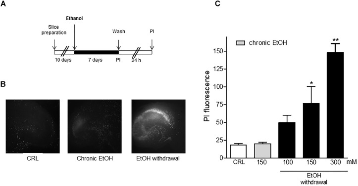
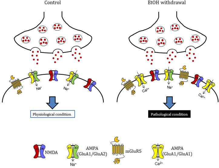
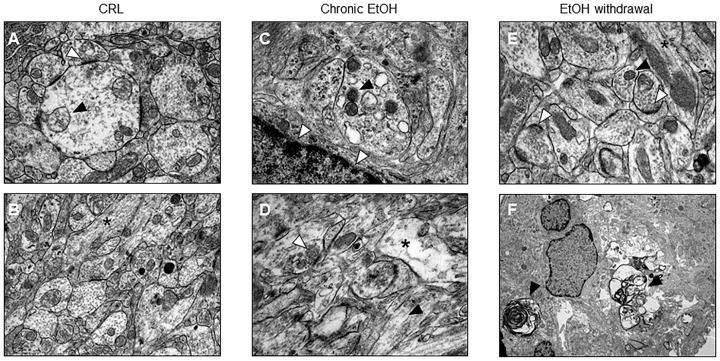
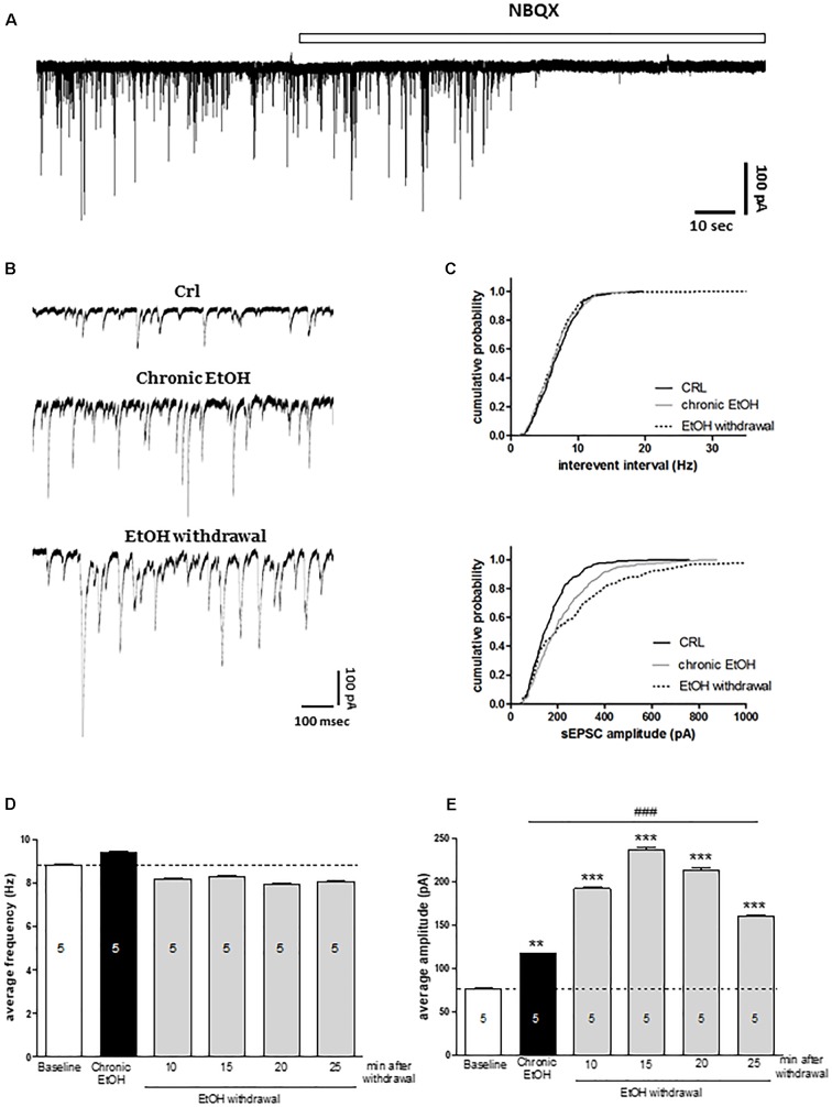
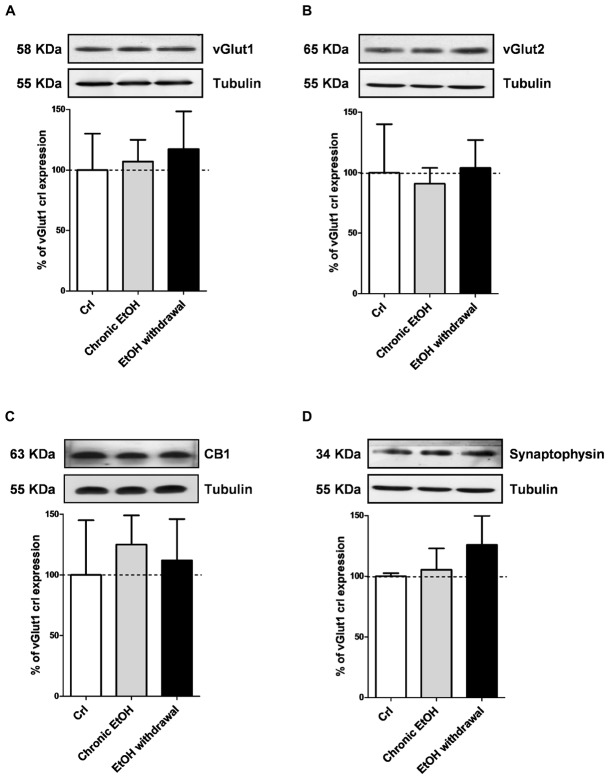
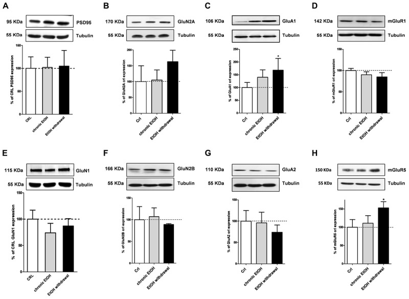
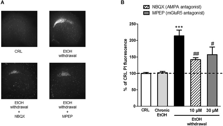
Long-term alcohol use can lead to alterations in brain structure and functions and, in some cases, to neurodegeneration. Several mechanisms have been proposed to explain ethanol (EtOH)-related brain injury. One of the most relevant mechanisms of alcohol-induced neurodegeneration involves glutamatergic transmission, but their exact role is not yet fully understood. We investigated the neurochemical mechanisms underlying the toxicity induced by EtOH dependence and/or withdrawal by exposing rat organotypic hippocampal slices to EtOH (100-300 mM) for 7 days and then incubating the slices in EtOH-free medium for the subsequent 24 h. EtOH withdrawal led to a dose-dependent CA1 pyramidal cell injury, as detected with propidium iodide fluorescence. Electron microscopy of hippocampal slices revealed that not only EtOH withdrawal but also 7 days chronic EtOH exposure elicited signs of apoptotic cell death in CA1 pyramidal cells. These data were supported by electrophysiological recordings of spontaneus Excitatory Post Synaptic Currents (sEPSCs) from CA1 pyramidal cells. The average amplitude of sEPSCs in slices treated with EtOH for 7 days was significantly increased, and even more so during the first 30 min of EtOH withdrawal, suggesting that the initial phase of the neurodegenerative process could be due to an excitotoxic mechanism. We then analyzed the expression levels of presynaptic (vGlut1, vGlut2, CB1 receptor, synaptophysin) and postsynaptic (PSD95, GluN1, GluN2A, GluN2B, GluA1, GluA2, mGluR1 and mGluR5) proteins after 7 days EtOH incubation or after EtOH withdrawal. We found that only GluA1 and mGluR5 expression levels were significantly increased after EtOH withdrawal and, in neuroprotection experiments, we observed that AMPA and mGluR5 antagonists attenuated EtOH withdrawal-induced toxicity. These data suggest that chronic EtOH treatment promotes abnormal synaptic transmission that may lead to CA1 pyramidal cell death after EtOH withdrawal through glutamate receptors and increased excitotoxicity.
长期饮酒会导致大脑结构和功能的改变,在某些情况下还会导致神经退行性变。已经提出了几种机制来解释乙醇(EtOH)相关的脑损伤。酒精诱导神经退行性变最相关的机制之一涉及谷氨酸能传递,但其确切作用尚未完全明确。我们通过将大鼠海马脑片在EtOH(100 - 300 mM)中暴露7天,然后在无EtOH的培养基中孵育接下来的24小时,研究了EtOH依赖和/或戒断所诱导毒性的神经化学机制。用碘化丙啶荧光检测发现,EtOH戒断导致剂量依赖性的CA1锥体细胞损伤。海马脑片的电子显微镜检查显示,不仅EtOH戒断,而且7天的慢性EtOH暴露都引发了CA1锥体细胞凋亡性细胞死亡的迹象。来自CA1锥体细胞的自发性兴奋性突触后电流(sEPSCs)的电生理记录支持了这些数据。用EtOH处理7天的脑片中sEPSCs的平均幅度显著增加,在EtOH戒断的最初30分钟内更是如此,这表明神经退行性变过程的初始阶段可能是由于兴奋性毒性机制。然后我们分析了在EtOH孵育7天或EtOH戒断后突触前(vGlut1、vGlut2、CB1受体、突触素)和突触后(PSD95、GluN1、GluN2A、GluN2B、GluA1、GluA2、mGluR1和mGluR5)蛋白的表达水平。我们发现,EtOH戒断后只有GluA1和mGluR5的表达水平显著增加,并且在神经保护实验中,我们观察到AMPA和mGluR5拮抗剂减弱了EtOH戒断诱导的毒性。这些数据表明,慢性EtOH治疗促进异常的突触传递,这可能在EtOH戒断后通过谷氨酸受体和增加的兴奋性毒性导致CA1锥体细胞死亡。