• 文献检索
  • 文档翻译
  • 深度研究
  • 学术资讯
  • Suppr Zotero 插件Zotero 插件
  • 邀请有礼
  • 套餐&价格
  • 历史记录
应用&插件
Suppr Zotero 插件Zotero 插件浏览器插件Mac 客户端Windows 客户端微信小程序
定价
高级版会员购买积分包购买API积分包
服务
文献检索文档翻译深度研究API 文档MCP 服务
关于我们
关于 Suppr公司介绍联系我们用户协议隐私条款
关注我们

Suppr 超能文献

核心技术专利:CN118964589B侵权必究
粤ICP备2023148730 号-1Suppr @ 2026

文献检索

告别复杂PubMed语法,用中文像聊天一样搜索,搜遍4000万医学文献。AI智能推荐,让科研检索更轻松。

立即免费搜索

文件翻译

保留排版,准确专业,支持PDF/Word/PPT等文件格式,支持 12+语言互译。

免费翻译文档

深度研究

AI帮你快速写综述,25分钟生成高质量综述,智能提取关键信息,辅助科研写作。

立即免费体验

扩展光片显微镜解析鸣禽大脑大部分区域的亚细胞结构。

Expansion Light Sheet Microscopy Resolves Subcellular Structures in Large Portions of the Songbird Brain.

作者信息

Düring Daniel Normen, Rocha Mariana Diales, Dittrich Falk, Gahr Manfred, Hahnloser Richard Hans Robert

机构信息

Institute of Neuroinformatics, University of Zürich/ETH Zürich, Zurich, Switzerland.

Neuroscience Center Zurich (ZNZ), Zurich, Switzerland.

出版信息

Front Neuroanat. 2019 Jan 31;13:2. doi: 10.3389/fnana.2019.00002. eCollection 2019.

DOI:10.3389/fnana.2019.00002
PMID:30766480
原文链接:https://pmc.ncbi.nlm.nih.gov/articles/PMC6365838/
Abstract

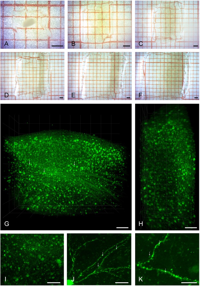
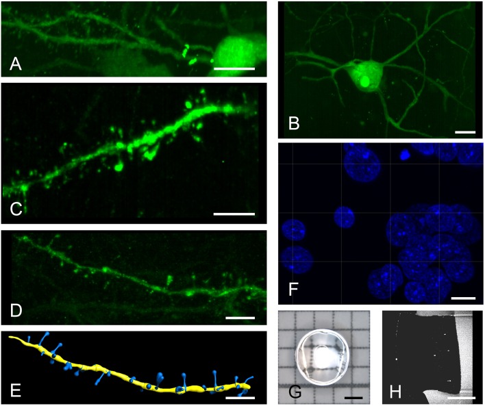
Expansion microscopy and light sheet imaging (ExLSM) provide a viable alternative to existing tissue clearing and large volume imaging approaches. The analysis of intact volumes of brain tissue presents a distinct challenge in neuroscience. Recent advances in tissue clearing and light sheet microscopy have re-addressed this challenge and blossomed into a plethora of protocols with diverse advantages and disadvantages. While refractive index matching achieves near perfect transparency and allows for imaging at large depths, the resolution of cleared brains is usually limited to the micrometer range. Moreover, the often long and harsh tissue clearing protocols hinder preservation of native fluorescence and antigenicity. Here we image large expanded brain volumes of zebra finch brain tissue in commercially available light sheet microscopes. Our expansion light sheet microscopy (ExLSM) approach presents a viable alternative to many clearing and imaging methods because it improves on tissue processing times, fluorophore compatibility, and image resolution.

摘要

扩展显微镜和光片成像(ExLSM)为现有的组织透明化和大体积成像方法提供了一种可行的替代方案。在神经科学中,对完整的脑组织体积进行分析是一项独特的挑战。组织透明化和光片显微镜技术的最新进展重新应对了这一挑战,并发展出了大量各有优缺点的方案。虽然折射率匹配可实现近乎完美的透明度并允许在大深度进行成像,但透明化后的大脑分辨率通常限制在微米范围内。此外,通常冗长且苛刻的组织透明化方案会阻碍天然荧光和抗原性的保留。在这里,我们在市售的光片显微镜中对斑胸草雀脑组织的大体积扩展脑区进行成像。我们的扩展光片显微镜(ExLSM)方法是许多透明化和成像方法的可行替代方案,因为它在组织处理时间、荧光团兼容性和图像分辨率方面都有改进。

https://cdn.ncbi.nlm.nih.gov/pmc/blobs/4081/6365838/455795b541ba/fnana-13-00002-g0002.jpg
https://cdn.ncbi.nlm.nih.gov/pmc/blobs/4081/6365838/172c08ad1f97/fnana-13-00002-g0001.jpg
https://cdn.ncbi.nlm.nih.gov/pmc/blobs/4081/6365838/455795b541ba/fnana-13-00002-g0002.jpg
https://cdn.ncbi.nlm.nih.gov/pmc/blobs/4081/6365838/172c08ad1f97/fnana-13-00002-g0001.jpg
https://cdn.ncbi.nlm.nih.gov/pmc/blobs/4081/6365838/455795b541ba/fnana-13-00002-g0002.jpg

相似文献

1
Expansion Light Sheet Microscopy Resolves Subcellular Structures in Large Portions of the Songbird Brain.扩展光片显微镜解析鸣禽大脑大部分区域的亚细胞结构。
Front Neuroanat. 2019 Jan 31;13:2. doi: 10.3389/fnana.2019.00002. eCollection 2019.
2
Tissue Clearing and Light Sheet Microscopy: Imaging the Unsectioned Adult Zebra Finch Brain at Cellular Resolution.组织透明化与光片显微镜技术:以细胞分辨率对未切片的成年斑胸草雀大脑进行成像
Front Neuroanat. 2019 Feb 14;13:13. doi: 10.3389/fnana.2019.00013. eCollection 2019.
3
Comparison of Transparency and Shrinkage During Clearing of Insect Brains Using Media With Tunable Refractive Index.使用具有可调折射率的介质对昆虫大脑进行透明化处理期间的透明度和收缩率比较
Front Neuroanat. 2020 Nov 20;14:599282. doi: 10.3389/fnana.2020.599282. eCollection 2020.
4
Whole-Brain Single-Cell Imaging and Analysis of Intact Neonatal Mouse Brains Using MRI, Tissue Clearing, and Light-Sheet Microscopy.利用 MRI、组织透明化和光片显微镜对完整新生鼠脑进行全脑单细胞成像和分析。
J Vis Exp. 2022 Aug 1(186). doi: 10.3791/64096.
5
Single-Step Fast Tissue Clearing of Thick Mouse Brain Tissue for Multi-Dimensional High-Resolution Imaging.一步法快速透明化厚鼠脑组织以进行多维高分辨率成像。
Int J Mol Sci. 2022 Jun 19;23(12):6826. doi: 10.3390/ijms23126826.
6
Comparison of Light-Sheet Fluorescence Microscopy and Fast-Confocal Microscopy for Three-Dimensional Imaging of Cleared Mouse Brain.光片荧光显微镜与快速共聚焦显微镜用于透明小鼠脑三维成像的比较
Methods Protoc. 2023 Nov 10;6(6):108. doi: 10.3390/mps6060108.
7
Tissue Clearing and Expansion Methods for Imaging Brain Pathology in Neurodegeneration: From Circuits to Synapses and Beyond.用于神经退行性疾病脑病理学成像的组织透明化和扩展方法:从神经回路到突触及其他。
Front Neurosci. 2020 Oct 5;14:914. doi: 10.3389/fnins.2020.00914. eCollection 2020.
8
[Getting an insight into the brain - new optical clearing techniques and imaging using light-sheet microscope].洞察大脑——新型光学透明技术及光片显微镜成像
Postepy Biochem. 2017;63(1):8-15.
9
Imaging the mammary gland and mammary tumours in 3D: optical tissue clearing and immunofluorescence methods.三维成像乳腺及乳腺肿瘤:光学组织透明化和免疫荧光方法
Breast Cancer Res. 2016 Dec 13;18(1):127. doi: 10.1186/s13058-016-0754-9.
10
3D Imaging for Cleared Tissues and Thicker Samples on Confocal and Light-Sheet Microscopes.共聚焦显微镜和光片显微镜对透明组织和较厚样本的三维成像
Methods Mol Biol. 2023;2593:143-161. doi: 10.1007/978-1-0716-2811-9_9.

引用本文的文献

1
Applications of Lightsheet Fluorescence Microscopy by High Numerical Aperture Detection Lens.应用高数值孔径检测透镜的光片荧光显微镜。
J Phys Chem B. 2024 Sep 5;128(35):8273-8289. doi: 10.1021/acs.jpcb.4c01721. Epub 2024 Aug 23.
2
Expanding boundaries - a cell biologist's guide to expansion microscopy.拓展边界——细胞生物学家的扩展显微镜指南。
J Cell Sci. 2024 Apr 1;137(7). doi: 10.1242/jcs.260765. Epub 2024 Apr 17.
3
Synergizing Algorithmic Design, Photoclick Chemistry and Multi-Material Volumetric Printing for Accelerating Complex Shape Engineering.

本文引用的文献

1
Tissue Clearing and Light Sheet Microscopy: Imaging the Unsectioned Adult Zebra Finch Brain at Cellular Resolution.组织透明化与光片显微镜技术:以细胞分辨率对未切片的成年斑胸草雀大脑进行成像
Front Neuroanat. 2019 Feb 14;13:13. doi: 10.3389/fnana.2019.00013. eCollection 2019.
2
Excitatory and inhibitory synapse reorganization immediately after critical sensory experience in a vocal learner.在发声学习者关键感官体验后立即进行兴奋性和抑制性突触重组。
Elife. 2018 Oct 25;7:e37571. doi: 10.7554/eLife.37571.
3
Expansion microscopy: enabling single cell analysis in intact biological systems.
协同算法设计、光点击化学和多材料体积打印技术加速复杂形状工程。
Adv Sci (Weinh). 2023 Sep;10(26):e2300912. doi: 10.1002/advs.202300912. Epub 2023 Jul 3.
4
A Model of Discovery: The Role of Imaging Established and Emerging Non-mammalian Models in Neuroscience.一种发现模式:成像技术在神经科学中已确立和新兴的非哺乳动物模型中的作用。
Front Mol Neurosci. 2022 Apr 14;15:867010. doi: 10.3389/fnmol.2022.867010. eCollection 2022.
5
ExSTED microscopy reveals contrasting functions of dopamine and somatostatin CSF-c neurons along the lamprey central canal.ExSTED 显微镜揭示了沿七鳃鳗中央管多巴胺和生长抑素 CSF-c 神经元的对比功能。
Elife. 2022 Feb 1;11:e73114. doi: 10.7554/eLife.73114.
6
Comparison of Different Tissue Clearing Methods for Three-Dimensional Reconstruction of Human Brain Cellular Anatomy Using Advanced Imaging Techniques.使用先进成像技术对人类脑细胞解剖结构进行三维重建的不同组织透明化方法比较
Front Neuroanat. 2021 Nov 11;15:752234. doi: 10.3389/fnana.2021.752234. eCollection 2021.
7
Expansion tomography for large volume tissue imaging with nanoscale resolution.用于大体积组织成像且具有纳米级分辨率的扩展断层扫描技术。
Biomed Opt Express. 2021 Aug 13;12(9):5614-5628. doi: 10.1364/BOE.431696. eCollection 2021 Sep 1.
8
Protein-retention expansion microscopy for visualizing subcellular organelles in fixed brain tissue.蛋白保留膨胀显微镜用于固定脑组织中观察亚细胞细胞器。
J Neurosci Methods. 2021 Sep 1;361:109285. doi: 10.1016/j.jneumeth.2021.109285. Epub 2021 Jul 7.
9
Application of SNAP-Tag in Expansion Super-Resolution Microscopy Using DNA Oligostrands.SNAP标签在使用DNA寡链的扩展超分辨率显微镜中的应用。
Front Chem. 2021 Apr 30;9:640519. doi: 10.3389/fchem.2021.640519. eCollection 2021.
10
Fast Retrograde Access to Projection Neuron Circuits Underlying Vocal Learning in Songbirds.快速逆行访问鸣禽发声学习中投射神经元回路。
Cell Rep. 2020 Nov 10;33(6):108364. doi: 10.1016/j.celrep.2020.108364.
扩展显微镜:在完整的生物系统中实现单细胞分析。
FEBS J. 2019 Apr;286(8):1482-1494. doi: 10.1111/febs.14597. Epub 2018 Jul 9.
4
A three-dimensional single-cell-resolution whole-brain atlas using CUBIC-X expansion microscopy and tissue clearing.采用 CUBIC-X 扩展显微镜和组织透明化技术的三维单细胞分辨率全脑图谱
Nat Neurosci. 2018 Apr;21(4):625-637. doi: 10.1038/s41593-018-0109-1. Epub 2018 Mar 5.
5
Expansion microscopy: development and neuroscience applications.扩展显微镜:发展与神经科学应用。
Curr Opin Neurobiol. 2018 Jun;50:56-63. doi: 10.1016/j.conb.2017.12.012. Epub 2018 Jan 6.
6
SnapShot: Tissue Clearing.快照:组织透明化。
Cell. 2017 Oct 5;171(2):496-496.e1. doi: 10.1016/j.cell.2017.09.025.
7
Superresolution expansion microscopy reveals the three-dimensional organization of the synaptonemal complex.超分辨率扩展显微镜揭示了联会复合体的三维结构。
Proc Natl Acad Sci U S A. 2017 Aug 15;114(33):E6857-E6866. doi: 10.1073/pnas.1705623114. Epub 2017 Jul 31.
8
Nanoscale imaging of clinical specimens using pathology-optimized expansion microscopy.使用病理学优化的扩张显微镜对临床标本进行纳米级成像。
Nat Biotechnol. 2017 Aug;35(8):757-764. doi: 10.1038/nbt.3892. Epub 2017 Jul 17.
9
EM connectomics reveals axonal target variation in a sequence-generating network.电子显微镜连接组学揭示了序列生成网络中轴突靶点的变异。
Elife. 2017 Mar 27;6:e24364. doi: 10.7554/eLife.24364.
10
Shrinkage-mediated imaging of entire organs and organisms using uDISCO.uDISCO 介导的整个器官和生物体的缩时成像。
Nat Methods. 2016 Oct;13(10):859-67. doi: 10.1038/nmeth.3964. Epub 2016 Aug 22.